Halloweenhas morphed into a holiday where people see how much it takes to scare them. Horror movies, haunted houses, dangerous pranks; people like to be scared.
What scares you the most– spiders, public speaking, death? These three are high on every list of common fears, but it wasn’t so long ago that another fear was in first place – taphophobia. Never heard of it? I bet that its mere definition will be enough to send a chill up your spine.
Manycultures built time delays into their death rites to make sure someone was truly dead. Greeks washed the dead….. and some would wake up. In more difficult cases, they would cut off fingers or dunk the bodies in warm baths. The custom of the Irish wake began with the Celts watching the body for signs of life. But mistakes were made, often in times of epidemic.
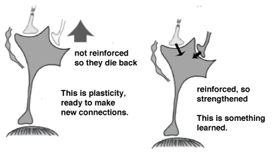
Thepicture at the side should help with this explanation, but I won’t give you all the gory details. Your muscle cells have systems that look like ratchets, using to proteins called myosin and actin which pull past one another to shorten (contract) the muscle fiber. The myosin is bound by ATP, which then hydrolyses to form ADP + P. When ADP + P is bound to myosin, it can reach out and bind to the actin.
In his book, Premature burial, and how it may be prevented, with special reference to trance catalepsy, and other forms of suspended animation, Tebb professed that he had found 219 cases of near premature burial and 149 live burials. He had some stunning stories of scratches on the lids of coffins and noises from newly filled graves.
Coma– In medicine, a coma is unconsciousness that lasts more than six hours and from which a person cannot be roused and will not respond to stimuli. Injury or inflammation of the cerebral cortex and/ or the reticular activating system in the brain stem can lead to coma. The things that can injure these structures are myriad, from traumatic injury, to drug overdose, to stroke or hyperthermia, etc.
To be successful, those folks above ground must have been very alert. A coffin has only about 20-40 minutes of air, so a person could go from dead to live to dead without the change being noted. To counteract this small window of time, Germany also built waiting mortuaries, where dead bodies could be held for longer periods of time. It came to be accepted that the only reliable sign death was putrefaction --- waiting mortuaries did not smell like flowers or fresh baked bread.
 |
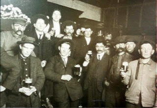
Miracle Max had his own methods for determining if someone was all dead or just mostly dead. They involved a bellows and Carol Kane’s voice. But the point is made, for centuries, people were just guessing if others werereally dead. There were few experts, and they were probably just comedians in make-up. |
Technically, taphophobia means “fear of graves” (taphos = tomb, and phobia = fear of), but its common use is “fear of being buried alive.” Premature burial is not an urban legend, incidents have been documented in nearly every society – and not all of them were just in the movies or books.
In the 1800’s and earlier, being dead was a lot like being a duck….. you know, if it looks like a duck, walks like a duck, and quacks like a duck….. The appearance of death was often enough to make a diagnosis and start going through their pockets.
As a good example of the wisdom of the age, George Washington had these last words, "Have me decently buried, but do not let my body be put into a vault in less than three days after I am dead…….., tis well." He wanted a sufficient amount of time to pass to ensure that he was in fact dead.
 |
The Irish wake probably originated in the leaving of the tomb unsealed for several days, just in case the dead person might wake. Later, stories came about concerning the lead in pewter tankards from which the Irish would drink. Lead poisoning could induce a state that resembled death. Sometimes, a wake is just another reason to raise a glass of ale. |
The hopes of preventing the spread of infection often lead to burying the dead before they were quite dead. I give you plague victim Eric Idle in Monty Python’s Search for the Holy Grail – “But I’m not dead yet…. I’m feeling much better.”
Even without epidemic, most people in the 18th, 19th, and early 20th centuries died at home, having never seen a doctor. If someone couldn’t hear a heartbeat or feel a pulse, then the patient was dead. But these were lay people, did they know how to feel for a pulse? Maybe they relied on another indicator of death - rigor mortis (rigor = stiffness, and mort = death).
In humans, rigor mortis begins 2-6 hours after death. Rigor is caused by a loss of ATP production. Follow me here--- no breathing, no oxygen; no oxygen, no ATP production. With no ATP, the muscle can’t relax. This may seem strange, since it takes ATP to contract a muscle in the first place.
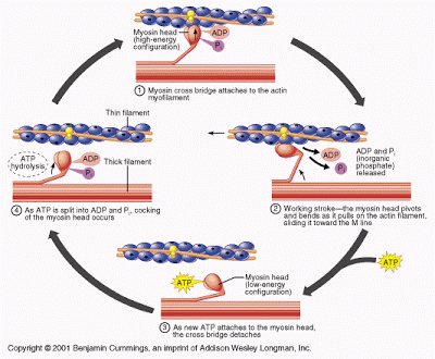 |
As described in the text, the thick filament (myosin) pulls itself along the thin filament (actin) by grabbing and releasing actin monomers. A single sarcomere (contractive subunit, ~100,000 in a muscle cell) has millions of myosin heads. They grab actin fibers that run on all sides of the myosin fiber. |
The ADP + P is released from the myosin and it flexes the head of the protein, which pulls it along the actin. When a new ATP is bound, the myosin lets go from the actin, and the cycle is repeated. Each muscle fiber in each cell has millions of myosin heads resulting in a contracted muscle.
In rigor, there is no more ATP, so the myosin doesn’t let go of the actin, therefore, no relaxation can take place. The muscles remain the length they were at death. After about 72 hours, the muscle proteins start to break down, rigor will lessens and the body will become limp again. But as we will see below, some conditions can mimic the signs of rigor, increasing the chances of premature burial.
In an effort to see how bad the situation was, the English reformer, William Tebb, in 1905 made a study of accidental premature burial. Tebb was quite the joiner; the weirder the society, the more he wanted to join or lead it. He worked with the Vegetarian Society, the anti-vivisection movement, the national Canine Defense League, and formed National Anti-Vaccination League in 1896.
 |
William Tebb’s book on premature burial was a best seller. You’d think he had a product to sell given the way he described some of the incidents. In one, Madame Blunden was buried in a crypt under a boys school. The next day, the students heard noises from below. They opened the tomb and coffin just in time to see her die from lack of oxygen. |
In her 1996 book, The Corpse: A History, Christine Quigley documents many instances of premature burial and near-premature burial (I LOVE the title). Skeletons were outside their coffins, sitting up in the corner of their vault after being opened years later. Others were found turned over in their caskets, with tufts of their own hair in their hands.
How might this happen? What conditions might make it look so much like you were dead that even your loved ones would let them plant you in the ground? The list is long and varied, but here are some of the more common things that can make you look dead:
Asphyxiation– anything that cuts off your supply of air can make you look dead once you fall unconscious – continuation of this condition leads to actual death. You look dead enough and won’t respond to external stimuli, so people assume you are dead. Close the coffin lid, and soon you really will be dead of asphyxia.
Catalepsy– Many things can bring on this catatonic state in which the muscles are rigid (like rigor mortis after death) and no pain is enough make you respond, one example is epilepsy. Hypnotists call their trances catalepsy (Greek for to grab and take down), but true catalepsy is much more severe and can last hours to days. Severe emotional trauma can also bring it on, so you can certainly be scared enough to look like you are dead.
 |
Catalepsy is denoted by muscle rigidity, so it can look like rigor mortis. But there is also waxy flexibility in some cases. The dead-looking not dead people can be posed, and they will hold the pose indefinitely. What little girl wouldn’t love a cataleptic doll for Christmas! |
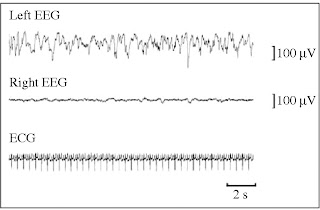
To show how medicine has changed, there is now a battery of assessments called the Glasgow coma scale (GCS) that are carried out on coma victims to assess their state and prognosis. In centuries past, you might look at them, hold a mirror under their nose, maybe lift and drop an arm….. bury them.
The GCS has traditionally been used in the hospital environment, but new evidenceshows that a prehospital GCS (assessment at scene or in route) can be just as accurate and may benefit treatment choice in pediatric traumatic brain injury patients. The study compared prehospital and emergency department GCS scores and showed that they were similar. They also compared outcomes with prehospital scores and showed a positive correlation. If assessment and treatment can be begun earlier, outcomes should improve.
Apoplexy– this not a very accurate term any longer, and has meant different things at different times. It can refer to bleeding within an organ or bleeding during a stroke. A stroke is very likely to leave survivors that look like they are dead, and are unresponsive. Nevertheless, there are stroke victims who regain consciousness.
Due to the above conditions, many people in the 1700’s and 1800’s made a hunk of change by promoting safety coffins and vaults. These might be as simple as attaching a rope to the hand of the deceased, and running this rope to the surface where it was attached to a bell.
In other coffins the alterations were more elaborate. There might be glass plates to view the face of the dead or a periscope to keep an eye on the corpse. Some thirty designs were patented just in Germany in the second half of the 19th century, including some that contained vibration sensors, and later… a telephone line.
 |
Waiting mortuaries were built in the 1800’s, mostly in Germany. Since the best sign of death was the beginning of the rotting process, these mortuaries were basically holding cells for bodies while nature took its course. If they didn’t start to smell, they had to look for fangs or a way to arouse them. |
Modern EEG and EKG have reduced the chance of premature burial or cremation, but mistakes do get made. In 2007, a Venezuelan man awoke during his own autopsy, and Quigley also writes of several modern instances of near-premature burial. Furthermore, the need for quick burial during epidemics has been replaced by the need for timely organ harvests – maybe they aren’t done with that kidney yet!
Next week we will take Halloween and death one-step further – could Halloween, or anything else for that matter, literally scare you to death?
Christopher Dibble (2010). The Dead Ringer: Medicine, Poe, and the fear of premature burial. Historia Medicinae
Nesiama JA, Pirallo RG, Lerner EB, Hennes H. (2012). Does a prehospital glasgow coma scale score predict pediatric outcomes? Pediatr Emerg Care. DOI: 10.1097/PEC.0b013e31826cac31
For more information or classroom activities, see:
Rigor mortis –
muscle contraction –