Biology concepts – internal anatomy, asymmetry, symmetry breaking, primary ciliary dyskinesia, situs solitus, situs inversus
But the heart is actually located in the middle of your chest, protected by the bony sternum. True, it’s tilted so some of it sticks out to the left side. This is why your left lung is smaller and has the cardiac notch (see this post). The reality is that some of the heart is located right along your midline and some of it is just barely in the left half of your thoracic cavity.
So if you want to cover your heart when the national anthem is played, put your hand over the center of your chest – but your going to look silly next to all those people who don’t really know their own anatomy. Or maybe they do; the heart isn’t always where the anatomy textbooks say it is supposed to be. And neither are the rest of the internal organs.
We’ve talked a lot in the previous weeks about internal and external asymmetries. Despite the fact that most animals are mostly bilaterally symmetric, there are parts of animals that just can’t be symmetric; we only have one of them. Unless they position themselves right along our midline and each half looks exactly like the other, internal asymmetry just isn’t possible.
Even for single organs that are on the midline, they're often not completely symmetric – see our posts on the brain (here and here). The heart itself isn’t symmetric, with the left heart ventricle being much thicker and a stronger pump than the right ventricle. We talked about the thyroid being larger on the right side; the pancreas crosses the midline but the head of the pancreas is much larger than the tail.
Your thoracic organs are indispensible – you might be able to live without one lung, but every try getting along without both of them? And forget about going without a heart; although the Grinch did fine for a while with one that was three sizes too small.
Paired organs aren’t necessarily symmetric either, but they are most often located symmetrically within their cavity. Each kidney is located pretty much the same distance from the midline. And even though your lungs are asymmetric, their location on each side of the thoracic cavity is fairly symmetric. Part of the advantage of paired organs (kidneys, lungs, adrenal glands, testes, ovaries) is that you have a back up if something goes wrong with one of them.
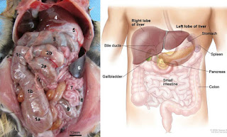
For single organs, there is no back up. If they go bad, you’re in trouble. You don’t have another source of insulin outside your pancreas, no other organ makes bile besides your gall bladder, and your spleen is crucial for removal of old blood cells and immune complexes. Yet, you can get along without some of your abdominal organs.
 |
If Hannibal Lecter had eaten just part of the census man’s liver and left him living, it is very possible that his liver would have grown regeneration. The hepatocytes start to split and regrow the mass. But they don’t have the signals to produce the original shape; they just form a functional mass of tissue. |
The injury often involves an automobile, but football players (American football) and rugby players have also lost spleens after tackles. One clinical case detailed a splenic rupture that didn’t occur until 70 days after an abdominal injury. You can get along without your gall bladder too, but you’ll have to read about that yourself - and don't eat potato chips while you read about it.
So how are our pieces fitted together – your heart points to the left of center, the liver and gallbladder are on the right. The stomach is on the left, and so is the spleen. The pancreas is about in the middle. For the digestive tract, the situation makes sense and is driven by the position of the stomach. Food exits the stomach to the right side, so all the things that help digest the food should be on that side – liver for glucose storage from small bowel, gall bladder for fats, head of pancreas for proteins and other things.
The normal arrangement of internal thoracic and abdominal organs (stomach, spleen and heart on left; liver pancreas, gallbladder on right) is called situs solitus. This is the picture you see in your anatomy textbooks and in bodies ripped open by movie werewolves and zombies. But sometimes the pictures aren’t right…. or left.
Some people have mirror image internal organs; what is usually on the right is found on the left, and what usually points to the left now points to the right. This situation is called situs inversus totalis. Luckily, the inversion of organs doesn’t really present a medical problem on its own. Since everything is reversed, the connections from organ to organ and from blood supply to organ are maintained. Sometimes there is a reason for the inversion, but other times it’s random.
A study from Norway indicated that the chance of situs inversus increases with maternal age, and it is also more common in cultures with small gene pools and higher incidence of inbreeding. However, it is not more common in twins than in multiple single births of a family.
In the cases of situs inversus that are not sporadic (random), what controls the differentiation of right and left and how does it go wrong?
 |
The nodal cilia rotate rather than beat. The third image shows how they tilt backwards while positioned in the posterior of each cell. |
The system goes back to the cilia that we talked about last Fall and Spring. The node cells that run along the midline of the embryo have monocilia(one cilium/cell). These are specialized cilia that don’t have the central pair of microtubules, instead they have a 9 + 0 configuration. They gyrate instead of beating.
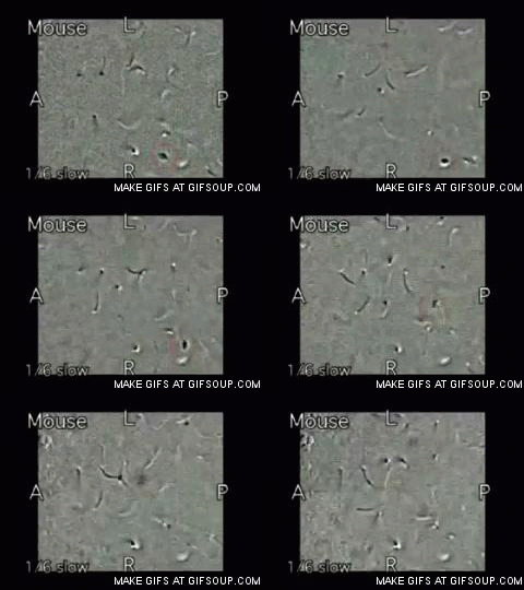
 |
Follow the debris to see the leftward flow of the fluid. |
The leftward flow is somehow responsible for turning on different genes in the embryonic cells that will be on the right and left sides, but people and other animals with a defect in nodal cilia are just as likely to exhibit situs inversus as situs solitus. It isn’t that non-functioning nodal cilia cause situs inversus, they just don’t promote situs solitus as they normally would.
It’s true that there is strong asymmetric gene expression on the left and right sides. The controllers are a couple of proteins related to an immune system protein called TGF-beta (reviewed here and here). The nodal protein is expressed only on the left side, while lefty protein is expressed on the right side. Nodal drives development of a left side, while lefty is a feedback inhibitor of nodal and therefore prevents a left side development on the right side. The asymmetry of nodal and lefty expression was supposedly driven by the concentration gradient of some morphogen generated by the cilia.
However, no one could find the morphogen. Meanwhile, it was discovered that mutations in another type of cilium also resulted in situs inversus. These cilia are immotile; they’re the sensory cilia we also talked about last fall. This led to the two cilia model to compete with the morphogen concentration hypothesis.
 |
In some people with primary ciliary dyskinesia, or immotile cilia syndrome, the mucociliary elevator isn’t operable, so they are subject to recurrent respiratory infections. |
All this was discovered by accident; different researchers were looking several different diseases. Primary ciliary dyskinesia (PCD) is a rare genetic disease where the motile cilia don’t move. About half the people with PCD have situs inversus, if they do then this PCD is called Kartagener’s Syndrome. Other researchers were looking at polycystic kidney disease (PKD). These people had a defect in immotile cilia in the kidneys, but also had a high incidence of situs inversus.
 |
In polycystic kidney disease (PKD), the defective cilia don’t sense flow and they trigger overgrowth of epithelium to form cysts. A normal kidney is on the right. |
For more information or classroom activities, see:
Situs inversus –
Liver regeneration –
Primary ciliary dyskinesia –
Polycystic kidney disease –


