Biology concepts – forms of DNA, history of DNA structure, triplex DNA, tetraplex DNA, protooncogene
Not every scientific success is driven by experimentation, trial, error, and eventual triumph. Sometimes, you just need to be paying attention. Oh, and be a little devious.
The other thing Watson and Crick had was an unwitting spy. Linus Pauling’s son Peter was a recent addition to the lab at Cambridge. Peter became friends with Crick and Watson. Through Peter, they knew Pauling was also working on a triple helix; they read his manuscript and knew it was flawed.
The B form of DNA is the one we see most often in biological systems. It is a right-handed helix with a major and minor groove that allows proteins good access to the DNA. On the other hand, the A form of DNA is more compact and occurs only when water is scarce. This doesn’t mean that your DNA changes form when you are dehydrated, it means that the A form is seen mostly in underhydrated crystals of DNA in the laboratory.
Z DNA is turning out to be more important than once thought. Z forms are important in transcription, as the reading of DNA to produce mRNA often induces a transient switch to the Z form. Z DNA is also important for the regulation of certain genes, including some genes important in preventing cancer. There exist some proteins that specifically bind Z-DNA in order to regulate the transcription of these genes.
Parvovirus B19 causes a common childhood disease called fifth disease (erythema infectiosum, or slapped cheek syndrome). The common name comes from the fact that it is categorized as the fifth of the childhood skin rash diseases – measles, german measles, scarlet fever, and another bacterial infection that has been dropped from the list.
Biffi G, Tannahill D, McCafferty J, & Balasubramanian S (2013). Quantitative visualization of DNA G-quadruplex structures in human cells. Nature chemistry, 5 (3), 182-6 PMID: 23422559
Feng L, Zhao A, Ren J, Qu X. (2013). Lighting up left-handed Z-DNA: photoluminescent carbon dots induce DNA B to Z transition and perform DNA logic operations. Nucleic Acids Research DOI: 10.1093/nar/gkt575
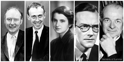 |
From left to right we have the main players in our little discussion of how science can be done without experimentation. Francis Crick, the man who never met a comb; James Watson, student of the greatest scientists of the time; Rosalind Franklin, denied a Nobel Prize because she died from cancer at the age of 36; Maurice Wilkins, Franklin’s boss who she treated like a red-headed step- child; and Linus Pauling, who won a Nobel Prize for discovering protein structure, and the Nobel Peace Prize for his work against nuclear weapon proliferation. |
James Watson and Francis Crick, along with Maurice Wilkins and Rosalind Franklin, worked out the structure of DNA in 1953. Years before, a Dr. Avery had shown that nucleic acids alone could be transferred between organisms to change their phenotype. This meant that it was the DNA, not the proteins, that were responsible for heredity.
Watson and Crick wanted to identify DNA's structure because, as is the case so often in biology, knowing the structure is crucial to knowing the function. The way DNA was put together would give clues as to how it passed on information to the daughter cells.
Watson and Crick were the exception in that they didn’t really do any of their own experimentation in this quest for the structure of DNA. They built some models based on other peoples’ data, and made some great insights that led them to the truth.
We know now that DNA is a double helix, but in the early 1950’s, no one had any idea about this. Rosalind Franklin had a structural picture of DNA that, when shown to Watson by Wilkins, immediately caused him to know that DNA was a helix.
The constituents and order of the building blocks of DNA (nucleotides) had been worked out by organic chemist Alexander Todd in the 1940’s– phosphate group, sugar, base. But were the phosphates on the inside of the helix or were the bases? And how were the different strands held together?
Watson and Crick thought DNA might be a triple helix. Don’t laugh, so did other eminent scientists, including Linus Pauling, the Wizard of Cal Tech, who would eventually be awarded not one, but two Nobel prizes.
 |
Alexander Todd showed that a nucleotide was composed of a phosphate, and ribose or deoxyribose sugar, and a nitrogenous base – in that order. This was some elegant work showing which moiety was bound to which. Pauling used this information to construct his triple helix model (right image), but he had the phosphates on the inside, and suggested that they were held together by hydrogen bounds – wrong on both counts. By the way, a nucleoside is just the same structure minus the phosphate group. |
About this same time, Crick and Watson were reminded of the conclusion of Chargaff that each cell contained the same amounts of A (adenine) and T (thymine), as well as the same amounts of G (guanine) and C (cytidine). Jerry Donohue, another addition from the land of Linus Pauling who liked to flap his lips, pointed out that A could bind to T through their hydrogens, and G could base pair with C.
You notice that nowhere have we talked about Watson and Crick’s data; so far they had only built a triple helix model with the phosphates in the center – and it was really wrong.
The final piece of the puzzle was a May, 1952 X-ray crystallography image of DNA made by Rosalind Franklin that was shown to Watson by her boss. Immediately, this image put to rest any doubts that DNA was a helix, and it gave accurate measurements for how wide the molecule was and the distance between complete turns.
Using this data, Watson and Crick returned to their model making and solved the puzzle in short order (by March, 1953). Their April 1953 paper was an exception in itself; it was only one page long. It contains the most understated sentences in the history of science since Alexander Fleming said, “Hey, all my bacteria are dead.”
The consistent base pairing of A and T or G and C led them to write, “It has not escaped our notice that the specific pairing we have postulated suggests a possible copying mechanism for the genetic material.” All this meant was that they realized that the DNA structure was a perfect explanation for how it replicates so that the genetic information is passed on to each new generation. Ho hum.
So that’s it - DNA is a double helix molecule with the bases on the inside. Well, not quite. I can think of many exceptions to these rules, but let’s talk about just a couple or three. DNA comes in at least three different double helices, A, B, and Z. This was apparent early in the studies of DNA, but only molecular biologists ever remember it.
 |
On the left are the three forms of DNA most often encountered. You see that the A form is more compact, has a deeper major groove and smaller rise for each turn as compared to the B form. The Z form occurs in the middle of the B form, when special repeats of bases are found. On the right are two X-ray images of DNA crystals. The left on is the A form, and the right one is the B form. The A form, being more compact, gives a poorly resolved image. You can’t blame Rosalind Franklin from opining that DNA wasn’t a helix based on the A form images that she first produced. |
The A form was the first form to be imaged by Rosalind Franklin. Since it was compact, it gave a muddied X-ray image, as seen in the picture. Information on water content and the length of each turn was also disturbing, and through of Watson and Crick for a while. The aha! image that Watson got a look at was Franklin’s attempt at B DNA, and it gave him all the information he needed to finish the model.
Z DNA does occur in nature, but usually not as the sole form of DNA. When certain runs of bases are encountered (called CpG, for runs of purine/pyrimidine), and when the salt concentration in the region is high, the DNA can locally switch to a left-handed turn helix. What we usually see is a B-Z-B region.
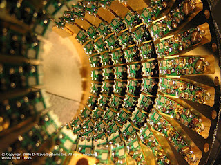 |
DNA computing is not a completely new idea. In 2003 and again in 2010, Israeli scientists built chips that computed using DNA – about 330 trillion operations/second! It all started in 1994, when a California scientist stored information in DNA to do a simple math problem. The new study using carbon nanodots will allow for even greater and faster computing power via light. |
Using this tendency of B DNA to switch to Z-DNA under the right conditions, some researchers are using carbon nanodots to create optical logic gates; they light up if bound to Z form, and don’t if bound to B form. By controlling the conditions and the sequence of small runs of DNA, you can turn the lights on and off, similar to the 1’s and 0’s of computers. You get it now – this has the potential to become a DNA-based nano-computer!
The A, B, and Z forms of DNA aren’t the only exception to the structure of this nucleic acid. These three are all double helices, but that doesn’t mean that all DNA exists as a double helix.
Some DNA is single stranded (ss). In every cell of every organism there is transient formation of ssDNA when it is replicated, transcribed, recombined, and repaired. SSDNA is also seen in some viruses, the best known and first discovered of these being the parvovirus.
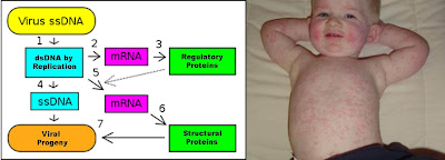 |
Single stranded DNA viruses enter a cell and their ssDNA becomes double stranded by using the hosts replication apparatus. Then the dsDNA is transcribed to make both types of proteins needed to make more viral particles. Meanwhile, one strand of the dsDNA is copied to ssDNA again so it can be packaged into the viral particle. On the right is the result of one ssDNA virus, parvovirus B19. It causes slapped cheek syndrome, with a skin rash on the trunk and limbs as well. |
Fifth disease is usually self-limiting, but new evidence is suggesting that there can be long term ramifications of a B19 infection. In 2013 alone, case studies have been published linking parvovirus B19 to acute kidney infections, neurologic complications, muscle cell death, and a purple tissue swelling called Wells Syndrome. All from a single strand of DNA.
On the other hand, some DNA exists in the form of three intertwined strands, called triplex DNA. Often used in the laboratory to manipulate gene expression, triplexes also form in cells on their own. The same protooncogene (c-myc) that we referred to in the section on Z DNA also has areas that form triplex DNA and work to control how much protein is made from this gene.
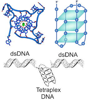 |
The top left image shows how duplex of DNA can rearrange to form a quadraplex. The top right cartoon shows how this tetraplex looks, forming three planar squares of interacting bases. The bottom image shows how a tetraplex unit can form within a dsDNA. This often occurs in the end pieces of our chromosomes (telomeres) and in the regulatory parts of some genes. |
Admit it, you laughed at Watson, Crick, and Pauling when you discovered that at first they all thought DNA was a triple helix. Who’s laughing now? Of course they still got the orientation wrong, with the phosphates on the outside. If you feel the need, go ahead and snicker at the guys with the three Nobel Prizes. How many have you got?
A newer discovery is quadruplex DNA; four strands come together to form a rectangle-like structure, where four bases bond together. It has been know for a few years that these complexes exist in the telomeres of mammals. Telomeres are on the ends of chromosomes and need special consideration to be replicated and preserved. The quadruplex structures aid in the preservation of our chromosome ends. This is important, as dysfunctions in telomere replication are thought to responsible for up to 85% of cancers.
Quadruplex structures are also being predicted and seen outside the telomeres. A new study used an antibody that recognizes quadruplex DNA to visualize and quantify these structures in living human cells. Their data shows that many DNA quadruplexes are associated with cell cycle progression, suggesting that manipulating them could become important in cancer treatment. And like clockwork, evidence also shows that the c-myc protocancer gene forms quadruplexes as well – is there any structure this gene won't form?
Next week we can continue our look at nucleic acids by looking at the exceptions to the rules of the building blocks, nucleotides. It’s not quite as easy as uracil (U) for RNA and thymine (T) for DNA. And why is U used only in RNA anyway?
For more information or classroom activities, see:
Search for the structure of DNA –
DNA activities –
Forms of DNA –
Triplex DNA –