Quantcast
Channel: As Many Exceptions As Rules
Viewing all 288 articles
Browse latest View live

Grin and Water Bear It

$
0
0
Biology concepts – tardigrade, cryptobiosis, anhydrobiosis, eutely, hyperplasia, hypertrophy, dry vaccine, dormancy

The original toughman contests were supposed to show us the
toughest of the general population. I’m not so sure how tough
it is to have a nickname like “Butterbean,” but maybe that
proves his toughness. On the other hand, do you get a cereal
box cover and a mohawk because your tough, or do you get
tough because you have a mohawk.
In the early 1980’s, a forerunner to the mixed martial arts craze was temporarily popular in the United States. Called the “ToughMan” contests, non-boxers would enter the ring for fights against other nonprofessional fighters. The rules were supposedly the same as in boxing, although many contests were run without proper supervision, and the tournaments sometimes required a participant to fight several times in one evening. Think legalized bar fights.

Famous participants in the toughman contests included “Butterbean” Esch – a 350 lb. boxer with one punch and an iron jaw, and of course, who can forget Mr. T. The contests are still held in many states, but MMA now has the majority of the fan base, even more than traditional boxing between trained fighters.

These guys were tough, but were they the toughest? Humans as a rule are weak for their size, scared of more things than they should be, and less inclined to fight to the death for a morsel of food or potential mate – well most are. So…..

Questions of the Day: What is the world’s toughest animal?

Ask a hundred people and you may get a hundred different answers. The bull elephant can fight off an entire pride of lions and can lift five tons. But can you really give the prize to an animals that is scared of a mouse?! Maybe they aren’t afraid of mice, but they will avoid them if possible, according to one of the more scientifically consistent episodes of Mythbusters.

A good second choice might be the honey badger. It supposedly knows no fear, and proves it by depriving lions of the prey they just killed. In one case, three honey badgers stole a entire carcass from seven lions! The South Africa National defense calls their armored personnel vehicles ratels, the afrikaans word for honey badger.

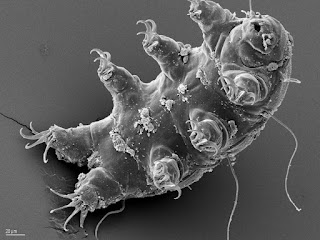
This is the toughest animal on Earth, although it may not
look it. The water bear looks more like a teddy bear,
although the claws might do some damage if you are a
bacterium or a protist. The mouth has stylets to puncture
plant cells and suck out the liquid nutrition.
But I will try to convince you that a type of bear is the toughest animal on the face of the Earth, a water bear to be specific. This animal has long claws on each foot and a mouth that takes up a good portion of its head. On the other hand, it's less than 1 mm long!

The water bear is more scientifically known as a tardigrade (latin for slow walker), a phylum that falls somewhere near arthropods and nematode worms. There are two classes (eutardigradia and heterotardigradia) and more than 900 species, but there may be some overlap in those descriptions.

The adult tardigrade will have 40,000 cells, and will never have more. What is more, everyspecies of tardigrade is matures with a specific number. This is called eutely (eu = good, and telos = end). Many lower organisms may be eutelic; their cells have a limited number of divisions, so they grow to that number and then stop.

It isn’t just whole organisms that might be eutelic, organs can be as well. For example, the nematode AscarisALWAYS has 162 neurons. The research model nematode C. elegans has exactly 959 somatic cells, although a 2011 study has shown that C. elegans can lose critical cell nuclei as they age – tell me about it. Other nematodes, rotifers, and gastrotrichs have also been shown to have cell constancy at the body and/or organ level.

Tardigrades do grow after they reach adulthood, just not by adding cells. Growth by additional cells is called hyperplasia (excess formation), while growth by existing cells becoming larger is called hypertrophy (excess nourishment). 

The gingiva around the teeth can overgrow in response
to some developmental disorders, but more often it is a
result of drugs given for epilepsy or other diseases. The
point here is that whether it is from hypertrophy
(increased cell size) or hyperplasia (increased cell
number), it looks the same. These are histologic
determinations and don’t really matter for clinical evaluation.
Prostate enlargement is often due to an increase in cells, hence the name benign prostatic hyperplasia, but hyperplastic growth doesn’t have to be pathologic. When you lose part of your liver, some can grow back by through hyperplasia. Likewise, hypertrophy is great when it is your muscles getting bigger, but not necessarily so good when your heart’s ventricles overgrow (ventricular hypertrophy).

Different tardigrade species are adapted to nearly every environment on Earth. They live in the Arctic and the Antarctic, in the mountains and the oceans, in the deserts and the jungles. All are found near water, some marine and some limnal(freshwater), some in the water and some just next to the water held in mosses or lichens.

But wherever you find them, you’ll find them in great numbers. The density of tardigrades can approach two million per square meter. Yellow crazy ants (Anoplolepis gracilipes) form supercolonies of incredible density, yet they can only muster about 2000 individuals per square meter. Haven’t heard of crazy ants? You will – look them up.

Tardigrade toughness doesn’t come from their pursuit of prey or their ability to fend off predators, but their willingness to live in conditions that would kill anything else, and I mean anything, else.

Cold, not a problem. Tardigrades can have liquid nitrogen (-346˚F/-210˚C) poured on them and they’re just fine. Heat – boil them for a couple of hours and then watch them lay eggs and go back to eating. Radiation isn’t a problem either; they can take 5700 grays of ionizing radiation without blinking.... well, they could if they had eyes. Humans curl up in a ball and die when exposed to 5 gray.

It is important to know how much radiation is absorbed
by the body, not just how much is in the air. The Gray,
named for Harold Louis Gray, is equal to 1 joule of energy
absorbed per kilogram of matter. Harold Gray is considered
the Father of Radiobiology. The old dose name was the
rad, and 1 Gray is equal to 100 rads. A chest X-ray is
typically about 0.0006 Grays.
Some tardigrades live in black smokers at the bottom of the ocean, yet most of them can take 6000x normal pressure in stride. To sum it all up, in 2007 the Russians fired tardigrades into space for 12 days (near absolute zero, total vacuum, cosmic radiation). They came back and starting having babies. Now that’s tough.

How do they manage these amazing feats? Basically – they die and then come back to life. Technically, it’s called cryptobiosis (hidden life), but death and self-resurrection is not a bad description. During cryptobiosis, metabolism is reduced by 1000x fold or even more, down to the level where there is NO detectable chemical activity.

There are five recognized types of cryptobiosis, based on the noxious environmental condition that triggers it – anhydrobiosis (without water), chemobiosis(chemicals), cryobiosis (cold), anoxybiosis (lack of oxygen), and osmobiosis (change in osmotic potential).

The primary form for tardigrades is anhydrobiosis. They drop their claws, retract their legs and roll up into a ball called a tun. 99% loss of water, roll up into “tun” this is important because it regulates the rate of evaporative water loss. At this level, they don't hold enough water for damaging reactions to take place or even enough water to form ice crystals. The water is replaced by a sugar called trehalose.

Trehalose production is similar to the way many organisms can protect their structures and biochemistry from environmental damage, but apparently scientists have just touched the surface of how tardigrades react to uncomfortable environments. A 2013 study indicates that there are many unidentified organic molecules present in the tuns of tardigrades that are not present in the organisms under normal physiologic situations.

The tun of a tardigrade is a very regulated structure.
When the claws are dropped and the legs retracted, the
tardigrade coils into almost a ball. In this situation, the
loss of water can be carefully controlled. The plates on
the back also form a protective armor for non-
environmental assaults – the spikes look imposing.
Amazingly, tardigrades are the only animal that can undergo all five types of cryptobiosis.  It's really like dying and coming back to life. Reviving from cryptobiosis can take a little while, usually the longer they have been in anhydrosis, the longer it takes to recover. They can’t survive this way forever either.

Earlier reports had professed that 120 year old tardigrades were revived from dried lichen and moss samples in the British Museum, and that decades old samples were just fine. But Dr. James Garey of the University of South Florida tells me that many of these reports have been called into question and cannot be repeated.

Dr. Garey’s estimate is that tardigrades can survive 1-5 years as a tun, with decreasing viability upon hydration after that. Still, could any other animal you know of be dead for five years, with no air, no water, high radiation, liquid nitrogen, and taunts about their size and lineage – and then come right back to life when the opportunity is right?

Cryptobiosis is quite different than dormancy. Dormancy doesn’t bring a huge change in physiology – like 99% dessication. Also, dormancy is preemptive while cryptobiosis is reactive. However, a very good 2011 review of tardigrade reactions shows that they can undergo both dormancy and crytobiosis – sometime simultaneously!

The question is – how do they survive the bad conditions WHILE they are forming the tun? It takes about 20 minutes for tun formation to occur, so it appears that many of the conditions they can endure require them to already be in the cryptobiologic state. They can survive the radiation whenthey are dessicated, they can survive boiling when they are dessicated. I don’t think it makes them any less amazing.

In early 2013, researchers from King’s College in England
developed a silicon mold with dissolvable sugar micro-
needles that can deliver a dry vaccine powder.  The system
would induce immunity through activation of skin immune
cells, would require almost no training to deliver, and no
refrigeration. The anhydrobiotic live vaccine is based on
tardigrade cryptobiotic features.
Tardigrades aren’t even considered extremophiles, since they are not designed to live in extreme environments. But this is precisely why I think they are so tough, because few of them are adapted to extreme conditions, but they can survive deadly situations anyway.

Can the exploits of this microanimal help humanity? You betcha. Tardigrades' ability to undergo anhydrobiosis has begun to influence the design of medicines. In third world countries, a lack of reliable refrigeration requires vaccines and medicines don’t need refrigeration, and can be reactivated upon ingestion.

Dry vaccines are a current goal, so the National Institutes of Health recently put out a call for proposals for research into more thermostable and reactivateable preparations. A late 2012 paper has identified a tablet form for deliver of some protein drugs, with reactivation of the molecules with saliva. This would be much better than the current reliance on hypodermics and refrigeration. So tardigrades are tough for themselves, and may fight for us as well.

Next week, just how do you describe the climate of your hometown? Biomes are scientific entities, but it seems we can't agree on what each looks like or is called.


Borde, A., Ekman, A., Holmgren, J., & Larsson, A. (2012). Effect of protein release rates from tablet formulations on the immune response after sublingual immunization European Journal of Pharmaceutical Sciences, 47 (4), 695-700 DOI: 10.1016/j.ejps.2012.08.014
 
McGee, M., Weber, D., Day, N., Vitelli, C., Crippen, D., Herndon, L., Hall, D., & Melov, S. (2011). Loss of intestinal nuclei and intestinal integrity in aging C. elegans Aging Cell, 10 (4), 699-710 DOI: 10.1111/j.1474-9726.2011.00713.x 

Guidetti, R., Altiero, T., & Rebecchi, L. (2011). On dormancy strategies in tardigrades Journal of Insect Physiology, 57 (5), 567-576 DOI: 10.1016/j.jinsphys.2011.03.003

 

A Linnaeus For The Biomes

$
0
0
Biology concepts – ecosystem, biome, naming systems, climate, chaparral, taiga, pyrophyte

Clint Eastwood was already a minor film star when he made the spaghetti westerns 
for director Sergio Leone. It is a genre he would return to as a director, with the
Oscar winning, Unforgiven. Notice the sparse landscape behind him. This was 
probably in Spain even though the film had an Italian director 
and crew. Chaparral biome is found throughout the Mediterranean region.
Clint Eastwood made a series of westerns in the mid-1960’s; nothing surprising about that. The Good The Bad, and The Ugly, For A Few Dollars More, A Fistful of Dollars; these were all directed by Sergio Leone, an Italian director.

Filmed on location, the landscapes were barren. There were rocks and dirt, bushes and the rare tree, buttes and canyons, coyotes and lizards – typical American southwest. But the movies were filmed in Italy and Spain! These were the spaghetti westerns.

The stories took place in the American southwest and northwestern Mexico, but they were actually half a world away. How can the countryside around the Mediterranean Sea look like the desert southwest in America? The climate, flora, and fauna of very different places can look very similar because they are the same biome, sooo…..

Question of the Day: What makes biomes the same or different and who decides which biome it is?

First of all, some of the desert southwest isn’t desert. It is a biome called chaparral (chapa = scrub in Spanish). Chaparral is the smallest of all the world’s biomes. A biome is a major ecosystem with a single climate, but perhaps more than one habitat or community. It’s a huge ecosystem, which in turn is made up of several smaller habitats. The major idea is that it is housed within a single climatic region.

In North America, the chaparral is found in California and maybe Arizona, at about 40˚ North latitude. In Spain and Italy, chaparral is found at just about the same latitude. In fact, all around the edge of the Mediterranean is very chaparral-like, including North Africa and much of Israel.

The distance from the equator with the tilt of the Earth means that 40˚ South latitude might have much the same climate, and we see that the Chilean chaparral, as well as similar biomes in South Africa and Australia are close to 40˚S.

Also called a Mediterranean scrub biome, chaparral is hot, dry, and liable to catch fire. Many of it plants depend on fire to disperse or activate their seeds (pyrophytes = fire-loving). Others have below ground growth that sprouts after a fire. We will have to do some posts on pyrophytes soon. However, lots of fire doesn’t mean chaparral is lifeless; it has 20% of the world’s plant species and many are endemic(only found in that climate).

A 2013 study in PNAS shows that fire is essential for regrowth
of habitats. The study found that a chemical is produced by
plants when they catch fire, called karrikins. This signaling
molecule settles in the soil after the fire and binds to the seeds
that are there. The binding protein was discovered to be KA12,
which then alters its shape and promotes germination of the
seeds right after the fire is over. Amazing.
One exception to the endemic nature of chaparral flora is the tumbleweed. Tumbleweed is known as any small round shrub that, when sufficiently tall, will be caught by the wind, torn off its roots and blown across the forbidding landscape. You can hardly watch a western without encountering at least one rolling tumbleweed.

But it’s all a lie! The tumbleweed proper is known as Russian thistle (Salsola tragus, probably several species). It doesn’t roll along because of the harsh and destructive nature of the chaparral; this is how it disperses it seeds. After flowering, the plant dries up and disengages from its roots on purpose. As it rolls, it drops seeds, like a trail of breadcrumbs never to be used to find a way home.

Heck, Russian thistle isn’t even from the chaparral biome. It's native to the steppe grasslands of Russia, a different biome altogether. Russian steppe is cold, and is wet, but it is windy, so it’s mechanism of seed dispersal works in chaparral. Therefore, it can survive in California, and Spain, and Italy, and Morocco, and Israel. Here we have a plant that is identified closely with one climate that comes from another.

Indecision and overlap abound when it comes to naming and defining biomes. For instance, what makes a chaparral a chaparral and not a desert? What makes it a chaparral and not a grassland?

For many ecologists, the difference is precipitation. Deserts get less rain, snow, fog, or humidity than chaparrals, which in turn get less of these things than grasslands. But others divide grasslands into tall and moist versus short and wet. So does the dry grassland still get more rain than the chaparral?

In the chaparral of Israel, different biomes can come as close together as different slopes of a canyon. A 2012 study has proposed that these canyon faces with completely different flora and climate should be used as “evolution canyons” where global warming can be monitored and changes in many different ecosystems can be tracked in a small place. And this is all supposed to be within the world's smallest biome?

This is one of the proposed “evolution canyons” in the Israeli
chaparral. Notice the different vegetation on one slope as
compared to the other. There are differences in rain,
temperature, and animal life – and yet it is all supposed to
be chaparral?
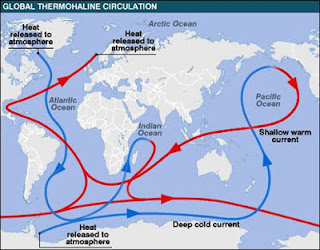
At the other extreme, the taiga is the world’s largest terrestrial biome (11% of land) and is quite wet. In North America, taiga is found north of 50˚ N latitude, and tundra begins as far south as 60˚ N. England and Scotland are situated at 50-58˚ N latitude, but they are nothing like taiga or tundra. Even though they are located at similar points on the Earth, they have a different climate because they benefit from the Atlantic Conveyor, an extension of the Gulfstream. This current pulls warm air up from the tropics and keeps Great Britain warmer than it would otherwise be.

At 50-60˚ S latitude, there is very little land. Almost all the way around the Earth at those latitudes there is nothing but ocean. So latitude isn’t everything when it comes to defining biomes. Tierra del Fuego and the southern part of Patagonia are located south of 50˚ S, but they are defined as neither taiga nor tundra.

As water travels around the world through the upper and lower
currents of the ocean, it picks up and releases heat. When it is
shallow and in the tropics, it arms and travels close to the
surface. As it cools and releases its heat to the atmosphere, it
drops deeper. The gulfstream ends in the Atlantic conveyor,
which keeps Great Britain warm. The warm water moves
faster, which is why it takes less time to sail from the US to
Europe but longer to go west.
You can tell by the descriptions above that many places around the world share general descriptions, so where does one biome end and a different one begin? This is a serious point of vagueness for me. Many people publish maps showing the biomes of the Earth, and no two of them agree fully. Look at the pictures published just below.

The major terrestrial biomes of the world include desert, tundra, taiga, deciduous forest, grassland, and tropical rainforest. But these lists are often incomplete or vague. Chaparral is often left off the list; it has climate very similar to desert, plants able to deal with desert-like heat, and plants that come from grassland biomes. What is even weirder, it is the only biome where the wet season is the same as the winter season. Overall, it’s an in between biome and muddies the waters, so it often falls through the cracks.

Biomes may be classified by climate (Holdridge scheme), which generally equates to latitude, but we have already seen exceptions to that. Another scheme, Whittaker’s biome-typing, works to classify regions based on temperature and precipitation. Other systems are based on some combination of these factors, but if they are using the same factors, why don’t they agree better?

This is a busy picture, but the different colors represent different biomes,
and vary from map to map. The thing to notice is the pattern is different
in each map; no one can agree on what biomes are where.
For these schemes, the discriminating factors are abiotic (non-living influences), so they do not take in the periodic movements of animals or the invasiveness of some species in defining their boundaries. But some abiotic factors are changing quickly; like temperature, while others take much more time to change (geography). Therefore, as conditions change, these biome designations will either diverge, or will come to represent a different flora and fauna. No naming system has got these sets of problems licked.

Another problem is in the naming itself. Who decides on the name, and what does it mean in different areas of the world? The World Wide Fund For Nature (WWF) has a naming system that tries to be specific and generic at the same time. What is often called rainforest is designated by WWF as either “tropical and subtropical moist broadleaf forest” or “tropical and subtropical dry broadleaf forest” depending on the amount of precipitation. Don’t really roll off the tongue, do they?

Locality plays a role in the naming problem. Mediterranean scrub biome - is it by the sea, not always. Is it only scrub brush, not always. So that name isn’t so good. But even around the Med it’s called different things – maquis in Italy, garriguein France, phyrygana in Greece and batha in Spain. In America, it’s the high chaparral– like the TV show. But in Chile, it’s called matorral (mata = shrub in Spanish). In South Africa, it’s the renosterveld or fynbos, but Australians know it as mallee scrub or Kwongan heath.

Carolus Linnaeus(1707-1778) developed a binomial system for naming in botany and zoology, specifically to alleviate the locality and organizational naming problems. But a system such of this was never developed for ecology. Why not?

All of this imprecise naming and description, and we’ve only touched on the terrestrial biomes. There's a whole set of problems attached to the aquatic biomes as well. Some definitions list only one biome in water, with different freshwater regions (ponds, lakes and such), and marine regions (oceans, reefs, estuaries).

The Tollund Man was found in a sphagnum peat bog in Denmark. The highly 
acidic peat tans the skin and the low oxygen condition preserves 
the clothes and hair. Only the phosphate in the bones is lost, so bones remain
in place, but not rigidly. You can see the rope around his neck. Autopsies in 
1950 and 2002 confirmed that he was hung rather than strangled. Tollund Man 
was most likely a sacrifice to the bog gods.
Other systems define each of the regions as a biome. This can also cause problems. One freshwater biome is a wetland – but what kind of wetland? There are bogs, swamps, marshes, fens (as in Fenway Park in Boston) and carrs(fens overgrown with trees). Each has a different type of feeder system, different majority floral and fauna, and even a different acidity. How could those all be the same biome?

Most bogs are found in boreal forests (taiga) in Great Britain and Scandinavia. They were thought of as sacred places where the gods lived, and where human sacrifices were often made. This is why you may find bog bodies, mummies of people who lived thousands of years ago. The acidic conditions and low oxygen preserved the tissues (but dissolved the bones usually).

So - are the bogs considered part of the taiga, or just habitats within a biome? Or are they their own biome within a biome? Not easy to decide. Biomes are essential for organizing the life on Earth, but the learning would be easier if we could find a Linnaeus for ecology.

Next week, can you have mold without mildew? Just what are they anyway?


Nevo, E. (2012). "Evolution Canyon," a potential microscale monitor of global warming across life Proceedings of the National Academy of Sciences, 109 (8), 2960-2965 DOI: 10.1073/pnas.1120633109
 
Guo, Y., Zheng, Z., La Clair, J., Chory, J., & Noel, J. (2013). Smoke-derived karrikin perception by the / -hydrolase KAI2 from Arabidopsis Proceedings of the National Academy of Sciences, 110 (20), 8284-8289 DOI: 10.1073/pnas.1306265110 

Mildew Broke The Mold

$
0
0
Biology Concepts – mold, fungus, powdery mildew, downy mildew, nosocomial infection, decomposer, parasite

Bill Cosby is one of the five funniest men ever. The other four, in
no particular order, are of course, Jonathan Winters, Steve Martin,
Groucho Marx, and Benny Hill. You might sub in Robin Williams
for Benny…. maybe.
I wish I had a nickel for every time my mother told me she was “sick and tired” of picking up after me, or of telling me to clean my room. I might have a lot of nickels, but she’d still have been right. Sick always goes with tired, as if having two descriptors makes it more believable.

Mold and mildew is another example of how one problem or explanation just isn’t enough. How many commercials have you seen for products that will rid your bathroom of mold andmildew? On the other hand, ever see a commercial for something that rids you of just mildew?

Question of the Day: Is mildew different than mold?

Molds are something we can identify with, we see mold on bread, oranges, and other things left around the kitchen for too long; they’re caused by many species of fungus.

Penicillin and many antifungal drugs come from molds, so they can be useful. We get a lot of our antibiotics from fungi because they are attacked by many of the same kinds of bacteria that attack us. The difference is that they have developed specific chemical defenses while we haven’t.

Fungi are closely related to animals, much more closely related to us than they are plants. This takes many people by surprise, especially since the internet is rife with websites that call fungus a form of plant life. Many fungi are decomposers, meaning that they gather their nutrients from dead organic material, but some are parasites of living things.

Mold fungi look very different from the mushroom fruiting bodies we normally picture when someone says fungus. However, they are very similar at the cellular level. Filamentous hyphae (singular form is hypha) are the key; these are the long lines of cells (filaments) that form the structure of the fungus before it forms fruiting bodies. In mold fungi, the fruiting bodies are usually spore-forming structures that are too small to be seen.

The hyphae form chains, some of spectacular length. When the hyphae branch, cross each other, form communications, and become sufficiently dense, they are called a mycelium. This is when you can see them. Even with mushroom fungi, it is the mycelium that lies just under the surface of the dirt that gathers the nutrients and connects the mushrooms together. Sometimes the mycelium is in your bathroom.

Fungal hyphae come as septate (top left) or coenocytic
(top right). Coenocytic are really multinucleate giant cells,
while the septa may or may not divide the cells completely.
The rhizoid is the projection that anchors the hypha to a
food source, and pulls in nutrients. In decomposing fungi,
the rhizoid attaches to something dead; in parasitic fungi,
they burrow into live tissue.
The bathroom is almost always a target for people selling you things to eradicate mold and mildew. Why the bathroom – humidity is key for mold growth. This is why they sell better breads in paper packaging. The paper allows the moisture to escape and keeps the bread mold free for a longer period of time.

Bathrooms are the perfect combination of heat and humidity for promoting mold growth. All that is needed is a surface that the mold fungus can colonize. The rhizoid of the fungus is a short hyphal structure that attaches the fungus to its substrate. It also has the ability to absorb nutrients from the substrate like the rest of the hyphae.

Ceramic tile is designed to have a very smooth, flat surface with few pores. This makes it hard for mold rhizoids to attach and stick to tile. But grout and caulk are more porous and irregularly surfaced, so mold is more likely to gain an attachment and colonize these surfaces. The black mold that you see in showers is most likely going to be in vertical or horizontal lines where the tiles meet.

There is more than one type of black mold; one is common, and the other is toxic. Stachybotrys chartarum is one of the most toxic species. It is often associated with wet drywall conditions. Non-toxic black mold is caused by many different species and is more common on caulk and grout.

While black mold is the most common version seen in the bathroom, pink mold is probably second most common. This is our first exception of the day, since pink mold isn’t mold at all. It’s a bacterium called Serratia marcescens, which produces a red or pink pigment called prodigiosin (more about this below). The slimy film in your bathroom is the bacterial colony that feeds on fatty residues and phosphates, things found in soaps and shampoos.
Toxic black mold (Stacchbyotrys species) can cause
internal bleeding, infertility, and respiratory problems.
No wonder they call it “sick building syndrome.” I
can’t imagine how someone could let it get this bad,
but it needn't be like this for it to be causing
you trouble. It could be under the paint or wallpaper.
Thank goodness, most black mold is not the
toxic variety.

In addition, the S. marcescens bacterium was first associated with nosocomial infections in the 1950’s. Nosocomium is the Latin word for hospital, so nosocomial infections are those that you contract while in the hospital. Before the advent of hygienic and antiseptic practices in the very late 1800’s, hospitals were the last place you wanted to be if you were sick. You entered with a hangnail and left on a slab from the plague you picked up there.

Today there are many marcescens strains that are resistant to anti-bacterial drugs. It is becoming a big enough problem that some cases of necrotizing fasciitis (flesh-eating bacterial infections) are being blamed on resistant S. marcescens strains. While not common, about 25% of people who contract flesh-eating disease will die from it, and many are otherwise healthy, if you overlook the bacterial infection that is destroying large parts of their body.

Although S. marcescens is a rare cause of this horrible disease, there are about 20 cases in the literature, brought about by such diverse things as a human bite, contamination of a portal for leukemia drug injection, and immunosuppression. Makes you think about cleaning your shower better, doesn’t it.

Joseph Lister was a British surgeon who thought it
might be a good idea to keep open wounds and
operating rooms clean. Listerine was named for him,
but not invented by him. Listerine was produced by
Jordan Wheat Lambert in St. Louis, MO. He traveled
to England to get Lister’s approval to use the name.
He thought it would sell better. Lambert’s son was a
financial backer of Charles Lindbergh’s transatlantic
flight; the St. Louis airport is named for him.
It’s not all bad news, prodigiosin pigment is a potential new, antibacterial, pain, antifungal, immunosuppressive and anticancer drug. A 2013 study showed that purified S. marcescens prodigiosin kills several drug-resistant bacteria strains. The researchers determined that prodigiosin is pro-apoptotic (it makes the cell kill itself) and is not affected by the multidrug resistance transporter, so it could be a great cancer drug too. Even bad guys have good days.

So now we know that your bathroom mold is only sometimes mold. What about the mildew part of, “mold and mildew?” When it comes to your bathroom cleaning, mildew is just a throw in, an advertising ploy.

Some mildews are indeed caused by fungi. Fungal mildews usually require organic surfaces to draw nutrients from. Paper will mildew, wood will mildew, so will drywall (wallboard, plasterboard) since it is paper backed. Clothes made of cotton or other natural fibers will mildew, as will leather.  But, unless you have a cotton shower curtain, the most likely place you’ll find mildew in the bathroom is your towels.

If mildews like this are fungi, what makes them different from molds? In most cases, it is the degree of growth. Mildews are just not quite as bulky as molds. If a mildew grows for a long time and achieves greater density and mass, it is often called a mold.

However, in the vast majority of cases, mildews are problems of plants, not bathrooms. Plant mildews come in two primary types, powdery mildew and downy mildew. Neither is likely to be found in your shower unless it is so dirty that you actually have plants growing from your grout.

Mildews require living cells to parasitize; they aren’t decomposers, but obligate parasites. Both powdery and downy mildew are problems of horticulture. While they may not directly kill crops, they can reduce yields and make them more susceptible to other infections. In the great majority of cases, fungi that cause powdery mildew are plant specific, there are thousands of species, each attacking a specific plant.

The physical difference between powdery and downy
mildew is seen above. Powdery is well, more powdery.
It looks fuzzier because it grows higher off the surface.
Downy mildew usually turns the leaf yellow or brown.
There are often plant specific, but in both cases above,
it's a grape leaf that is infected.
There are wheat mildews, grape mildews, melon mildews etc. and each may require a different treatment. The hyphae attach to the leaf or the fruit and suck out the nutrients they need. This can’t be good for the plant.

If powdery mildew is a fungus, but only grows on living organisms, what does this make fungal infections of humans; are they more like molds or mildews? There was an incident in Maine in 2005 after it rained for many days in a row. Doctors started seeing fungal growths in the ears of inhabitants (would they be called Maineiacs?), primarily in their outer ears. This was fungal and was a form of human mildew, and overgrowth of normal fungal flora.

What about athlete’s foot? Is athlete’s foot, toenail fungus, or trench foot really just foot mildew? I suppose it just wouldn’t be polite to tell someone they were mildewing, so we call them fungal infections.

Another exception - downy mildew isn’t even a fungus. The causative organisms are oomycetes, a type of false fungus. Having “mycete” in the name makes it sound like they are fungi, and they used to be categorized with the fungi. New naming systems have altered what is or isn’t a fungus based on shared DNA.

This is a coccolithophore, a member of the chromalveolata, the
supergroup that also includes downy mildew oomycetes. They
live in the oceans and die in the trillions each day. The plates
are made from calcium carbonate, so they fall to the bottom
and become limestone or chalk. They might end up as
drywall that could mildew. It’s a circle of life sort of thing.
Now the fungi have been joined to animals in the supergroup Unikonta (one flagellum). You see now why some many fungal drugs are useful for us, fungi are more closely related to us than they are to plants.

Under some new phylogeny schemes, downy mildew organisms are completely unrelated to fungal powdery mildew organisms, which are only distantly related to the organisms that cause some molds. The Tilex people have no idea what a box of worms they’ve opened.

Next week, let's examine your fingernails and toenails. If they are made from the same thing as hair, why are they so tough?


Elahian, F., Moghimi, B., Dinmohammadi, F., Ghamghami, M., Hamidi, M., & Mirzaei, S. (2013). The Anticancer Agent Prodigiosin Is Not a Multidrug Resistance Protein Substrate DNA and Cell Biology, 32 (3), 90-97 DOI: 10.1089/dna.2012.1902
 
Rehman, T., Moore, T., & Seoane, L. (2012). Serratia marcescens Necrotizing Fasciitis Presenting as Bilateral Breast Necrosis Journal of Clinical Microbiology, 50 (10), 3406-3408 DOI: 10.1128/JCM.00843-12


Tough As Nails

$
0
0
Biology concepts – keratin, crystalline form, cornification, regeneration, stem cells

Horns are made from keratin, just as are nails, hooves, and hair. 
Believe it or not, this is a real picture of a condition called 
a cutaneous horn. It is a tumor of keratin producing cells and 
can be benign or malignant. They can be removed surgically, 
which makes me wonder why it is still on her head.
Thankfully, the cancerous ones are usually wider than they are long, 
so this example is probably benign.
The first time I remember being amazed by biology was when I found out that our fingernails and hair were made of the same thing. My hair is (was) red, but my nails are pretty much transparent and colorless. Hair is thin and is broken easily - you should see my hairbrush. But fingernails and toenails are tough. How can they be made of the same material?

Question of the Day – How do fingernails grow to be so tough and why do they grow at all?

You use your fingernails everyday, but do you take the time to think about how amazing they are? It’s true that nails are made of a protein called keratin, just like the dead layers of your skin and your hair, but there are differences too.

You’d be surprised to find out what else they do. Your fingers and toes are basically tools; they grasp, push, pull, and gather information. Switching back and forth between gross and fine movements and manipulations requires that your fingers and toes be able to take a lot of punishment, but at the same time maintain themselves for delicate work.

Your nails protect the ends of your fingers and toes during work that could injure them, this keeps them in good shape for the precise things we need to do all day long. But nails are tools as well. They can slip under surfaces and pry them up, and they can act as a hard surface against which you can apply pressure.

This idea of pressure is important for fingers especially. You push down with your fingertip to deliver a precise amount of pressure. The nail provides a flat, rigid surface against which you can measure the pressure and fine tune your work. Nerves abound beneath the nail, you know that from trimming it too short or from having a splinter driven beneath it. Ouch.

But nails have other jobs as well. Did you know that your nails can be good indicators of your overall health? Healthy nails are smooth and have no ridges, although as you get older you may notice more vertical lines. Yellow nails may indicate a malignancy or fluid in your lung spaces (called pleural effusions). Pitted nails might indicate a connective tissue disease (like psoriasis or scleroderma).

Hippocrates was a Greek physician. His importance to the
profession of medicine is evident – new physicians take the
Hippocratic Oath, promising first to do no harm. In other
words, don’t make things worse. One of his great
contributions was to make the connection between an
outward sign (clubbing) and an internal disease. In this case,
it is usually something wrong in the chest (heart, lungs,
upper GI).
Clubbing of nails as a sign of lung or heart disease was recognized by Hippocrates 2300 years ago. However, clubbing is just an anatomic variation 60% of the time and reflects no underlying disease. What you need to look out for is a change from non-clubbed to clubbed fingers and toes. For a good review, read a 2012 review by Tully et al., you’ll be surprised at what you can learn from your nails.

Even more surprising is the job your nails could do for you if you happen to get the tip of your finger or toe chopped off. Unlike starfish and lizards, we don’t generally have the ability to regenerate lost anatomical units or tissues. One exception is the ends of our digits. If you get a portion of your finger cut off, it might grow back, nail and all. But it must be just the tip, some of the nail must be left. If not, all you can do is learn a new way to type.

A 2013 study has shown that nail beds contain stem cells that can grow back the skin, muscle, and even bone of the digit. A specific signaling pathway (Wnt) is key, and when blocked, there is no regeneration. But if you stimulate the Wnt pathway, you can get regeneration beyond the nail bed. This may be huge for future regeneration of lost limbs in humans.

So these are the things your nails do for you, but we haven’t tackled the question of how they can be tough enough to carry out these tasks.

The white semicircle at the base of your nails is the lunula (luna, as in moon). This is where your nails grow from and the mass of cells that produce the nail is called the matrix. Above the matrix, but below the nail is the cuticle. This connects the nail to the finger and hurts like heck if your cut into it while trimming.

The nail and the hair are made from keratin filaments joined
together. However, in nails they are wide masses of shorter
fibrils and in hairs they are fewer but longer. You can see that
both hair and nails have a matrix that produces the keratinizing
cells, a cuticle, and the new cells push the older cells along to
make the nail or hair longer.
The cuticle and matrix are white because the melanocytes there are inactive, so there is no pigment produced. The matrix cells divide and the new cells produce a lot of keratin protein. The older cells fill with keratin and die, and the new cells push them out of the way – this means toward the end of your finger. This is how your nails grow.

But our fingernail doesn’t look like individual cells. You are sloughing millions of skin cells each day, but for a nail the dead cells all mass together. Nails only wear away or must be trimmed. Individual cells are not lost. The solidity of the nail comes from the connecting of the dead cells together by junctions between the cells called desmosomes, and by the interlocking of the cells like jigsaw puzzle pieces. But there’s more.

Individual keratin protein filaments also become connected so that the entire mass of keratin becomes one solid structure. This is called cornification, like the stratum corneum (the outer, dead layers) of your skin.

One of the two main forms of keratin in your nails iscrystalline keratin, which is rigid, stronger, and has an ordered structure - like a tinker toy cube. Transmitted light is less likely to strike an atom and bounce back when the atoms are all lined up, so this is why many crystalline lattices appear translucent. Precious gems have crystalline forms.

The other keratin is more gel-like and connects the different filaments of crystalline keratin together.  There are crosslinks that join glutamine and lysine amino acids in one keratin filament to those in many other filaments; the crosslinking is performed by an enzyme called transglutaminase. There are billions more of these crosslinks in nails as compared to those in dead skin or in hair.  This is why nails are much stronger than skin or hair.
Transglutaminase has become a favorite of chefs. They
can put different cuts of meat together to forma solid
mass. Also called “meat glue,” transglutaminase can be
used to fuse ground or cut meat into something sold as
a single piece. Health officials worry about this because
it takes outside surfaces that may have been
contaminated an puts them on the inside, which can
promote bacterial growth.

So if this is how nails grow, and it is the same for all your nails, why do they grow at different rates? The average rate of growth is about 3.5 mm (0.14 inch) each month (influenced by genetics, age, and health), while toenails grow about half has fast (1.5 mm or 0.06 inch/month). Toenails are thicker and more rigid, does it take longer to make their cornified structure?

Not really. The answer has more to do with the way the body responds to your behaviors and the environment. Live cells in the matrix produce the keratin before the cells die and are joined together as the nail. Being alive means that they need nutrients and oxygen – things carried in the blood.

Anything that increases the blood flow to an area will allow for faster cell grow and division. This includes heat; your superficial vessels dilate to release excess heat to the environment when your body is in a warmer environment.  Dilated vessels hold more blood, so this would mean more growth in those areas. This is why your fingernails grow faster in summer than in winter. They do – you mean you don’t keep track of how often you trim your nails?

This is pianist Liu Wei from China. He lost his arms at
the age of ten when he was electrocuted. I wonder if
his toenails now grow faster than mine. It’s amazing
what people can do with their feet. Tisha Unarmed is
a fantastic video blog where she shows you how she
all her daily chores using her feet. You should check
it out.
There are other things that increase blood flow and nail growth rate. Activity is a big determinant. This is why your fingernails grow faster than your toenails. Muscular movements of the fingers are nearly constant during the day. There is little we do that doesn’t involve moving hands and fingers. All that muscular movement requires oxygen, so blood flow is increased – and the nails grow faster.

This idea is reinforced by the fact that fingernails grow faster on your dominant hand. More use, more blood, more growth. For people that have lost the use of the their arms and learn to write, eat, brush their teeth, etc. with their toes – do their toenails grow faster than their fingernails? If they don’t have fingers, you can’t compare the growth rates of their toenails versus their fingernails, but I bet their toenails grow faster than average.

Blood flow is also increased by trauma; part of the swelling when you whack your thumb with a hammer is due to increased blood flow to the area in an effort to start the healing process. This will also make your nails grow faster. Many scientists believe that everyday uses of fingers, tapping, typing, prying, etc., are all types of microtrauma, so the more you use your fingers, the more blood flow you are inducing.

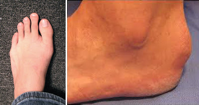
On the left is Morton’s toe, also called Greek foot. Is my big toe
short, or is my second toe long? On the right, I just decided to
show another deformity I have, called Haglund’s deformity. It
is a bony bump on my heels, and makes it hard to buy decent
hiking boots. Neither picture is my foot by the way; nobody
wants to see that mess.
The one determinant I don’t understand - fingernails and toenails on longer digits grow faster. Your index finger’s nail grows faster than your pinky nail. Is it due to usage? Maybe, but then why do longer toenails grow faster- are you using them that much more? My second toe is longer than my big toe, a condition called Morton’s Toe.  When I hike, my second toe pushes off the ground last, so maybe it is bearing more weight and doing more work. I can’t test it though, since my second toenails are always running into the front of my boots and the nails are always splitting and falling off.

By the way, since your nails come from the division of live cells, they only grow while you are alive. The old tale about hair and nails growing after you die is untrue. It may have started because other tissues lose water and retract after death, but proteinaceous (meaning made of protein) structures like hair and nails do not contract. Therefore, they may appear to have grown a little bit after death.

Next week - ever wonder why your grass grows back after you mow, but that tree you cut down probably won't? Believe it or not, it is related to your fingernails!


Takeo, M., Chou, W., Sun, Q., Lee, W., Rabbani, P., Loomis, C., Taketo, M., & Ito, M. (2013). Wnt activation in nail epithelium couples nail growth to digit regeneration Nature DOI: 10.1038/nature12214

Tully AS, Trayes KP, & Studdiford JS (2012). Evaluation of nail abnormalities. American family physician, 85 (8), 779-87 PMID: 22534387

Go Prune the Grass?

$
0
0
Biology concepts – meristem, monocot, dicot, herbaceous, arborescent, xylem, phloem

Knots can form in trees for different reasons. When you
prune a limb back and a callus grows over it in a few
years, this may form a loose knot. Even limbs that are
still there begin to be swallowed by the expanding
trunk; this would be a tight knot. The knot on the right
is not usual.
Summer brings warmer weather, outdoor activities, and yard work. Tree trimming is rare, but mowing the lawn is a weekly chore. I like mowing in April, May, and June, but by August and September, I’m over it.

Question of the day – Why does the cut grass grow back, but when you prune a tree limb, it stops growing from that end?

You cut the ends of the blades of grass and they grow back in a week or so. But have you ever seen a limb regenerate on a tree? It would be unusual; if you cut the limb off, the tree forms a knot as the trunk grows around the cut stub.

The difference lies in the kinds of plants grass and trees are, and how these types of plants grow. Most trees are dicotyledon (di = two, and cotyledon = embryonic leaf) plants, while most grasses are monocotyledons(mono = one). The differences between these types of plants are many, but the names come from the various embryonic forms they take in their seed.

Monocots generally do not overwinter well, are herbaceous (without woody stems), have fibrous root systems, and have flowers with petals in multiples of three. The leaves of monocots usually have veins that are parallel to the direction of growth, and the leaves are most often long and thin. They way they move water and nutrients (in the xylem and phloem, respectively), is constructed in small islands, with many bundles of xylem and phloem spread throughout the stem.

Dicot plants, on the other hand, are often perennial plants that survive winters well. They can herbaceous or arborescent (woody), and their transport systems (phloem and xylem) are arranged in concentric circles. Long, deep taproots are more common in dicots, as are flowers with petals in groups of four or five and broad leaves with networked veins.

Monocots and dicots have similar structures (leaves, flowers, stems, etc.) but they arrange them differently and they have different properties. One thing they have in common is that their parts are generated from similar cells, in structures called meristems.
On the left is a cutaway of a bud. The arrow points to the meristem
tissue from which all other cells develop. On the right is a
photomicrograph of the meristem, showing the different structures
that form from it. The meristem sits on top of, and is surrounded
by, the structures it formed previously (larger leaf primordial and
axillary buds). The procambium will become the xylem and phloem.

A meristem (meristos= divisible, and em = the ending from xylem and phloem) is vascular tissue that can divide; they are the source of all new cells in growing plants. Meristem cells act much like stem cells; they each have the potential to become many different types of cells. However, I could not find evidence that the older word, meristem, was the source of the term stem cell.

All plants have meristems, both monocots and dicots - no exceptions, if you can believe it. The smallest flowering plant, Wolffia globosa, has meristems, though the whole plant is only a millimeter long. Some meristem features are found only in dicots and others only in monocots, one type of meristem that both plant types share is the apical meristem.

Shoot apical meristems make new stems, shoots, leaves and other above ground structures. The pluripotent cells of the meristem divide and differentiate to become the cells that are needed. The structure of the typical meristem is shown in the picture to the above.

In grasses, there are shoot apical meristems, although they may sometimes be called intercalary meristems. They are located at the base of the leaves and the stem, and allow for re-growth of the leaf after it is grazed by herbivores…. or mowed down by Harry Homeowner. Therefore, blame the evolution of low placed meristems as an adaptation to deal with prehistoric cows and goats for your need to mow each week.

Node meristems are forms of intercalary meristems. Growth can
continue to take place from each node. On the left is bamboo, a
good example of a monocot grass with nodal meristems.
Grasses may also have nodal meristems; these allow for growth between the nodes, as well the addition of leaves and such. Bamboo is a grass in which you can see the nodes very well (see picture), and growth from each node is one of the reasons why bamboo can grow so quickly. While bamboo may feel like wood, it is a monocot like other grasses, and therefore is not what we consider true wood.

On the other hand, most trees are dicots. Their shoot apical meristems are located at the ends of their limbs and top of the trunk. As the tree grows, limbs branch off from the trunk, each forming from a different shoot apical meristem.

The apical meristems at the ends of the limbs and top of the trunk take the shape of buds in the spring. Each bud will become a short twig and leaf. In the fall, the leaf falls off, the tree goes dormant, and the next spring a new bud forms on the end and the process is repeated.

This is how a branch becomes longer. If you prune back a limb, rarely will it grow from the pruned end – you cut the meristem cells away! The limb may grow from one of the branch points behind where you pruned, but not from the cut end.

Trunks get taller and limbs get longer from these apical meristems, but there are also axillary (lateral) meristems produced when a limb branches or a leaf grows from the twig. However, most of these meristems do not support growth; they remain dormant.

The meristem at the top of the trunk and the end of the limb are dominant; they produce plant hormones to prevent growth from the meristems below them (on the trunk) or closer to the trunk (on limbs). This is why many trees have a single, central trunk. If that meristem is lost, one or more may become dominant and several trunks may form.

Auxin is the hormone that induces and maintains dormancy in lateral meristems, while cytokinin hormones promote growth from the laterals. For a good review of their roles, see the paper of Muller and Leyser from 2011. Other plant hormones are relevant for induction and emergence from dormancy induced by winter for all meristems. Abscisic acid (ABA) induces winter dormancy while gibberellins break the dormancy. A 2012 study has defined gene expression patterns during short and long-term cold spells and how they affect the ABA/gibberellin ratio and how short cold periods alter ABA concentrations.
When a dominant apical meristem is lost, a dormant
axillary (lateral) meristem may take over to become the
new trunk. However, sometimes more than one may try
for dominance, as in this conifer located near my home.

A new study has investigated how fruits can also induce dormancy in meristems. So much energy is put into fruit development that energy must be stolen from plant growth. The fruits themselves produce auxin to save energy by stopping meristem function. This is why fruit trees have alternating years of fruit vs. growth; when they make fruit they don’t grow, and when they grow, they don’t make much fruit.

In some cases, if the shoot apical meristem is lost, the lateral meristem on a branch can become the dominant shoot apical meristem and the branch will begin vertical growth as a new trunk. However, this isn’t always the case (see picture to the right).

These are the ways trees get taller and fuller, but it's also related to how they get thicker. Apical meristems give rise to a thin line of cells that form the procambiumand the secondary meristem all around the periphery of the trunk or limb.

Just as apical meristems form buds and leaves when the winter breaks, so do secondary meristems form new vascular tissue all around the periphery each growing season, this tissue is called the vascular cambium. We see their work as the rings that can be used to age a tree (dendrochronology). Twigs don’t stay twigs because each year their secondary meristems make them thicker.

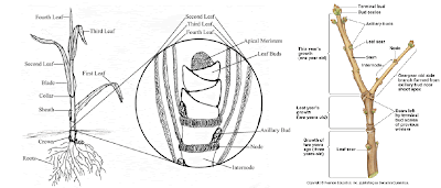
A cartoon of grass is on the left, showing that the meristem is
located low on the plant. On the right is a twig of a dicot tree
growing from the end, several buds may form during one
growing season, but if you cut them off, growth stops.
So now we know why you have to mow your grass – you don’t cut the low-placed monocot meristems when you mow, but dichotomous trees that are trimmed back lose their shoot apical meristems, therefore they don’t re-grow limbs. But, as always there are exceptions.

Exception one - some things we call trees are actually monocots, not dicots. If you look at the wood from a palm tree or a cordyline, you can see the small, dispersed groupings of vascular tissue that are typical of monocots. The reason they grow taller and appear more like trees is that they produce more lignan than other monocots, so they can grow taller. Lignan is the stiffest of the plant structural tissues and provides the resistance to gravity and wind.

Palm trees also have secondary mersitems, similar to the vascular cambium in dicot trees that add girth. However, palm secondary meristems just add more vascular bundles, not the rings of vascular tissue you see in dicot trees.

The second exception has to do with regrowth of trees even when the complete limb or trunk is cut away. There are meristems that get buried by the expanding wood layers of the trunk or when a limb grows thicker.

Basswood is one example of a tree that grows much
better from stump sprouts than from seeds. Almost all
basswood is harvested from clumps of trees that have
sprung up from a previous stump. Basswood rarely
grows a full tree from a seed, which makes me wonder
why they still make seeds.
In some cases, a new trunk may sprout from a dormant meristem in a cut stump. This is called “stump sprouting” or epicormic growth (epi= on top of, and cormal= trunk stripped of its boughs). In botanical terms, epicormic has come to mean growth from a previously buried, dormant meristem. This type of growth occurs most in hardwoods and has become important for regeneration of wood in the forestry industry, but it has also been used to help propagate Christmas trees (which are softwood conifers).

Regeneration of whole trees by stump sprouting is based on loss of inhibitory chemical signals and stimulation of stem cells, much like last week’s discussion of the stem cells in your nail matrix and how they can regenerate the tips of your fingers. Maybe soon we will catch up to plants and be able to grow new limbs.

Next week, one final summer post question before we start investigating exceptions to rules again. We know that prokaryotes come in various shapes, but why - what has shape got to do with survival. And just what shapes are possible anyway - would you like to see star-shaped bacteria?


Smith HM, & Samach A (2013). Constraints to obtaining consistent annual yields in perennial tree crops. I: Heavy fruit load dominates over vegetative growth. Plant science : an international journal of experimental plant biology, 207, 158-67 PMID: 23602111 

Leida C, Conejero A, Arbona V, Gómez-Cadenas A, Llácer G, Badenes ML, & Ríos G (2012). Chilling-dependent release of seed and bud dormancy in peach associates to common changes in gene expression. PloS one, 7 (5) PMID: 22590512

Müller D, & Leyser O (2011). Auxin, cytokinin and the control of shoot branching. Annals of botany, 107 (7), 1203-12 PMID: 21504914
 

How Prokaryotes Shape Up

$
0
0
Biology concepts – morphology, eubacteria, coccus, rod, bacillus, spirilla, spirochete, prosthecae, halotolerant,

The standard menu of prokaryotic shapes may seem boring, but they
really perform specific functions. The underlined types are those we
hear about most often, perhaps you’ve heard of “bacillus” more often
called “rod.” Bacillus is actually the name of one genus of rod-shaped
bacteria, but the name has taken over in many circles, like Kleenex for
facial tissue. Most likely, all the forms evolved from rods, and you can
see how this might be possible from the cartoon.
Most prokaryotes are small - really small. At best they show up as small dots in most microscopes. But we know they have definite shapes. The question is, is the shape of a bacterium an important detail?

Question of the Day – What shapes can prokaryotes take and are their shapes important?

We are taught in school that bacteria have three shapes; spheres, rods, and spirals, but there are actually many more. The second part of our question is just as important to think about. Does it matter what shape a bacterium takes? Do you really think it’s random… really? After all the things we have discussed in this blog?

A 2007 paper summarized the evidence for why scientists believe the morphology (shape) of bacteria must be important. Here's a brief summary. One - genera of bacteria will always show the same subset of shapes. Why spend the energy to maintain the genetic blueprint if it isn’t important? We have talked before about how genes that are not necessary are allowed to drift, but morphology genes don’t drift.

Two - changes in environmental conditions or pressures will bring changes in the morphology of many prokaryotes. What is more, the same change will be bring the same morphology alteration again and again. This implies both regulation and specific functions for different shapes. Shape must be important if it is worthy of controlled regulation.

And three - archaea and eubacteria are very different, as different as you and a pine tree, but they tend to fall within the same types of morphologies. Each representative morphology must be adaptive (important for survival and propagation) if they turn up again and again in very unrelated organisms.

This and other papers by Dr. Young also discuss the ways morphology confer advantages. Our morphology discussion should also include size. Small bacteria usually have shorter generation times. This is important because faster developing bacteria usually out-compete larger ones for limited resources and nutrients. Therefore, most bacteria are very small.

The typical classroom answer for small size is that bacteria do not have intracellular transport systems, so all movement of nutrients and important molecules must be by diffusion. A big cell means too slow a transit time and death. Also, being small is a good way to maximize surface area to volume, so lots of room is given for possibly contacting food, while keeping diffusion time fast.

These are valid reasons to be small, but size is just as important in reducing the chances of being eaten. There probably hundreds of thousands of different single-celled eukaryotes that feed exclusively or mostly on bacteria. But bacteria are cannibals as well. They'll eat other bacteria, and if times are bad enough, may feed on their own kind.

To avoid being the midnight snack of some protozoan, bacteria have several choices. You can be small and fast, or you might opt to become huge - too large to ingest or even be recognizes as food.

There is a lot going on in this collage. On the left is the aggregates that
cocci can form based on the plane in which they divide. The only form
possible for rods is the strepto- form since they always divide through
their short side. The pictures on the right are to illustrate the size
difference between E. coli and T. nambibiensis. If E.coli were the size of
a tic tac candy, the crater seen below could not hold T. nambibiensis.
Bacteria can also join forces by attaching to one another and become too large to consume, either in long chains, large masses, or complex biofilms. Would you pull out a fork and spoon and try to choke down an elephant in the wild – what about a herd of elephants all stuck together?

But back to shape - what might different shapes do for different bacteria? Let’s look at some amazing prokaryotic shapes in terms of several factors: nutrient acquisition; predators; cell division; attachment; dispersal; motility; and differentiation of function.

Coccus
Cocci (the plural of coccus, from the Greek kokkus = berry) are round bacteria. Spherical is a safe shape since it gives the maximum surface area for a given volume. However, spherical doesn’t necessarily mean small. Thiomargarita namibiensis is a spherical bacterium, but it is the second largest prokaryote we know of. If an E. coli cell was the size of a tic tac, T. namibiensis would have a diameter a bit larger than the Barringer Meteor crater in Arizona (see picture above).

Rod
The rod is probably the oldest prokaryotic morphology. It is most probable that cocci were short rods that kept getting shorter, and that other shapes we will talk about are also modifications of rods. So give the rods (of which E. coli is one) the respect they deserve – it may seem mundane later when we talk about weird shapes, but the rod is the mother of all shapes.

Rods show that motility comes into play as a reason for shape. The rod shape, longer than wide, is the fastest mover in response to chemical signals (chemotaxis, chemo = chemical and taxis = arrangement), the chemical trail left by a potential meal for example. Becoming longer and thinner is also a good way to increase apparent size (reduce predation) and provide more surface area (for food collection).

Spirals
Spiral shaped bacteria are faster through viscous fluids – so this shape is probably an adaptation to allow movement in different fluids. Many spiral bacteria live in environments thicker than water, so moving faster than predators would be important.

The spiral shape of spirilla is usually thicker and flatter than that of spirochetes, and another difference is that spirochetes often have different attachments for their flagella (whip-like oars for movement).

Predation may also play a different role in spiral shape development. Arthrospira platensis (a cyanobacteria) grows as a spiral.  It also known as spirulina, a potential superfood, but promoters usually say it’s a blue-green algae, not a bacterium.

Spirulina is eaten by a protist that can turn left on its long axis up to six times to ingest the A. platensis. Low and behold - A. platensis can reverse it spiral direction in the face of predation so that the ciliate would have to spin right to eat it, and it can’t do that. 

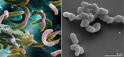
On the left is Caulobacter crescentus, a crescent shaped prokaryote. It
takes two forms, a swarming crescent with flagella for times of
plenty and a stalked crescent for times when food is scarce. In some
parts of the image, you can see the stalk. Atopobium rimae is on the
right, a coccobacillus. A. rimae is a constituent of normal oral flora,
but can cause periodontal disease as well. Its looks remind me of a
microscopic Easter egg hunt.
Coccobacilli

I call these bacteria elliptical; one nice example is Atopobium rimae. They look like footballs, not quite a rod (bacilli are one genus of rod shaped bacteria), and not yet reduced to a sphere.

Many coccobacilli are pathogenic, including the organisms that cause chancroid STDs, brucellosis, pneumonia, infectious blindness, bacterial flu, and whooping cough. However, a link between shape and disease causation escapes me. I haven’t found any evidence that someone has even asked the question. Maybe you can.

Crescent shaped
These are rod shaped bacteria that have become curved. Vibrio bacteria are an important group of crescents (named because they looked like they vibrated as they moved)….. oh, and because they cause a lot of disease.

The crescent shape can be important for movement. Vibrio alginolyticus swims forward just fine, but when it encounters a flat surface – an area where food might be gathered – it swims backward. Its crescent shape keeps it bumping into the flat surface. This keeps it longer in the area of food. In some crescents, a mutation making them straight means that they lose their motility and ability to find food, so it must be important.

Triangular and square
Yes, there are prokaryotes that look very much like triangles or squares. I am listing them together because the thing they have most in come is that they are usually halophiles(halo = salt, and philic = loving) or are halotolerant.
Both of the bacteria above are halophilic, meaning they love salt.
Haloarcula japonica (left) and Holoquadratum walsbyi (right)
for perfectly shaped to form contiguous sheets of cells. This
helps them float parallel to the surface of the water they live in.
These are archaeal extremophiles, but it is interesting that
archaea and bacteria use the same sets of shapes.

The triangular example I have for you is Haloarcula japonica, so named because it was discovered in a Japanese salt evaporation field. Another member of the same genus is H. quadratum, which you might guess, is square.

But it isn’t just this genus that take on definite geometric shapes, there is another salt-loving arachaean called Haloquadratum walsbyi that is also square. Mind you, these are not cubiodal bacteria, and the H. japonicais not a triangular prism. They are very flat; H. walsbyi, for example, is 5 µm square but only 0.1 µm thick. The same can be said for the triangular H. japonica.

They all tend to grow as flat masses, like little floating mosaics. Their flat shapes keep them buoyant and floating parallel to the water’s surface. Their geometry then provides the largest surface toward the sun, in order to pick up the most heat and energy. Sounds logical to me.

Star
These are my favorites, the stars of the bacterial world. The far left is
Stella vacuolata. Those bubbles in the middle are gas – yes, bacteria get
gas too. The middle photomicrograph is Prosthecomicrobium.
Prostheacae are the arms, and are right in the name, even though these
are very short prosthecae. On the right is Ancalomicrobium adetum. Its
prosthecae are much longer and are used to deter predators, amongst
other things. There is even one very long rod bacterium that has a
star cross section.
It must take quite a bit of regulation to build and maintain a star shape. The ones I have seen come in a couple of varieties. Some are definitely star-shaped because of their cell body. Stella vacuolata is star-shaped, as are Prosthecomicrobium species. The reason for a star-shaped adaptation--- I don’t know - patriotism maybe?

There are also bacteria that have projections from their cell body that make them look like fireworks as they explode; see the picture of Ancalomicrobium adetum. The projections are called prosthecae(Greek for appendage), and can serve many functions. They can increase surface area without increasing mass for better diffusion. They can make a bacterium large enough to not be eaten. They may also catch more water, to help non-motile bacteria be moved along by the current.

Other
We have only touched the surface of the morphologies that prokaryotes can assume. There are others that look like Y’s (bifid bacteria), some that look like connected lollipops, some that look like segmented worms, and at least one that builds a net of connected tubules near black smokers at the bottom of the ocean (Pyrodictum abyssi).

Among the many other shapes possible for bacteria, the bifids are
interesting (left). Certain proteins are produced only in the ends of
the bacteria, and if they need more of that protein, especially for
attachment, one way to get it is to have more ends. On the
right is an archaea found at the bottom of the ocean. The fine lines
are actually tubular projections with fine nets of bacteria cell
inside. The cell bodies are the nodular areas seen nonsymmetrically.
Just how do prokaryotes construct and control their shape? This is an active area of research and may be important for medicine. We have seen that shape affects function and survival, so new antibiotics might just work to turn rods into cocci or stars into blobs.

A 2011 paper shows that zinc metal is essential for proper morphology. Zinc is an important part of several proteins that control which DNA is read to make proteins (zinc finger transcription factors), so this is evidence that discrete controls are in place to define a bacterium’s morphology.

A 2013 study shows that E. coli rod shape is determined mostly by its cell wall. If the cell wall was removed, it took 4-6 generations for the rod shape to be recovered. If mutations in certain lipoproteins or penicillin binding proteins were present, the bacteria progeny would always remain spherical. These genes are not even used in producing the cell wall, so it is apparent that many genes are needed just to maintain cell shape. My current research concerns the ability to bend rod bacteria into tiny balloon animals.

Next week we will start our series of exceptions and core concepts for the year. We begin by looking at the elements of life - there's more than you think, and then we'll look at the four types of biomolecules. 


Ranjit DK, & Young KD (2013). The Rcs stress response and accessory envelope proteins are required for de novo generation of cell shape in Escherichia coli. Journal of bacteriology, 195 (11), 2452-62 PMID: 23543719

Bayle L, Chimalapati S, Schoehn G, Brown J, Vernet T, & Durmort C (2011). Zinc uptake by Streptococcus pneumoniae depends on both AdcA and AdcAII and is essential for normal bacterial morphology and virulence. Molecular microbiology, 82 (4), 904-16 PMID: 22023106 

Young KD (2007). Bacterial morphology: why have different shapes? Current opinion in microbiology, 10 (6), 596-600 PMID: 17981076


Life is Elemental

$
0
0
Biology concepts – elements, biomolecules, biochemistry, trace elements, selenocysteine, stop codon

The blue whale is the often 30 m (100 ft) long and can reach a
mass of more than 175 tons (160,000 kg). As such, it is the
largest animal ever to grace the face of Earth. Yet, it owes it
shares its intricate biochemistry with even the smallest
organisms on the planet. The commonality is due to the
chemical elements that make up the biomolecules of all living
things. A few elements are used in most biological compounds,
and many elements are used in just a few. This is today’s story.
A blue whale is the largest animal on the face of the Earth - ever. You could swing a tennis racquet while standing inside a chamber of its heart (left). Built very differently, the watermeal plant is the size of a grain of salt. Comparing the two organisms at the macrolevel is like comparing lug nuts and twinkies, or pink and Darth Vader. But looks are often deceiving.

At the genetic level, about 50% of the genes from whales and watermeal are exactly the same, coding for the same structural proteins or enzymes. At a biochemical level there's even more similarity; even if the gene products are different most of the processes that huge whales and tiny flowering plants carry out are exactly the same.

They are so similar for one overarching reason, and that reason points out an amazing commonality. Both the world’s largest animal and the world’s smallest flower come from a common ancestor. It may have been many moons since their family had that argument at the summer picnic that drove them apart forever, but they are still related nonetheless.

And since they have a common ancestor, they are going to harbor many of the same traits as that ancestor – including the ways they carry out the reactions and functions in their cells. The totality of the molecules that are present in an organism and how they interact to perform different jobs is termed an organism’s biochemistry.
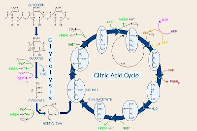
Biochemistry is the chemical reactions that take place in living
organisms, like glycolysis and the citric acid cycle shown above.
Though different organisms may have subtle differences in the
proteins or even eliminate some of the steps, the overall
pathways are conserved across all life on earth. Look at the
molecules, the elements are common in availability and
common to all life. This is evidence of evolution and why
biochemistry is shared so completely.

Biochemistry refers to how information flows through organisms via biochemical signaling and how chemical energy flows through cells via metabolism. All life on Earth uses basically the same biochemistry since we all came from a common ancestor – to the best of our knowledge.

Organisms on Earth have similar biochemistry in part because they use the same types of macromolecules. Life as we know it is based on the interactions (biochemistry) of lipids, carbohydrates, proteins and nucleic acids. Each of these macromolecules is amazing and contains many exceptions, so we will deal with each in next few posts.

Whales and watermeal, all life for that matter, is organic(Greek, pertaining to an organ) since their biochemistry is based on carbon, but there many exceptions to our important molecules being organic. What is the most abundant molecule in living things? Water. Is water organic? No.

What creates the electrochemical gradient that fires our neurons? Sodium, chloride, and potassium. Are they organic? No. So the next time someone makes a joke about being a carbon-based life form, you can say you are just partly organic, and then let them ponder whether you are some kind of cyborg.

So what do the macromolecules have in common that is related to the biochemistry of life? They are made up of the same chemical elements. In fact, most all biomolecules are made up of just five or fewer different elements; carbon (C), hydrogen (H), oxygen (O), nitrogen (N), phosphorus (P), and sulfur (S).

Carbon’s importance lies in its ability to bond to many different elements, and because it can accept electrons in a bond or donate electrons to a bond. Carbon can bond to four different elements at the same time. This increases the possibility of complexity and is one reason our molecules are based on carbon. The situations are similar for oxygen, sulfur, nitrogen, and phosphorous.

One of the lesser abundant elements is sulfur since it is used in proteins as a structural element mostly, although it shows up in bone and other skeletal materials as well. Still, the average adult male (80 kg/175 lb) contains about 160 grams of sulfur; this would be about a salt shaker’s worth.

Carbon is the basis of life on Earth because of its ability to form single,
double, and triple bonds, and because it can bond with so many
different elements. On the left is the top right corner of the periodic
table, showing carbon and silicon in the same column (family).
Elements in the same family have similar chemical properties, so
scientists believe that life on other planets could be based on silicon.
This is how we got the look and feel of the alien in the Sigorney Weaver
movies of the same name. But, silicon is more abundant than carbon,
so why don’t we look like the alien?
Only two of the twenty common amino acids that make up proteins contains sulfur (methionine and cysteine). But don't minimize its importance just because it is present in only two of the protein building blocks. The sulfurs in proteins often interact with one another, determining the protein's three-dimensional structure. And for proteins, 3-D structure is everything  - their function follows their form.

Sulfur is important in other ways as well. Some bacteria substitute sulfur (in the form of hydrogen sulfide) for water in the process of photosynthesis. Other bacteria and archaea use sulfur instead of oxygen as electron acceptor in cellular metabolism. This is one way organisms can be anaerobic(live without oxygen).

In a more bizarre example, sea squirts use sulfuric acid (H2SO4) in their stomachs instead of hydrochloric acid – just how they don’t digest themselves is a mystery. Just about every element has some off label uses; we could find weird uses for C, H, O, N, and P as well. Heck, nitric oxide (NO) works in systems as diverse as immune functions and vasodilation (think Viagra).

So these are the “elements of life” – right? Well, yes and no, you can’t survive without them, but you also can’t survive with only them. There are at least 24 different elements that are required for some forms of life. Two dozen exceptions to the elements of life rule – sounds like an area ripe for amazing stories.

Some of these exceptions are called trace elements, needed in only small quantities in various organisms. It may be difficult to define “trace” since some elements are needed in only small quantities in some organisms, but in great quantities (or not at all) in others. Take copper (Cu) for instance. Humans use it for some enzymatic reactions and need little, but mollusks use copper as the oxygen-carrying molecule in their blood (like we use iron).

Let’s start with a list is of the exceptions; a list will allow you to do some investigating on your own to see how they are used in biologic systems.

Aluminum (Al)        0.0735 g
Arsenic (As)              0.00408 g
Tyrian Purple, or royal purple, is a dye made from the bodies of several
mollusks from the eastern Mediterranean. The spiny dye snail (left) is
one such mollusk that produces the purple dye from its hypobrancial
mucus glands. The dye is based on a bromine-containing compound that
the snails use to protect their eggs from microbial predators (right) and
for hunting. The dye was prized because instead of fading with time and
sun exposure, it actually became brighter. Used as early as 1500 BCE by the
Phoenicians, Tyrian Purple was worth its weight in silver for two
thousand years.
Boron (B)                   0.0572 g
Bromine (Br)            0.237 g
Cadmium (Cd)          0.0572 g
Calcium (Ca)             1142.4 g
Chlorine (Cl)             98.06 g
Chromium (Cr)        0.00245 g
Cobalt (Co)                 0.00163 g
Copper (Cu)               0.0817 g
Fluorine (F)               3.023 g
Gold (Ag)                     0.00817 g
Iodine (I)                     0.0163 g
Iron (Fe)                      4.9 g
Magnesium (Mg)      22.06 g
Manganese (Mn)       0.0163 g
Molybdenum (Mo)   0.00812 g
Nickel (Ni)                   0.00817 g
Potassium (K)           163.44 g
Selenium (Se)             0.00408g
Silicon (Si)                   21.24 g
Sodium (Na)               114.4 g
Tin (Sn)                        0.0163 g
Tungsten (W)            no level given for humans  
Vanadium (V)            0.00245 g
Zinc (Zn)                      2.696 g

You can see that for each element I gave a mass in grams. This corresponds to the amount that can be found in an 80 kg (175 lb) human male. But don’t confuse the mass found with the mass needed.

Barium (Ba) isn’t used in any known biologic system, yet you have some in your body. It is the 14th most abundant element in the Earth’s crust, so it can enter the food chain via herbivores or decomposers and then find its way up to us. You probably have a couple hundredths of a gram in you right now.

Bromine (Br) is a crucial element for algae and other marine creatures, but as far as we know, mammals don’t need any. In fact, this brings up an interesting thing about chemistry. Chlorine is integral for human life, just about anything that requires an electrochemical gradient will use chlorine, to say nothing of stomach acid (HCl).

However, chlorine gas is a chemical weapon that will burn out your lungs (and did in WWI). Bromine gas is very similar to chlorine gas - so elements that are useful as dissolved solids can be lethal as gasses.

How about something supposedly inert, like gold (Ag)? We use it for jewelry because it is rare and supposedly it doesn’t cause allergy (wrong - see this previous post). But some bacteria have an enzyme for which gold is placed in the active center. Gold is rare, so why would it be used for crucial biology? Most elements in biology are more common.

Finally, we should describe a couple of the uses of non-standard elements:

Selenium in proteins is important for stopping damage from
oxygen, but in case you don’t think that is important enough,
how about insulin function. From the cartoon above, you can
see that selenoprotin function affects insulin responsive
elements (IRS) that in turn control DNA function, cell survival
(Akt), and carbohydrate management.
Selenium is a rare element, being only the 60thmost common element in the Earth’s crust. Yet, without 0.00408 grams of selenium on board, a human is only so much worm food. Selenium is only essential for mammals and some higher plants, but it performs a unique role in those organisms.

In a few proteins, particularly glutathione peroxidase, selenium will take the place of sulfur in certain cysteine amino acids. Selenocysteineis an amazing exception because it is not coded for by the genetic code! Instead, the stop codon, UGA, (a three nucleotide run which calls for protein production to stop), is modified to become a selenocysteine-coding codon.

The selenocysteine amino acid changes the shape of the protein, and is found to be the active site for proteins such as glutathione peroxidase and glutathione S-transferase. These enzymes are crucial for cellular neutralization of reactive oxygen molecules that do damage by reacting with just about any other cellular biomolecule.

So selenocysteine is an endogenous biomolecule that is important for protecting our bodies – as important as the antibiotics we use from other organisms. But a 2013 study shows that some antibiotics (doxycycline, chloramphenicol, G418) actually interfere with the production of selenocysteine proteins by inhibiting the modification of the UGA codon. In many cases, the amino acid arginine is inserted instead of selenocysteine, reducing the functionality of the enzymes. Yet another reason to not overprescribe antibiotics.

One last exceptions - silicon is important for many grasses. Remember, this is silicon, the element; not silicone the polymer used in breast implants and caulk; and not silica, the mineral SiO2. Silicon is taken up by grasses of many types; crops, weeds, and water plants (although silicon in grasses may take the form of silica).  

Silicon (top) is an element that is used in many ways, including
in computer chips. Silica is a combination of silicon and oxygen
(middle) which is part of many products as well, including the
lightest material on Earth, aerogel, used in NASA projects.
Silicone is a rubbery material (bottom) that is used in caulks and
in many other things, including creepy movie prosthetics.
In some grasses, the inclusion of silicon makes them less likely to be victims of herbivory (being grazed on by herbivores). Herbivores avoid high silica-containing grasses because they aren’t digested well. A 2008 study showed that this reduced digestibility is related to silicon-mediated reduction in leaf breakdown through chewing and chemical digestion.

Another protective function of silicon in grasses was illustrated by a 2013 study. In halophytic (salt-loving) grasses that live on seashores, increased silicon uptake resulted in increased nutrient mineral uptake, and increased transpiration, the crucial process for water movement through the plant.

In addition, these plants have better salt tolerance in the presence of increased silicon, even though they already have specific mechanisms for reducing the damage that could be induced by such high salt concentrations. Silicon reduced the amount of sodium element found in the saltwater grasses. Pretty important for an element that is considered non-essential.

Next week, let’s start to look at the biomolecules made from C, H, O, N, P, and S. Proteins are macromolecules made up of amino acids, and amino acids are exceptional.

Tobe R, Naranjo-Suarez S, Everley RA, Carlson BA, Turanov AA, Tsuji PA, Yoo MH, Gygi SP, Gladyshev VN, & Hatfield DL (2013). High error rates in selenocysteine insertion in mammalian cells treated with the antibiotic doxycycline, chloramphenicol, or geneticin. The Journal of biological chemistry, 288 (21), 14709-15 PMID: 23589299
 
Mateos-Naranjo E, Andrades-Moreno L, & Davy AJ (2013). Silicon alleviates deleterious effects of high salinity on the halophytic grass Spartina densiflora. Plant physiology and biochemistry : PPB / Societe francaise de physiologie vegetale, 63, 115-21 PMID: 23257076

For more information or classroom activities, see:

Elements of life - 

Trace elements in diet –

Trace elements in plants –

What is biochemistry –

Sulfur –

Bromine –

Selenium/selenocysteine –

Silicon based life –


So Many From So Few

$
0
0
Biology concepts – protein, amino acids, non-standard amino acids, peptide bond


Severe dietary protein deficiency leads to distinct
symptoms, and if not resolved, death. Called kwashiorkor
(Ghanan word meaning “disease from second born”),
the deficiency leads to changes in osmotic potential in
the bodies cells as compared to their blood.
Hypoalbuminemia (low levels of the blood protein
albumin) lead to fluid leaving the vessels and accumulating
in the abdomen, called ascites. This often occurs when infants
stop nursing (like when a second child is born); they take in
enough calories but not enough protein.
Heterotrophic organisms, including us humans, must consume protein in order to survive. Meat is a great source, by far the best protein source per unit mass and the best for obtaining necessary protein subunits (amino acids). If you look at complete protein sources compared to caloric intake, four of the top five foods are: turkey/chicken; fish; pork chops; and lean beef.

Tofu comes in sixth and soybeans are seventh. This is why humans have sharp canine teeth – we're meat eaters. You can live happily (well, somewhat happily) as a vegetarian; you just have to work much harder at it.

So why is protein so important? How about, because it is one of the four major biomolecules and without it you die a horrible death? Sounds like a good reason to me.

Proteins reside in every cell of every living organism, from prokaryotes to your favorite uncle. There isn’t a job in a cell that proteins don’t have their hands in; proteins even perform numerous tasks at the extracellular level. Heck, that spider web hanging from your dusty Stairmaster is made of protein!

From prokaryotes to spiny echidnas to rosebushes, let’s look where proteins are involved in life. Proteins provide the structure from which cells hold their shape and onto which they build a membrane. Proteins do the talking, providing chemical signals and ways to sense chemical signals.

Proteins do the dirty work; as enzymes they put molecules together, cut them apart, and change their parts around. And most times, they make these reactions happen faster than they would otherwise and without being used up in the process.


Enzymes are specific for a very few molecules (called
substrates). Enzymes have a particular shape, and this
allows the correct substrate to bind and be acted on;
called the lock and key system. Notice that the
enzyme itself is not altered by the reaction, so it can
work again on another substrate molecule. However
there are exceptions – suicide enzymes are inactivated
by their own action, so they only work once.
Proteins allow for movement, like the contractile proteins in your muscles or the proteins that make up flagella and cilia. Proteins even act as defenders of the cell, as antibodies and myriad other immune molecules.

A typical cell may contain 10 billion protein molecules. However, not every cell has the same proteins. Many proteins are necessary for every cell, but others have specialized functions needed in only some cells. The exception is unicellular organisms. Their one cell must be able to produce every kind of protein they might ever need.

Space is at a premium, so cells can’t waste room on proteins that aren’t needed right now. Therefore, making protein must be efficient, tightly regulated, and fast. Over 2000 new protein molecules are made every second in most cells, while some proteins exist only to destroy unneeded or old proteins.

Humans can make about 2 million different proteins, but we only have about 25,000 genes that code for them. We accomplish this by having some genes produce many different proteins, just by changing the parts of the gene used. These alternative splice variant proteins may have different functions even though they come from the same gene. For example, the cSlo gene is required for hearing, and each one of the 576different splice variants is responsible for sensing a different frequency. Biology is just so dang efficient.

Now that you know how important proteins are, let’s find out what they are. Proteins are polymers (poly = many, and mer= subunit) made up of bonded amino acid mers. Proteins come in many sizes; the TRP-Cage protein of gila monster spit is a polymer of only 20 amino acids, while the titin protein of your connective tissue is over 38,000 amino acids long.

Maybe we'll dig into the degeneracy of the genetic code when we talk about nucleic acids, but for now let’s just accept that DNA triplets code for different amino acids, and the order of the codons determines the order in which amino acids are linked to form a specific protein. The order of the different amino acids is the key. Why? I’m glad you asked.

Amino acids (or aa’s) are all small molecules made up of carbon, hydrogen, oxygen, nitrogen, and sometimes sulfur – five of last week’s “elements of life.” It’s the arrangement of these elements that makes an amino acid. Refer to the picture below for a visual aid. The central carbon is bound to four other things (often called moieities). One is simply a hydrogen. Another is an amino group (contains the nitrogen). The third is a carboxylic acidgroup. Get it? amino acid.


While not the most exciting images, these cartoons should
help you understand the structure of the amino acid (left)
and the building of the proteins (right). Each amino acid has
the same structure, except for whatever the R group might
be. The amino end of amino acid 2 is joined to the carboxylic
acid of amino acid 1. The next peptide bond would be between
the carboxy end of amino acid 2 and the amino end of amino
acid 3. Notice how water is created each time a peptide bond
is made.

The fourth group is what makes each aa different. Called an R group, this side chain can be small or big, neutral or charged, and gives the aa its properties. The R stands for something, but that story is just too long.

In glycine, the R group is merely another H, but in tryptophan it contains complex rings. We have talked about how tryptophan is the least used amino acid; it is bulky and introduces big bends in the peptide. We’ll show that bends, kinks and other interactions between aa’s are important for the protein function.

Most organisms can make all the amino acids they need, but mammals are the exception. We have abandoned (genetically) pathways for making some aa’s, so we must get them from our diet. These are the essential amino acids, of which there are nine if you are healthy. Tryptophan must acquired by all animals – good thing plants still have the recipe.

Ribosomes (made of proteins and nucleic acids) link the individual aa’s together in the order demanded by DNA via the mRNA. The bond that connects them is called a peptide bond, and is a “dehydration” or “condensation” reaction.

Look at the amino acid picture again; the peptide bonding process kicks out water, ie. dehydration (de = lose, and hydro– water). Water forms from seemingly nowhere, like condensation on your mirror. See how fitting the names are?

When in a protein chain (also called a peptide), the order of aa’s is called the protein’s primary (1˚) structure. The primary structure in turn dictates the secondary (2˚) structure, which is a folding of small regions of the protein based on the interactions of the side chains of closely associated amino acids.

In turn, the folding of small regions brings together aa’s from farther apart, and they fold up based on their interactions. This is the tertiary (3˚) structure of the protein. If a protein needs more than one peptide chain to be functional, the shape that those different chains form when they interact is called the quaternary (4˚) structure.

These cartoons can help you picture how an individual amino
acid can affect the structure of an entire protein. In the
secondary structure cartoon, there are two basic forms that
the nearby amino acids can form, helices and sheets, other
parts will form no patterned form at all. The tertiary and
quaternary cartoons are for hemoglobin, showing how non-
amino acids may be involved (heme), and how the
individual peptides fit together.

The hemoglobin that carries oxygen in our red blood cells is made up of four protein subunits. Why is this important – because what the protein does in life is completely dependent on its three dimensional shape. Lots of aa’s means lots of potential shapes. This is in itself one of the greatest exceptions, since one of the basic tenets of biology is “form follows function.” But with proteins, function follows form.

For the greatest number of possible combinations and shapes, it’s lucky that DNA codes for 20 aa’s. Or are there more? Proteinogenic aa’s are those that can be added into a growing peptide chain, and there are actually 22 of them. The two exceptions are selenocysteine (like cysteine with selenium substituting for sulfur) and pyrrolysine(like lysine with a ring structure added to the end).

We talked last week about the functions of selenocysteineand how it can be incorporated into a peptide even though there isn’t a normal mRNA codon dedicated to it. Pyrrolysine is similar in that it becomes coded for after the modification of what is usually a stop codon, in this case UAG (a signal to add pyrrolysine is located after the UAG codon).

Pyrrolysine is used by methanogenic (methane producing) archaea and bacteria. It's important in the active site of the enzymes that actually produce the methane. New research is showing that more organisms than previously believed use pyrrolysine. A 2011 study identified more than 16 archaea and bacteria with pyrrolysine coding mRNA modifications, but it looks like there may be more.

While the mammalian titin protein is the largest protein
known (38,136 amino acids), there is a close second in a
bacterium called Chlorobium chlorochromatii CaD3. The
gene has been found for a protein of 36,000 amino acids,
but we don’t know yet of the protein is actually made. In
archaea, the halomucin protein from the square prokaryote
Haloquadratum walsbyiis 9,200 amino acids but is exported
to protect the organism from its extreme environment.

A 2013 study indicates that the typical modification of the mRNA that occurs 100 bp downstream of the UAG stop codon isn't even there in some pyrrolysine-coding genes. One hypothesis is that in genes without the modification, the UAG sometimes acts as a stop codon and sometimes incorporates a pyrrolysine. Therefore, there are truncated (prematurely stopped) and full-length versions of the protein in the cell, and the relative number of each can be affected by local conditions and stressors.

In this paper, the authors have developed a different predictor, which doesn’t rely solely on the presence of the modification. Using it, they have identified many new candidate genes in archaea and bacteria that could be using pyrrolysines. Here’s my question – all organisms use selenocysteine, but it seems only arachaea and a few bacteria use pyrrolysine. Why did it go away in higher organisms? Can it only be used for methane production? Please, no methane production jokes.

Pyrrolysine and selenocysteine are coded for by mRNA and are added to proteins, so we definitely have 22 aa’s, but could there be more? You betcha. There are over 300 non-standard amino acids, but that isn’t such a big deal. Remember the definition of amino acid; a central carbon with a hydrogen, a carboxylic acid, an amino group, and something else attached. It isn’t a wonder there are many of them.


Bacteria kill bacteria all the time. They make their own
antibiotics, called bacteriocins, by modifying short peptides
so that they interfere with cell wall synthesis in other strains.
To do this, they modify amino acids in peptides to non-standard
amino acids, including lanthionine and 2-aminoisobutyric acid.
Those that contain lanthionine are called lantibioticsand are
hot commodities right now.
A few non-standard aa’s can be found in proteins, like carboxyglutamate which allows for better binding of calcium, and hydroxyproline, crucial in connective tissue function. These are formed by modifying the amino acids already added to the growing peptide chain.

Other non-standard aa’s are produced as intermediates in other pathways and are not used in proteins. The list of them is great and their functions are even greater, but some act as neurotransmitters, others are important in vitamin synthesis, especially in plants. Still think life uses just 20 amino acids?

Next week we can finish up proteins. Life is very selective with the form of its amino acids – except when it isn’t.


Theil Have C, Zambach S, & Christiansen H (2013). Effects of using coding potential, sequence conservation and mRNA structure conservation for predicting pyrrolysine containing genes. BMC bioinformatics, 14 PMID: 23557142

Gaston MA, Jiang R, & Krzycki JA (2011). Functional context, biosynthesis, and genetic encoding of pyrrolysine. Current opinion in microbiology, 14 (3), 342-9 PMID: 21550296


For more information or classroom activities, see:

Dietary proteins –

Functions of proteins –

Standard amino acids –

Peptide bond –

Protein structure –

Non-standard amino acids -

Three Lefts Make A Right

$
0
0
Biology concepts - chirality, homochiral, enantiomer, stereoisomer, racemic mixture, biofilm, antimicrobial peptide, protein, amino acid


Geordi was the chief engineer on Start Trek; The Next
Generation, but I was partial to Scotty on the original.
They both work on the matter-antimatter reactors that
powered everything from the warp drive to the phasers.
Don’t laugh – NASA is working on a warp drive as we
speak, and we have been able to produce antimatter for
years. Not sure how antimatter will solve our energy
problems though, it takes much more energy to produce
it than we get from a matter-antimatter reactor.
Today we’ll start with a story that may seem to have nothing to do with biology. Hopefully we can draw a parallel later.

For every type of every subatomic particle there is an opposite particle. There are protons and anti-protons, electrons and anti-electrons (positrons), neutrons and anti-neutrons. Together, they make up matter and antimatter– think Star Trek.

So why is our universe made of matter and not antimatter? It turns out that when matter meets antimatter, they obliterate one another. This is bad – Scotty was always trying to prevent a matter/antimatter catastrophe on the Enterprise.

When the universe was young, there was antimatter and matter; lots of annihilations. Scientists hypothesize that there were a few more molecules of matter than antimatter, so when the fireworks were over, only matter remained; so matter matters.

What’s this got to do with protein exceptions, our subject for last week and this? We’ll get to that, a little background first. Remember that the shape of the protein is important for its function, and its shape is dependent on the amino acid order and the structure of those amino acids.

Look at your hands. They’re mirror images of one another, but no matter how you try, you can’t make your left glove fit exactly on your right hand. Unlike a ball reflected in a mirror where the two images will be superimposable on one another, there is no way to do this with say….. your left and right shoes.


If you reflect your right hand in a mirror, you get an
image exactly like your left hand. But your right and
left hands can’t be superimposed – try it, turn one over,
turn the other over, turn them around the over
direction, detach one – you still can’t do it. It is the same
with chiral molecules. The different groups come at you
or away from you, and once you reflect them, no two can
be superimposed – try it, at least one group will be
pointing the wrong direction.
Molecules like this in chemistry are called chiral; the amino acid central carbon (chiral carbon) has four different groups, so no flipping will make the two mirror images look exactly the same. The two different amino acid variations are called stereoisomers, specifically, enantiomers.

The different enantiomers (enantio = opposite) have different chemical properties. Without getting into too much chemistry (nobody wants that), different enantiomers cause light waves to rotate in different directions.

One version of a molecule called gluteraldehyde rotates light to the right (dextrorotary or D, dextra = right), while the other is levorotary (levo = left). Amino acid enantiomers are comparable to the structure of gluteraldehyde, so amino acids that parallel the two gluteraldehydes are assigned a D- or L- label, ie. L-alanine has a structure similar to the gluteraldehyde enantiomer that rotates light to the left.

This is a bit of a misnomer because light rotation depends on many factors. In fact, many amino acids labeled L-type because of their similarity to L-gluteraldehyde actually rotate light to the right –that little factoid won’t be on anyone’s final exam.

Most proteins fold on their own, and they fold the same way every time. But what might happen if the protein sometimes used a certain L-amino acid and sometimes the D-version? The two resulting proteins would fold differently and therefore have different possible functions. Heaven forbid!

To avoid this, nature has devised an ingenious solution - only L-amino acids are used. This assures that all proteins of a specific type assume the same shape, and it turns out that proteins are most stable when they are made of all L- or all D-amino acids. This is the rule of homochirality.


Polarized light has practical implications. Your 3-D movie
glasses may be plane polarizers. The light from the screen
is a mix of right and left polarized light. One lens lets in
one, while the other lets in the opposite light. This splits
the image into two images, one for each eye, and they are
separated by a distance. This gives a stereoptic image, just
like your eyes seeing something in real space.
This is one of the best-known rules of biology, right behind the “form follows function” we talked about last week. But we saw that proteins were exceptions to that rule, so there must be exceptions to this one as well.

One question before the exceptions -just why did life opt for all L-proteins instead of all D-proteins? Do they annihilate one another when they come into contact, like matter and anti-matter? Thankfully, no.

Were there more L-amino acids available on the early Earth so life made a choice and stuck with it? Maybe – this is one of the hypotheses currently being investigated. It’s also possible that some life developed as D-protein makers, but they were out-competed somehow and we descended from the L-protein winners. It isn’t as easy a question as the physics matter/antimatter issue - more on this biology exception next week. There are so many more exceptions in biology as compared to physics – that’s why I love life more than physics itself.

The most mundane is exception to homochirality is the one amino acid that isn’t chiral. Glycine’s R group is just a hydrogen, so the central carbon has two bound hydrogens, and the mirror images can be superimposed. There’s no L-glycine or D-glycine, just glycine. Don’t worry, the rest of the exceptions are better.

It turns out that the rule of homochirality is more of a guideline - there are examples of important D-amino acids (D-aa) in plants, animals, and prokaryotes. We don’t have time or space to go into many of them, but I will highlight two exceptions that are simply amazing. Let’s start with the bacteria since they own the planet, and we probably inherited our D-amino acid uses from them.

Bacteria use D-amino acids in various ways. First, new research shows that as bacterial numbers go up and resources (food) go down, D-aa-containing proteins might prepare bacteria for the bad times ahead. A 2009 study shows that Bacillus subtilisand Vibro cholerae make large amounts of D-aa as they age, including D-tryptophan, D-tryosine, D-phenylalanine, and D-leucine. As the amounts increase, they start to have an effect on the bacterial cell wall.


A gram positive bacterium has a thick cell wall, which
includes peptidoglycan. The glycan parts are NAM and
NAG. The NAM has a tetrapeptide attached to it, and this
is where the D-aa can be used. This strengthens the cell
wall and makes it even thicker. The lipoteichoic acid in
the outer layer is where change to a D-aa prevents
defensins from sitting in the cell wall and disrupting
the buried plasma membrane.
The D-aa are incorporated into the growing peptidoglycan, the elastic and stress-bearing component of the cell wall, and they also regulate enzymes that control the thickness and structure of the peptidoglycan. By putting D-aa into the cell wall, the bacteria make themselves strong for the lean times ahead.

What is more, a 2010 study showed that in B. subtilis and S. aureus, the increasing numbers of bacteria and their increasing concentration of D-aa’s leads to a breakdown of the extracellular matrix that holds all the bacteria together (the biofilm). D-aa's were able to prevent biofilm formation and degrade existing biofilm, again preparing the bacteria to go off on their own as resources dwindle.

S. aureus also uses D-aa-proteins to avoid being killed. We have antimicrobial peptides (AMP) on our skin and mucosal surface that are always looking to kill bacteria. They often work by poking holes in the bacterial membrane. However, a 2013 studyshows that by switching out L-alanine to D-alanine in its cell wall, S. aureus can render the AMP’s ineffective. The D-alanine gives the protein a different shape, so the AMP's can’t fit in and do their job. Smart bacteria.

Animals have gotten into the act, especially the gastropod mollusks, ie. some snails and sea hares (marine slugs). D-tryptophan is their exception of choice. The cone snails (genus Conus), the most predatory and venomous of the marine snails, use D-tryptophan in the active peptides of their venom, called contryphans. Large cone snails have been known to kill humans, but the role of the D-tryptophan in the venom activity is not yet known.


The cone snails use many peptide venoms to incapacitate
their faster prey; they move like snails don’t ya know. The
siphon has the black stripe and tests the water for prey.
Then the proboscis below (kind of pinkish orange at the
tip) sends out the radula that envenomates the victim.
The sea hare, Aplysia kurodai, also uses D-tryptophan in a cardioexcitatory neuropeptide called NdWFamide (it speeds up the slug’s heartbeat). This protein has also been found in terrestrial slugs, so it may be that many mollusks are D-aa users.

How about us? Do humans use D-amino acids? Sure we do. But there are tricksters here. D-alanine is found in all mammals, but we don’t know what it might be doing. Its levels change with the time of day (circadian cycling), so who know what it might be controlling. A 2013 study set out to determine what drove the circadian changes. They tested several things: changing diet or fasting didn’t matter; enzymes that degrade D-alanine didn’t change levels of function either. But when they studied germ-free mice, the D-alanine levels didn’t change.

The scientists determined that it’s our gut bacteria making D-alanine, and circadian changes in intestinal absorption rates is the reason that the D-alanine levels fluctuate. It doesn’t mean that D-alanine isn’t doing something, but it sure had us fooled for a while.

Mammals don't stop there; D-serine and D-aspartate are so important, we have special enzymes called racemases whose job it is to convert their L-forms to D-forms (racemic mixtures contain both D- and L-versions of a molecule). The most amazing exception must be D-serine. However, we will see that there is a Goldilocks effect to D-aa’s, you don’t want too much or too little.

A certain brain neuron receptor (called NMDAR, important for learning and memory) is activated by an amino acid called L-glutamate, but it needs help from either glycine or D-serine to set off the electrical impulse. It turns out that in Lou Gehrig’s disease (amyotrophic lateral sclerosis), lower motor neurons die because they undergo aberrant excitation. In genetic cases of ALS, patients have too much D-serine!


Most patients with ALS are diagnosed after age 50 and
live about five years. Stephen Hawking- the Nobel
winning cosmologist – was diagnosed at 21 and has
lived with the disease for 50 years! Hawking has round
the clock care, for the only muscles that work for him
are breathing, swallowing an eye movement. Lucky for
him, most people’s breathing and swallowing are not
spared and this is how they die. He uses his eye
movement to run his computer.
A 2012 study showed that D-amino acid oxidase (DAAO) is mutated in these patients and doesn’t do its job of breaking down D-serine. Mice with a mutated DAAO were shown to have decreased lower motor neurons and more ALS signs.

So too much D-Ser is bad, but how about too little? Many studies have shown that schizophrenia patients have low levels of D-Ser. It might be that DAAO is too active, or perhaps a D-Ser racemase is inactive, or maybe there is just too little L-Ser to make D-Ser from – we don’t know yet. 

A 2013 study has also implicated D-aspartic acid in schizophrenia. D-Asp can replace L-glutamate in activating NMDA receptors, and schizophrenic patients have low D-Asp levels in the brain and blood. The D-Ser and D-Asp data implicate glutamergic receptor activation in schizophrenia, so much work is underway to find ways to increase these D-aa’s in our brains - the very things that the rules say we shouldn’t be using in the first place. But let’s not raise them too much, no one wants ALS!

Next week, we switch our attention to carbohydrates, the energy sources in our cells. Every cell on Earth is designed to make ATP from glucose - except for those cells that ONLY use fructose.


Lam H, Oh DC, Cava F, Takacs CN, Clardy J, de Pedro MA, Waldor MK. (2009). D-amino Acids Govern Stationary Phase Cell Wall Re-Modeling in Bacteria Science, 18 (325), 1552-1555 DOI: 10.1126/science.1178123

Kolodkin-Gal I, Romero D, Cao S, Clardy J, Kolter R, Losick R. (2010). D-Amino Acids Trigger Biofilm Disassembly Science, 328 (5978), 627-629 DOI: 10.1126/science.1188628
Sasabe J, Miyoshi Y, Suzuki M, Mita M, Konno R, Matsuoka M, Hamase K, Aiso S. (2012). 

D-amino acid oxidase controls motoneuron degeneration through D-serine. Proc Natl Acad Sci U S A. , 109 (2), 627-32 DOI: 10.1073/pnas.1114639109 

Simanski M, Gläser R, Köten B, Meyer-Hoffert U, Wanner S, Weidenmaier C, Peschel A, & Harder J (2013). Staphylococcus aureus subverts cutaneous defense by d-alanylation of teichoic acids. Experimental dermatology, 22 (4), 294-6 PMID: 23528217

 Errico F, Napolitano F, Squillace M, Vitucci D, Blasi G, de Bartolomeis A, Bertolino A, D'Aniello A, & Usiello A (2013). Decreased levels of d-aspartate and NMDA in the prefrontal cortex and striatum of patients with schizophrenia. Journal of psychiatric research PMID: 23835041

For more information or classroom activities, see:

Chirality –

Bacterial cell wall –

Biofilm –

Antimicrobial peptides –

D-amino acids –

Amyotrophic lateral sclerosis –

Schizophrenia -


Sugars Speak In Code

$
0
0
Biology concepts – carbohydrates, monosaccharides, hexose, glycocode, starch, glycogen, carbohydrate linkage, bacterial persisters, fructolysis


Refined sugar is produced from two main sources, sugar cane (37
different species of grass from the genus Saccharum, bottom right),
and sugar beet (Beta vulgaris, top right). Sugar cane accounts for
80% of the sugar produced today. The cane or the beets are ground
and the sugary juice is collected with water or on its own. To refine
the sugar, which still has molasses from the fiber, is processed with
lime or soda and evaporated to produce crystals. The color is
removed by activated charcoal to produce the white sugar we most
often see (top middle). Brown sugar is sugar in which the molasses
has not been removed and still coats the crystals (bottom middle).
Unprocessed sugar from cane is shown on the bottom right, while raw
sugar (not whitened) is on the top right.
It would be hard to argue that without sugars, none of us would be here. Glucose provides us with short and medium term storage of energy to do cellular work, but would you believe that certain parts of reproduction use a completely different energy source. All hail fructose!

Sugars are better termed carbohydrates, because they are basically carbon (carbo-) combined with water (-hydrate). The general formula is Cn(H2O)n; for instance, the formula for glucose is C6H12O6.

The simplest sugars are the monosaccharides (mono = one, and sacchar from the Greek = sugar. They can be composed of 4-7 carbons, called tetroses (4 carbon sugars), pentoses (5), hexoses (6), and septoses (7).

Things aren’t so simple though, even for the simple sugars. Let’s use the hexoses as an example, although what we say will also apply to the other sugars. We said the formula for glucose is C6H12O6, so that makes it a hexose. Is it the only hexose – heck no! Hexoses can be aldoses or ketoses, depending on their structure (see picture). Even more confusing, -OH groups can be located on different carbons making them act different chemically.


This chart is a brief introduction to the complexities of simple
sugars. They can vary in the number of carbons (triose vs.
pentose vs. hexose. They can also vary in their structure even
if they have same number of carbons (glucose vs. galactose).
Yet another difference can come in their reactive group on the
end, being either a ketone group (ketoses) or an aldehyde
group (aldoses).
There are actually 12 different hexoses – some names you know; glucose, fructose, or galactose. Others are less common; idose, tagatose, psicose, altrose, gulose – you won’t find those in your Twinkies. Then there are the deoxysugars, carbs that have lost an oxygen. Fucose is also called 6-deoxy-L-galactose, while 6-deoxy-L-mannose is better known as rhamnose.

If this wasn’t difficult enough, stereoisomers again rear their ugly head, as it did last week with the proteins. Hexoses have three (ketoses) or four (aldoses) chiralcarbons each so hexoses can have eight or 16 stereoisomers! Every isomer may act differently from every other; this allows for many functions. But wait – there’s more trouble when we start linking sugars together.

Simple sugars can be joined together to build disaccharides (two sugars), oligosaccharides (3-10), and polysaccharides (more than 10). The subunits are connected by a hydrolysis reaction. Just like with the amino acid linkages in proteins, a water molecule is expelled when two sugars are joined together. Sucrose (table sugar) is a disaccharide made up of a glucose linked to a fructose.

Just where the linkage takes place is also important. Our example again can be glucose. Many glucoses can be linked together with an alpha-1,4 linkage. Long chains of glucoses linked in this way are called starch or glycogen, based on the different branching patterns they show. Mammals store glucoses as glycogen, while plants store them as starches.


Amylose is one type of starch, amylopectin being another.
They are different from celluloses only by the way the sugars
are linked together. You can see that in starch the CH2OH
group are all on the same side, while in cellulose they alternate.
This may seem like a small difference, but we can digest only
starch (or glycogen, which has the same type linkages),
not cellulose.
Humans can digest both starch and glycogen because we have enzymes that can break alpha-1,4 linkages. But if you change the chemical shape of the bond (see picture) to a beta-1,4 linkage, the glucose polymer becomes cellulose.

Plants make a lot of cellulose for structure, but even though it is made completely of glucose, humans can’t digest it at all! Ruminate animals can digest cellulose, but it takes some powerful gut bacteria to help out, and one of the side effects is a powerful dose of methane. Cows are the greatest source of methane on the planet!

We have talked about carbohydrates as energy sources, but pretty much every biological function and structure in every form of life involves carbohydrates.

Carbohydrates are important structural elements. Cellulose, thousands of beta-1,4-linked glucoses, help give plants their rigidity, especially in non-woody plants, but in woods as well (linked together by lignin). As such, cellulose is by far the most abundant biomolecule on planet Earth.

Chitin is another structural carbohydrate. Chitins make up the spongy material in mushrooms, and the crunchy stuff of insect exoskeletons.  You don’t get much more structural than keeping your insides inside.

Carbohydrates are often part of more complex molecules as well. Nucleic acids like RNA and DNA have a five-carbon ribose or deoxyribose at the core of their monomers. Glycolipids and glycoproteins (glyco- from Greek, also means sweet) are common in every cell. Over 60% of all mammalian proteins are bound to at least one sugar molecule.

The different sugar-linked complexes are part of the glycome (similar to genome or proteome), including oligo- and polysaccharides, glycoproteins, proteoglycans (a glycoprotein with many sugars added), glycolipids, and glycocalyxes (sugar coats on cell surfaces). None of these carbohydrate additions are coded for by the genetic code, yet a great diversity of glycomodifications are found on most structures of the cell.


The carbohydrate code is still a mystery to us. The glycosylation can be
linked together by N-type or O-type linkages, the order of the sugars
can vary, the numbers of each type of sugar can vary, and the branching
can vary. Every difference adds to the complexity of the code and can
direct a different message to the cell or the molecules with which
these glycans come into contact.
The diversity and complexity of these added carbohydrates is highly specific and highly regulated – this is the glycocode or carbohydrate code. Yet, we haven’t even come close to breaking the code, i.e., what series of what sugars means what.

The glycocode is important for cell-cell communication, immune recognition of self and non-self, and differentiation and maturation of specific cell types. Dysfunction in the glycocode leads to problems like muscular dystrophy, mental defects, and the metastasis of cancer – we better get cracking on the code breaking.

In the middle of 2013, a new method was developed for detecting the order and branching of sugars on different molecules. This method uses atomic force microscopy (AFM) to actually bump over the individual sugars on each molecule and identify them by their atoms, even on live cells. I’m proud to say that my father-in-law played a role in developing AFM for investigation of atom distributions on the surfaces of solid materials, mostly superconductors.

The glycome is even more diverse because different types organisms make different sugars. One thing I find interesting is that mammals don’t make sucrose. No matter what we mammals do, we won’t taste like table sugar when eaten – more’s the pity. I wonder what a sweet pork chop might taste like.


Proof that many foods have sugars – the Maillard reaction. That gorgeous
browning of your bread or steak comes from a chemical interaction
between the sugars and amino acids of the food. In the process, hundreds
of individual different compounds are made, each with a different flavor
profile. The example in the chart above is for caramelizing onions. Each
food and its chemical make up produces a different set of Maillard
products. You roast your coffee beans for the same reason. This is why
Food Network always suggests ways for you to get great searing and
browning of food.
We use sucrose as sugar because it is relatively easy to obtain from the plants that do make, like sugarcane or sugar beets. Fructose (often called fruit sugar) is actually sweeter on its own; almost twice as sweet as sucrose and three times as sweet as glucose.  This explains why so many sweetened foods are full of high fructose corn syrup (go here for our previous discussion of high fructose corn syrup).

We all know that organisms use glucose as an energy source, first through its breakdown to pyruvate via glyceraldehyde -3- phosphate (G3P) in glycolysis; the pyruvate then travels through the citric acid cycleto produce enough NADH and NADPH to generate a lot of ATP. But fructose can be used as well.

Fructose undergoes fructolysis, different from glycolysis only in the fact that one more step must be taken to generate G3P (adding the P to G3 is done by the enzyme trioskinase). In humans, almost all fructose metabolism takes place in the liver, as a way to either convert fructose to glucose to make glycogen, or to replenish triglyceride stores – so be good to your liver.

The big exception is how important fructose is in mammalian reproduction. Spermatozoa cells use fructose as their exclusive carbohydrate for production of ATP while stored in the testes. This fructose comes not from the diet but the conversion of glucose to fructose in the seminal vesicles.

Why use a different carbohydrate source just for sperm? Seminal fluid is high in fructose, not glucose. Perhaps this is a factor in seminal fluid viscosity. If this problem is solved using fructose, then the cells swimming in it would probably switch evolve to use it as an energy source.

I asked Dr. Fuller Bazer of Texas A&M about this and he pointed out that fructose can be metabolized several different ways, and some of these lead to more antioxidants and fewer reactive oxygen species - it would be important to leave sperm DNA undamaged, especially since we have previously talked about how they are more susceptible to oxidative damage.

Bazer also pointed out that unlike glucose, fructose is not retrieved from tissues and put back into circulation. Once it’s sequestered to the male sexual accessory glands, it would stay there. Still lots to be learned in this area.


Fructose is sweeter than glucose. Sucrose is one glucose joined to one
fructose, so the ratio is 50:50. In most honey, the fructose:glucose ratio
is about 55:45, so it is often sweeter than table sugar. Since it is higher
in fructose, some people liken it to high fructose corn syrup, but there
are many compounds in honey that also help the immune system, etc.
However, recent evidence is showing that some honey is being diluted
with high fructose corn syrup and some bees are being fed HFCS. The
benefits from true honey are then lost.
A 2013 study shows that maternalintake of fructose can also affect reproduction. Pregnant rats fed 10% fructose in their drinking water had significantly fewer babies, but a greater percentage of the offspring were male (60% versus 50%). The fructose did not arrest female embryos from developing or have a sex-specific effect on sperm motility, suggesting that the sugar has a direct effect on the oocyte that increases the chances of being fertilized to produce a male. Weird.

Using sugars other than glucose may be a big deal for mammals, but bacteria can thrive on many different sugars. E. coli can process glucose, but if other sources of sugar are around, they will switch over in a heartbeat – if they had a heart. E. coli has a whole different set of genes for lactose metabolism, found in something called the Lac operon. The operon gets turned on only if lactose is present and glucose is not.

The ability for bacteria to use other sugars might save us as well. Some bacteria can just shut down their metabolism if antibiotics are present and just hangout until the drugs are gone. These are called persister organisms, and they are different from antibioticresistant bacteria. A 2011 study showed that if you give sugar in combination with some kinds of antibiotics, the persisters just can’t resist the sweet treat and will not shut down their metabolism. The antibiotics then become effective. Using sugars we don't metabolize, like fructose or mannitol, ensures that they will be around to help kill the bacteria. Amazing.

We have just brushed the surface of sugary exceptions. Next week we will see how nature first selected a single type of sugar to use in biology, and then went right out and broke its own rule.


Gunning AP, Kirby AR, Fuell C, Pin C, Tailford LE, & Juge N (2013). Mining the "glycocode"--exploring the spatial distribution of glycans in gastrointestinal mucin using force spectroscopy. FASEB journal : official publication of the Federation of American Societies for Experimental Biology, 27 (6), 2342-54 PMID: 23493619

Gray C, Long S, Green C, Gardiner SM, Craigon J, & Gardner DS (2013). Maternal Fructose and/or Salt Intake and Reproductive Outcome in the Rat: Effects on Growth, Fertility, Sex Ratio, and Birth Order. Biology of reproduction PMID: 23759309

Allison KR, Brynildsen MP, & Collins JJ (2011). Metabolite-enabled eradication of bacterial persisters by aminoglycosides. Nature, 473 (7346), 216-20 PMID: 21562562 


For more information or classroom activities, see:

Testing for carbohydrates in foods –

Structures of carbohydrates –

Glycocode/carbohydrate code –

It’s Not Just Our Tooth That’s Sweet

$
0
0
Biology concepts – homochirality, carbohydrates, chiral discrimination, glycoside, H antigen


It isn’t just biomolecules that show chirality. There is also
chiromorphology, like snail shells that usually turn to the right
(dextral, or D-). There are factors in early embryonic
development that cause the body and shell to be right handed
in most gastropod species, yet other species are left handed.
There are also instances where a right-handed species will
produce a left-handed individual, so shell collectors have to be
on the look out for abnormal individuals.
A couple of weeks ago we talked about how, in most cases, life uses exclusively the left-handed enantiomers of amino acids to make proteins.  This homochirality is also see in the sugars we talked about last week, but in this case, mostly D-sugars are utilized in biological systems.

What isn’t amazing is that it happens to be L- for amino acids and D- for carbohydrates; the fact that they’re different is no big deal. Evolution just wants the parts to fit together, so if an enzyme evolved to use D-sugars, it’s not a surprise that the D-sugar would be favored in the pathway then now on. 

But it might not have been random either. No one knows for sure, but hypotheses abound for how homochirality in these biomolecular monomers was established.

One 2009 paper was concerned with the maintenance of homochirality rather than its establishment. Dr. Soren Toxvaerd stated that if you don’t believe life as we see it today occurred in a singular event, then it must have developed over a long period of time. Evidence indicates that small changes in the self-assembly of biomolecules took place over at least thousands of years.

If life took a long time to develop, then prebiotic (before life) earth must have been fairly stable in terms of enantiomer concentrations. But we know that homochiral solutions will turn to racemic mixtures (containing both L- and D- enantiomers) in a short time, days for amino acids and just hours for sugars. So how could the environment have been stable enough for life to develop over time?


One possible hypothesis about the establishment of homochirality
was put forth in 2010 by Koji Tamura, PhD in the Journal of
Cosmology. Put very simply, RNA may have developed before
proteins. RNA evolved to use only D-ribose because a mixture
would have been a symmetry violation. The action of D-ribose
would have been driven toward L-amino acids because of shape
problems with attaching D-amino acids to tRNAs. Now prove it.
Louis Pasteur, he of bacteria-free milk and germ theory, may have shown us the way. He discovered chiral discrimination. Racemic mixtures, under the right conditions, will separate into pools of homochirality. There is an energy gain and stability to packing homochiral molecules together; the other enantiomer will be excluded. This could help explain life using one enantiomer only.

What is more, hydrothermal vents and black smokers have just the needed conditions for both chiral discrimination and for self-assembly of biomolecules. Interesting huh? Think it’s a coincidence that black smokers harbor some of the oldest archaea on Earth? We may owe our very existence to plumes of superheated water and the xenophobia of enantiomers.

Lastly in this area, it may be that sugars and amino acids selected each other for homochirality. Glyceraldehyde is 1) highly discriminate for its enantiomers, 2) was present in large amounts in prebiotic oceans, 3) is used in self-assembly of many biomolecules, and 4) D-glyceraldehyde very much likes to bind to L-serine. So a slight excess in either one of these could have helped select for the other, and if this was stable, it could have caught on like “Gangnam Style.” This may be why life uses mostly D-sugars and L-amino acids and why I know the name Psy.

Now that we have delved into the mire that is maintenance of homochirality in sugars, let’s look at the rule breakers. D-sugars aren’t the only game in town.

Bacteria, oh bacteria! Once again, they lead the way in rule breaking. Last week we discussed how E.coli can generate ATP from several different sugars - glucose, lactose, etc. It takes different enzymes to metabolize each sugar, so if they are going to invest the energy in maintaining those genes and making those enzymes, there better be a good reason.


Paracoccus species 43P  has been shown to have an L-glucose
metabolic pathway. This organisms is very closely related to
Paracoccus denitrificans. P. denitrificans is believed to be the
organism that was engulfed to become the eukaryotic
mitochondrion. It closely resembles the mitochondrion, and
although random genes needed for aerobic respiration have
been found in many prokaryotes, P. denitrificans is the only
prokaryote in which all the necessary genes have been found.
A 2012 study tried growing soil bacteria on medium that contained only L-glucose as an energy source. One species of bacterium, Paracoccussp. 43P, was able to metabolize L-glucose to pyruvate and glyceraldehyde-3-P, and then make use that for ATP production. The researchers discovered an L-glucose-specific dehydrogenase enzyme, and this enzyme was active in the fluids from broken up paracoccus cells. The process is similar to one in E. coli, but here it is L-glucose specific.

Mammals can’t manage as well as some bacteria; we can’t metabolize L-glucose at all. However, that doesn’t mean it can't work for us. L-glucose has been proposed as an artificial sweetener, especially for type II diabetics. One form of L-glucose can stimulate insulin release, so this would be doubly good for type II diabetics. Unfortunately, L-glucose costs 50% more than gold; therefore, don't look for it next to the Truvia anytime soon.

One, but only one, study has been published showing rats metabolized L-fructose and L-gulose, but not L-glucose. From 1995, the authors waited until the end of the paper to explain that the metabolism was being carried out by the rodents gut bacteria, not by the rats themselves. No wonder it was only one paper.

Just because we can’t metabolize L-sugars doesn’t mean that we mammals are left out in the cold. Some sugars are used in the L-form even if they aren’t broken down to make ATP. The most egregious example of this is a hexose sugar called L-altrose. Why is it different than some other exceptions here? Because altrose doesn’t even occur in nature as a D-sugar; only the L-form has ever been found. It was first isolated in 1987 from a bacterium called, Butyrvibrio fibrisolvens, which is found in the GI tract of ruminate animals (cows and such).


Ruminants are mammals that have more involved digestive
strategies. Ruminants have many types of GI bacteria to help
them break down tough plant material; it isn’t surprising that
some of them can use nonstandard carbohydrates in their
physiology.  “Ruminating” is the act of re-chewing food that
has been partially softened by bacterial action in the first
compartment of the stomach, and then brought back to the
mouth as “cud.” I ruminate on ideas all the time, but I think I
will stop – I’m going to call it “further thought” from now on.
Ruminates go the extra mile. They digest longer and work on food harder, using bacteria to help with much of the work. Therefore, it isn’t strange to note that L-altrose has also been seen in another ruminate bacterium, Yersinia enterolitica. Remember though, this altrose isn’t being used in energy production; it's found in their outer cell wall glycoprotein, LPS (lipopolysaccharide).

It turns out that L-sugars are common in bacterial LPS. I found examples from several different bugs, including L-quinvose (6-deoxy-L- glucose), L-rhamnose, and L-fucose (6-deoxy–L- galactose).

When it comes to L-sugars, plants can get into the act as well. Rhamnose  (6-deoxy-L-mannose) occurs in nature, and can be isolated from several plants of the genera Rhamnus and Uncaria, including Buckthorn, poison sumac, and many other plants.

Rhamnose from plants takes the form of a glycoside. There’s there word again, glyco-. A glycoside in general terms is any molecule bound to a sugar. In plants, attaching sugars to create glycosides is a common way to inactivate molecules so that they can be stored for later use. When needed, the sugar residues of glycosides are cleaved away by special enzymes and then the protein, enzyme, lipid, etc. becomes active.


Digoxin (or sometimes digitalis) are cardiac glycosides from
foxglove plants. They are used to treat atrial rhythm or heart
failure problems. First used by William Withering in 1785,
digitalis is said to be the first of the modern day therapeutics.
But it can kill you too, both the plants and the drugs. A nurse
was sentenced to 18 life sentences after he was convicted of
killing more than 40 patients with digoxin.
Glycosides can be differentially regulated because there are many sugars that can be used, and several different possible linkages for each sugar/substrate combination. Therefore, cells can precisely control just when and where the glycosides are activated. This may allow cells to function for longer periods of time, but isn’t the reason that rhamnose and fucose (both L-sugars) are being included in obscenely expensive anti-aging creams.

Some evidence suggests that rhmanose and fucose can inhibit the activation of the elastase enzyme in skin cells. Elastase is known to increase in expression and activity as skin cells in culture divide several times. Therefore, companies want you to believe that rhamnose will keep your skin from looking old. Forget that keratinocytes in a petri dish bear as much resemblance to your skin as Watchmen does to Hamlet.

That was a bit sarcastic, but the cosmetic industry is a pet peeve of mine. And while I’m exposing my soul, I might as well admit to being a bit of a speciesist. I like the exceptions best when they involve Homo sapiens, so the last exception for today has to do with our own uses for a deoxy-L-sugar, fucose. I must admit that several uses of fucose apply to many mammals, but being the speciesist that you know I am, I ignore them to focus on humans.

Fucose (6-deoxy–L-galactose) is crucial for the turning of an unloved spermatozoa and a lonely oocyte into very premature teenager. Both the development and maturation of gamete cells and the development of the embryo depend on the recognition and communication of surface molecules that include fucose. But wait, there’s more.


The H antigen is linked to the red blood cell through a fucose
residue, but not in the “h” antigen mutant. Because of this, it
is not recognized for modification to the A or B antigen, and
the typical H antigen is not there to prevent development of
the H antibody.
Fucose is also a component of many glycans, including substance H. Also called the H antigen, this molecule is a precursor to the A and B antigens found on red blood cells. For people with A, B, or AB blood, the H antigen is modified to become the mature A or B antigen, but in people with O blood, the H antigen doesn’t mature and remains an H. Therefore, principal factors in every human’s development and physiology are determined in part by a sugar that we shouldn’t be using – according to the rules anyway.

However, not all is goodness and light when it comes to fucose. Some folks have a mutation in their H antigen gene that prevents its maturation to the A or B antigen. All cells would have the mutant H antigen, called h. This is different from being type O (meaning not having any A or B antigen, but still having the H antigen).

The hh or Oh blood type is called the Bombay type, and is very rare. Bombay individuals can donate blood to anyone, regardless of blood type (because they do not express any antigen to be attacked). However, because they make A, B, and H antibodies, they can receive blood only from another person with Bombay blood type. Since Bombay occurs about three times in a million births – good luck with that search for blood.

Let’s tackle the nucleic acids and their exceptions starting next week. By training I am a molecular biologist; I know an exceptional number of nucleic acid exceptions.


Shimizu T, Takaya N, & Nakamura A (2012). An L-glucose catabolic pathway in Paracoccus species 43P. The Journal of biological chemistry, 287 (48), 40448-56 PMID: 23038265

Toxvaerd S (2009). Origin of homochirality in biosystems. International journal of molecular sciences, 10 (3), 1290-9 PMID: 19399249
 
For more information or classroom activities, see:

Bombay blood type –

Glycosides –

Racemization –
http://journalofcosmology.com/SearchForLife108.html

RNA Takes First Place

$
0
0
Biology concepts – nucleic acids, DNA, RNA, central dogma of molecular biology, ribozyme, RNA world hypothesis


The Library of Congress in Washington DC was designed as a 
showplace as well as a repository. The main reading room looks as
much like a museum or a cathedral as it does a library. If I could
figure out how to get away with it, I would live in the LOC.
Did you know that there are more than 155.3 million informational items (books and such) in the Library of Congress? Established in 1800 with 3000 volumes, the library was originally housed in the Capitol Building. Unfortunately, all the books were lost when the British fired Washington in 1814. No worries, the LOC then purchased Thomas Jefferson’s personal library of over 6500 books and set up shop in new building, although not the 1892 designed library that exists today (left).

In a way, you can think of the molecular workings of the cell like the Library of Congress. You need information storage – these are the books. In each book (chromosome or parts of a chromosome) contain the instructions (genes) needed to make products (proteins) the cell may need.

Each time you want to make a certain molecule, you must consult the book (chromosome) that has the correct instruction page (DNA gene). But you may be making many copies of your product in a short period, so one book might not be enough.

You could keep many copies of each book, maybe thousands, but this would take up too much room. The LOC already covers 2.1 million sq. feet (and that’s just one main building). What if you needed 1500 copies of One Good Turn (and interesting book about the history of the screw and screwdriver) because at some time or another, 1500 people wanted to learn how to build a square screwdriver?

To avoid this need for extra space, you make copies of pages (mRNA) from the books (chromosomes) that can be taken out of the library (nucleus) and used for making the products. Each time you want a product, a translator (tRNA and ribosome) must be used. This converts the copied instructions (mRNA) into a usable product (protein).

When one or several translations have been made, the copied instructions start to tear and get worn, and finally break down. Good thing we still have the original copy of the book stored in the nucleus… I mean library. We can go back and make more copies later if we need them. Humans are amateurs, we only have about 25,000 sets of instructions stored in 46 books, nowhere near the 155.3 million of the LOC.


The central dogma of molecular biology says that DNA is replicated to
DNA, so daughter cells get a full set of instructions. DNA is also
transcribed to mRNA, which is a copied message of the instructions to
build one protein. Finally, the mRNA acts as a code that is translated
into an amino acid polymer – a protein. HIV and other retroviruses
laugh at the central dogma, going the opposite direction, RNA to
DNA. Retrotransposons laugh at HIV, as they can do all that and more.
Cells take this library/nucleic acid analogy further. Sure, they have DNA, mRNA, and tRNA so that they can carryout the central dogma of molecular biology --- DNA goes to mRNA goes to protein (via tRNA and rRNA), but they have so much more. Just as there are many kinds of information storage at the LOC--- books, images, recordings, manuscripts, pamphlets, there are different kinds of nucleic acids as well.

Ever here of small nuclear RNAs, or micro RNAs, or plasmid DNAs for that matter? We have talked about plasmids as extrachromosomal pieces of DNA that can code for genes, especially antibiotic resistance genes in prokaryotes.

But the list of RNAs is far more impressive. There are regulatory RNAs that control gene expression (whether or not a protein is made from a gene), RNAs that control modification of other RNAs or work in DNA replication. There are even RNAs that are parasitic, like some viral genomes (RNA viruses) and retrotransposons.

Of these, retrotransposonsmay be the most interesting. A transposon is a piece of DNA that can jump around from place to place in the chromosomes of a cell. Barbara McClintock won a Nobel Prize for identifying transposable elements were responsible for the different colors of corn kernels in maize.


Ancient viral RNA got inserted into plant and animal genomes. The
retrotransposon can be transcribed to mRNA, and then could be
reverse transcribed back into DNA or translated into protein. The
DNA can then insert itself anywhere in the genome. Since several
mRNA transcripts can be made from one transcribed retrotransposon,
and since several pieces of DNA can be reverse transcribed from just
one mRNA, we have the potential for millions of retrotransposons in
the genome – and that’s exactly what we have found. The bottom
cartoon shows HIV. Since reverse transcription makes more mistakes
than DNA replication, many more mutants can be produced. This is
one reason HIV is so hard to treat – it’s always changing.
Retrotransposons use the library analogy to fill the shelves with hundreds of copies of themselves. If plant nuclei were like libraries, up to 80% of their book pages would be retrotransposons!

In and of themselves, retrotransposons represent an exception in nucleic acids. They are mRNA sequences that can turn back into DNA. Transcription is the process of using DNA to produce an mRNA, so going the opposite direction is called reverse transcription. This is also what retroviruses like HIV do.

In the case of retrotransposons, the chromosome held copies will be transcribed to an mRNA, and some of those copies might be translated into protein. Other copies will be reverse transcribed back to DNA by an enzyme called reverse transcriptase and will insert themselves somewhere in the genome (see picture).

In this way, retrotransposons can make more copies of themselves and end up all over the chromosomes of the organism. Mutation occurs at a higher rate in reverse transcription than in DNA replication because reverse transcriptase makes more mistakes than replication enzymes. This is why HIV is so hard to treat; it mutates so often that drug design can’t keep up with the changes in the viral proteins.

So how can the same mRNA sometimes be translated, and other times end up in a new place on the DNA? A 2013 study has investigated how one type of retrotransposon manages these different outcomes. The BARE retrotransposon of plants has just one coding sequence for a protein, but the study results show that it actually makes three distinct mRNAs from this one piece of DNA.


Sam Kean is the author of The Violinist’s Thumb, a very readable
book on molecular biology. He goes through how fruit flies were
recruited to disprove DNA heredity and ended up as the strongest
evidence for it; how DNA is linked very strongly to linguistics and
math; and how Stalin tried to breed a race of half human - half
chimps. This is in addition to showing how most DNA on Earth is
descended from viruses.
One transcript (mRNA) is modified so it can be translated but cannot be reverse transcribed. The second transcript is packaged in small bundles to be reverse transcribed later back to DNA. The third transcript type is smaller and actually houses the bundles of mRNAs to be reverse transcribed. So this retrotransposon balances itself between making protein and inserting itself into new places in the genome.

If plants have so much nucleic acid in the form of retrotransposons, could these be the remnants of ancient viral infections? You betcha, and it doesn’t stop with plants. In his fascinating book, The Violinist’s Thumb, Sam Kean lays out a compelling argument that most human DNA is actually just viral nucleic acid remnants, much of it being mutated versions of old RNAs.

Old RNA is probably the best way to describe all nucleic acids, because the generally accepted view of the evolution of life on Earth is that everything started with RNA. This called the RNA world hypothesis and professes that the job that DNA does now was first done by RNA.

The hypothesis also says that what those that protein enzymes now do - cutting things up, putting things together, and modifying existing structures - was originally done by RNAs as well, called catalytic RNAs.

We have evidence for this hypothesis, specifically, we know of many RNAs that have enzymatic activity. Called ribozymes (a cross between ribofor RNA, and zyme for enzyme), some RNAs carry out enzymatic roles in our cells and the cells of every eukaryote and prokaryote ever analyzed for their presence.


Ribozymes, a form of catalytic RNA, are present in most cells. They come
in two flavors based on what someone thought their secondary structure
looked like – the hammerhead or the hairpin. Scientists aren’t the most
imaginative when it comes to naming things. They both sit down on an
RNA where they recognize their specific sequence, and make a cut in the
strand. In the cartoon, N stands for any nucleotide, and X stands for
unknown. On the right side is a diagram showing how one ribozyme can
act again and again to cleave RNAs.
So now we are aware of two exceptions when it comes to the central dogma of molecular biology and RNA – 1) RNA can be converted back into DNA and 2) RNA can act like an protein enzyme.

One essential ribozyme function is the synthesis of protein. The ribosome (a riboprotein because it is made up of many RNAs and proteins) translates the codons of mRNA into a sequence of amino acids. It uses the RNA to link the individual amino acids together via peptide bonds. I’d say that’s essential.

Other ribozymes work on themselves. Many mRNAs, when first copied from DNA have sequence within them that is not used in the final product. These are called intervening sequences (or introns), and are cut out (spliced) as part of the transcript processing. Group I and II introns are self-splicing. They fold over on themselves and cause their own excision from the RNA of which they are part!

Group I introns can be found in the mRNAs, rRNAs, and tRNAs of most prokaryotes and lower eukaryotes, but the only place we have found them so far in higher eukaroytes are the introns of plants and the introns of mitochondrial and chloroplasts genomes.  Yet more evidence for the plastid endosymbiosis hypothesis.

If the RNA world hypothesis is to be strengthened, we must find a catalytic RNA that can replicate long strings of RNA “genes.” If RNA was both the storage material and the enzymatic material, there must have been an RNA-dependent, RNA polymerase that was itself a piece of RNA. An RNA replicase has not been found, probably because life moved on to using DNA as the long-term repository of genetic information, But we should be able to make an RNA replicase as a proof of concept.


The RNA world hypothesis is an idea of how early life on Earth transmitted
information and carried out functions. RNA did everything, stored info.,
replicated itself, and carried out enzymatic activity. A – E represent a
possible sequence, although no times can be assigned yet. According to this
theory – the last thing that developed was enzymatic proteins – but new
evidence suggests that proteins were important for the development of
tRNAs so they must have been around earlier. Step B is an area of interest,
as scientists are trying to make an RNA that could replicate any RNA, even itself.
A few ribozymes can polymerize a few nucleotides into short RNAs. The problem is that we need to show that there is an RNA that could replicate long strings of RNA that could then go on to have biological function. Until 2011, the best we’d produced was a ribozyme (called R18) that could polymerize just 14 ribonucleotides.  

Then a study was published showing that a modification of R18 could synthesize much longer strings and could replicate many different RNA templates. In this publication, the authors could synthesize ribonucleic acids of 95 bases, almost as long as the R18 replicase itself. Another study has shown that some catalytic RNAs can self-replicate at an exponential rate, making thousands of copies of themselves while still having catalytic function.

It seems that the RNA hypothesis is getting stronger, but there remain some hurdles.
A July, 2013 study shows that primitive protein enzymes (called urenzymes, where ur = primitive) activate tRNAs much faster than do ribozymes. These primitive proteins date to before the last common ancestor, so they have been around nearly as long as life itself. tRNA urenzymes suggest a tRNA-enzyme co-evolution, providing evidence that catalytic proteins and the conventional central dogma were important in early life – a result that does not support the RNA world hypothesis. I’m glad – the hunt goes on.

In the next weeks, let’s take a look at nucleic acid structures and their building blocks. Think DNA is double stranded? – not always. Think A, C, G, T, and U are the only nucleotides life uses? – not even close.


Chang W, Jääskeläinen M, Li SP, & Schulman AH (2013). BARE Retrotransposons Are Translated and Replicated via Distinct RNA Pools. PloS one, 8 (8) PMID: 23940808

Li L, Francklyn CS, & Carter CW (2013). Aminoacylating Urzymes Challenge the RNA World Hypothesis. The Journal of biological chemistry PMID: 23867455

Ferretti AC, & Joyce GF (2013). Kinetic properties of an RNA enzyme that undergoes self-sustained exponential amplification. Biochemistry, 52 (7), 1227-35 PMID: 23384307


For more information or classroom activities, see:

Nucleic acids –
Central dogma of molecular biology –

Types of RNA –

Retrotransposons –

RNA world hypothesis –

Catalytic RNA (ribozymes) –

DNA is As Easy As A, B, Z

$
0
0
Biology concepts – forms of DNA, history of DNA structure, triplex DNA, tetraplex DNA, protooncogene


From left to right we have the main players in our little discussion
of how science can be done without experimentation. Francis Crick,
the man who never met a comb; James Watson, student of the
greatest scientists of the time; Rosalind Franklin, denied a Nobel
Prize because she died from cancer at the age of 36; Maurice
Wilkins, Franklin’s boss who she treated like a red-headed step-
child; and Linus Pauling, who won a Nobel Prize for discovering
protein structure, and the Nobel Peace Prize for his work against
nuclear weapon proliferation.
Not every scientific success is driven by experimentation, trial, error, and eventual triumph. Sometimes, you just need to be paying attention. Oh, and be a little devious.

James Watson and Francis Crick, along with Maurice Wilkins and Rosalind Franklin, worked out the structure of DNA in 1953. Years before, a Dr. Avery had shown that nucleic acids alone could be transferred between organisms to change their phenotype. This meant that it was the DNA, not the proteins, that were responsible for heredity.

Watson and Crick wanted to identify DNA's structure because, as is the case so often in biology, knowing the structure is crucial to knowing the function. The way DNA was put together would give clues as to how it passed on information to the daughter cells.

Watson and Crick were the exception in that they didn’t really do any of their own experimentation in this quest for the structure of DNA. They built some models based on other peoples’ data, and made some great insights that led them to the truth.

We know now that DNA is a double helix, but in the early 1950’s, no one had any idea about this. Rosalind Franklin had a structural picture of DNA that, when shown to Watson by Wilkins, immediately caused him to know that DNA was a helix.

The constituents and order of the building blocks of DNA (nucleotides) had been worked out by organic chemist Alexander Todd in the 1940’s– phosphate group, sugar, base. But were the phosphates on the inside of the helix or were the bases? And how were the different strands held together?

Watson and Crick thought DNA might be a triple helix. Don’t laugh, so did other eminent scientists, including Linus Pauling, the Wizard of Cal Tech, who would eventually be awarded not one, but two Nobel prizes.


Alexander Todd showed that a nucleotide was composed of a
phosphate, and ribose or deoxyribose sugar, and a nitrogenous
base – in that order. This was some elegant work showing which
moiety was bound to which. Pauling used this information to
construct his triple helix model (right image), but he had the
phosphates on the inside, and suggested that they were held
together by hydrogen bounds – wrong on both counts. By the way,
a nucleoside is just the same structure minus the phosphate group.
The other thing Watson and Crick had was an unwitting spy. Linus Pauling’s son Peter was a recent addition to the lab at Cambridge. Peter became friends with Crick and Watson. Through Peter, they knew Pauling was also working on a triple helix; they read his manuscript and knew it was flawed.

About this same time, Crick and Watson were reminded of the conclusion of Chargaff that each cell contained the same amounts of A (adenine) and T (thymine), as well as the same amounts of G (guanine) and C (cytidine). Jerry Donohue, another addition from the land of Linus Pauling who liked to flap his lips, pointed out that A could bind to T through their hydrogens, and G could base pair with C.

You notice that nowhere have we talked about Watson and Crick’s data; so far they had only built a triple helix model with the phosphates in the center – and it was really wrong.

The final piece of the puzzle was a May, 1952 X-ray crystallography image of DNA made by Rosalind Franklin that was shown to Watson by her boss. Immediately, this image put to rest any doubts that DNA was a helix, and it gave accurate measurements for how wide the molecule was and the distance between complete turns.

Using this data, Watson and Crick returned to their model making and solved the puzzle in short order (by March, 1953). Their April 1953 paper was an exception in itself; it was only one page long. It contains the most understated sentences in the history of science since Alexander Fleming said, “Hey, all my bacteria are dead.”

The consistent base pairing of A and T or G and C led them to write, “It has not escaped our notice that the specific pairing we have postulated suggests a possible copying mechanism for the genetic material.” All this meant was that they realized that the DNA structure was a perfect explanation for how it replicates so that the genetic information is passed on to each new generation. Ho hum.

So that’s it - DNA is a double helix molecule with the bases on the inside. Well, not quite. I can think of many exceptions to these rules, but let’s talk about just a couple or three. DNA comes in at least three different double helices, A, B, and Z. This was apparent early in the studies of DNA, but only molecular biologists ever remember it.


On the left are the three forms of DNA most often encountered. You
see that the A form is more compact, has a deeper major groove and
smaller rise for each turn as compared to the B form. The Z form
occurs in the middle of the B form, when special repeats of bases are
found. On the right are two X-ray images of DNA crystals. The left on
is the A form, and the right one is the B form. The A form, being more
compact, gives a poorly resolved image. You can’t blame Rosalind
Franklin from opining that DNA wasn’t a helix based on the A form
images that she first produced.
The B form of DNA is the one we see most often in biological systems. It is a right-handed helix with a major and minor groove that allows proteins good access to the DNA. On the other hand, the A form of DNA is more compact and occurs only when water is scarce. This doesn’t mean that your DNA changes form when you are dehydrated, it means that the A form is seen mostly in underhydrated crystals of DNA in the laboratory.

The A form was the first form to be imaged by Rosalind Franklin. Since it was compact, it gave a muddied X-ray image, as seen in the picture. Information on water content and the length of each turn was also disturbing, and through of Watson and Crick for a while. The aha! image that Watson got a look at was Franklin’s attempt at B DNA, and it gave him all the information he needed to finish the model.

Z DNA does occur in nature, but usually not as the sole form of DNA. When certain runs of bases are encountered (called CpG, for runs of purine/pyrimidine), and when the salt concentration in the region is high, the DNA can locally switch to a left-handed turn helix. What we usually see is a B-Z-B region.


DNA computing is not a completely new idea. In 2003 and again
in 2010, Israeli scientists built chips that computed using DNA –
about 330 trillion operations/second!  It all started in 1994, when
a California scientist stored information in DNA to do a simple
math problem. The new study using carbon nanodots will allow
for even greater and faster computing power via light.
Z DNA is turning out to be more important than once thought. Z forms are important in transcription, as the reading of DNA to produce mRNA often induces a transient switch to the Z form. Z DNA is also important for the regulation of certain genes, including some genes important in preventing cancer. There exist some proteins that specifically bind Z-DNA in order to regulate the transcription of these genes.

Using this tendency of B DNA to switch to Z-DNA under the right conditions, some researchers are using carbon nanodots to create optical logic gates; they light up if bound to Z form, and don’t if bound to B form. By controlling the conditions and the sequence of small runs of DNA, you can turn the lights on and off, similar to the 1’s and 0’s of computers. You get it now – this has the potential to become a DNA-based nano-computer!

The A, B, and Z forms of DNA aren’t the only exception to the structure of this nucleic acid. These three are all double helices, but that doesn’t mean that all DNA exists as a double helix.

Some DNA is single stranded (ss). In every cell of every organism there is transient formation of ssDNA when it is replicated, transcribed, recombined, and repaired. SSDNA is also seen in some viruses, the best known and first discovered of these being the parvovirus.


Single stranded DNA viruses enter a cell and their ssDNA becomes
double stranded by using the hosts replication apparatus. Then the
dsDNA is transcribed to make both types of proteins needed to make
more viral particles. Meanwhile, one strand of the dsDNA is copied to
ssDNA again so it can be packaged into the viral particle. On the right
is the result of one ssDNA virus, parvovirus B19. It causes slapped
cheek syndrome, with a skin rash on the trunk and limbs as well.
Parvovirus B19 causes a common childhood disease called fifth disease (erythema infectiosum, or slapped cheek syndrome). The common name comes from the fact that it is categorized as the fifth of the childhood skin rash diseases – measles, german measles, scarlet fever, and another bacterial infection that has been dropped from the list.

Fifth disease is usually self-limiting, but new evidence is suggesting that there can be long term ramifications of a B19 infection. In 2013 alone, case studies have been published linking parvovirus B19 to acute kidney infections, neurologic complications, muscle cell death, and a purple tissue swelling called Wells Syndrome. All from a single strand of DNA.

On the other hand, some DNA exists in the form of three intertwined strands, called triplex DNA. Often used in the laboratory to manipulate gene expression, triplexes also form in cells on their own. The same protooncogene (c-myc) that we referred to in the section on Z DNA also has areas that form triplex DNA and work to control how much protein is made from this gene.

The top left image shows how duplex of DNA can rearrange to
form a quadraplex. The top right cartoon shows how this
tetraplex looks, forming three planar squares of interacting
bases. The bottom image shows how a tetraplex unit can form
within a  dsDNA. This often occurs in the end pieces of our
chromosomes (telomeres) and in the regulatory parts of
some genes.

Admit it, you laughed at Watson, Crick, and Pauling when you discovered that at first they all thought DNA was a triple helix. Who’s laughing now? Of course they still got the orientation wrong, with the phosphates on the outside. If you feel the need, go ahead and snicker at the guys with the three Nobel Prizes. How many have you got?

A newer discovery is quadruplex DNA; four strands come together to form a rectangle-like structure, where four bases bond together.  It has been know for a few years that these complexes exist in the telomeres of mammals. Telomeres are on the ends of chromosomes and need special consideration to be replicated and preserved. The quadruplex structures aid in the preservation of our chromosome ends. This is important, as dysfunctions in telomere replication are thought to responsible for up to 85% of cancers.

Quadruplex structures are also being predicted and seen outside the telomeres. A new study used an antibody that recognizes quadruplex DNA to visualize and quantify these structures in living human cells. Their data shows that many DNA quadruplexes are associated with cell cycle progression, suggesting that manipulating them could become important in cancer treatment. And like clockwork, evidence also shows that the c-myc protocancer gene forms quadruplexes as well – is there any structure this gene won't form?

Next week we can continue our look at nucleic acids by looking at the exceptions to the rules of the building blocks, nucleotides. It’s not quite as easy as uracil (U) for RNA and thymine (T) for DNA. And why is U used only in RNA anyway?


Biffi G, Tannahill D, McCafferty J, & Balasubramanian S (2013). Quantitative visualization of DNA G-quadruplex structures in human cells. Nature chemistry, 5 (3), 182-6 PMID: 23422559

Feng L, Zhao A, Ren J, Qu X. (2013). Lighting up left-handed Z-DNA: photoluminescent carbon dots induce DNA B to Z transition and perform DNA logic operations. Nucleic Acids Research DOI: 10.1093/nar/gkt575


For more information or classroom activities, see:

Search for the structure of DNA –

DNA activities –

Forms of DNA –

Triplex DNA –


Is There Anything Fat Can’t Do?

$
0
0
Biology concepts – lipid, phospholipid, eicosanoid, sterol, unsaturation, omega fatty acids, essential fatty acids, fluid mosaic model, lipid droplets, myelination

Last week we talked about fat, but fats are just one of the many lipids in living organisms. Lipids are the fourth of our four biomolecules; we’ve already discussed proteins, carbohydrates and nucleic acids. Lipids may be last, but they're certainly not least.

Basically, all fats are lipids, but not all lipids are fats. The players are varied both in structure and function. Fats are three fatty acids + glycerol, but lipids are any of a group of organic compounds, including fats, oils, waxes, sterols, and triglycerides. The unbroken rule is that all lipids are insoluble in water, soluble in nonpolar organic solvents, and oily to the touch.

This cartoon shows the plasma membrane of a typical
cell. Water on the outside (exoplasmic face) and inside
(endoplasmic face) next to the outer and inner leaflets
of the bilayer. The hydrophobic and hydrophilic regions
are set by the structure of the phospholipid, shown bigger
on the right. The little + shows where the head molecule
is, and the _ sign is where the phosphate is (hence the
name phospholipid). The two fatty acid tails can be the
same or different, here they are same (count the
carbons in grey).
  

All lipids are made from fatty acids – although only about half are recognizable as fatty acid products. Fats are a glycerol molecule with three fatty acids attached, but phospholipidsare usually a glycerol with just two fatty acid chains. The third glycerol position is taken by the head molecule of the phospholipid. The common heads are choline, ethanolamine, and the amino acid serine. Yet another non-protein function of an amino acid.

Phospholipids spontaneously form lipid bilayers (see picture) and spherical hollow spheres, which are perfect for making cell membranes. The heads are hydrophilic (hydro = water, and philia = loving) while the fatty acid tails are hydrophobic (phobia = fear). This makes for a potent cell membrane, keeping the large water-soluble molecules inside the cell because they won’t pass through the hydrophobic tail region. Notice how the two hydrophilic regions face water, while the two hydrophic portions of each layer hide on the inside.

The cell membrane isn’t made of only lipids; there are thousands of different proteins that perform many functions – signaling, receiving chemical messages, acting as channels and transporters for different molecules that can’t make it through the membrane on their own. There are also some other lipids in the membrane that we will talk about in a minute.


Sphingosine is the backbone for sphingolipids, but it has
other functions as well. S1P stands for sphingosine-1-
phosphate, and important molecule in membrane signaling.
You can see some of the players here, Akt, Rac, Src and
other second messengers are phosphorylated and
phosphorylate other molecules to pass the message
along. Here the message is about whether the cell should
move (migrate). The shown pathways are just the
start of the response.
Of course there are exceptions within the phospholipids. Triglycerides and phospholipids are built on a glycerol backbone, but sphingolipids are based on a molecule called sphingosine. Sphingomyelin (the head is choline) is considered a phospholipid because it functions similarly, but it is structurally different and has only one fatty acid tail.

Sphingolipids also sit in membranes, but they are usually asymmetric. This means that there are usually more of these in the outer layer of the phospholipid bilayer, also called the outer leaflet. From here they participate in a lot of the signaling through the membrane and in protecting the membrane from damage. When the cell is damaged and needs to commit suicide (apoptosis), divide, reduce its function (senescence), or differentiate (change what type of cell it is), part of the signaling will go through the sphingolipids.

The fatty acids that make of the hydrophobic tails of phospholipids and sphingolipids can vary; even  the two fatty acids on a single phospholipid can be different. One of the most common phospholipids is DPPC – dipalmitoylphosphatidylcholine. This means the head molecule is phosphocholine, the backbone is glycerol, and the two fatty acids are both palmitic acid, a fatty acid with no double bonds (saturated) and a chain of 16 carbons.

Palmitic acid itself is designated as C16:0, meaning it has 16 carbons and zero double bonds; oleic acid is C18:1, and so on. Fatty acids of two to 36 carbons have been found in nature, with many variations as to the number of double bonds.


These are typical fatty acids found in most cells. Palmitic
is one of the most common in the fatty acids of membranes.
You see that the more double bonds (all cis- here) they
have, the number after the colon, the more they bend.
Arachidonic acid is a veritable contortionist. Erucic is one
of the lesser common fatty acids, it is important in Lorenzo’s
oil, a treatment for adrenoleukodystrophy, and the subject
of the movie of the same name.
Oddly, fatty acids with an odd number of carbons are rare in mammals. Our system of producing fatty acids works by adding carbons two at a time, so the products are usually even numbered. Breaking down fatty acids is also done two at a time; oxidation of odd chain fatty acids requires three extra enzymatic steps. That’s a waste of resources, so we stick to even numbers.

But there are exceptions. Ruminant animals have about 5% odd chain fatty acids, much more than other mammals. Why would this be?

What if I told you that bacteria and plants have lots of odd chain fatty acids? Ruminants (cows and such) have lots of bacteria in their numerous stomachs; the microbes help break down the plant cellulose of the ruminants’ plant diet. Naturally, some of these odd number FAs get incorporated into the animals’ tissues. A 2012 review pieced together the evidence so that changing levels of odd chain fatty acids in the stomachs of cows could be used to predict rumen health. More of C17:0 for instance, might indicate a rumen acidosis.

Free fatty acids (FFA) are attached to nothing, and rarely found on their own. In fact, they can be toxic in high concentrations. However, when we break down triglycerides to burn for energy, the fatty acids are released as FFA, so a low level can be found in the blood. Does that mean it's bad to lose weight too quickly; could you release so much FFA that you toxify yourself? Exercise might be dangerous!


Free fatty acids are also referred to as unesterified fatty acids.
It is an ester bond that attaches them to the glycerol molecule
in a triacylglycerol (fat) molecule. Here is one pathway where
they work to increase sugars in the blood (hyperglycemia).
This is one way FFA are said to lead to obesity and type II
diabetes. They also have effects on the insulin producing cells.
FFAs can be trapped in cells and membranes. FFAs are broken down through a process called lipid peroxidation, releasing free oxygen radicals that can damage the cell. This is thought to be important in development of obesity-related type II diabetes, since the insulin producing beta cells of the pancreas are targeted by FFAs. I don’t know why the FFAs target the beta cells – that could be your ticket for a Nobel.

Here comes an exception - heart and skeletal muscle actually preferto use FFAs as energy sources as compared to glucose! This may be due to the higher energy storage potential of fatty acids, these high energy demanding tissues can get more bang for their buck by using fatty acids. The difference is that these fatty acids are usually bound to albumin in the blood, so they are not toxic like free fatty acids.

We humans can make most of the fatty acids they need. But, as with amino acids, there are a couple that we must get from our diet. These are the essential fatty acids, linolenic (C18:3n-3) and linoleic (C18:2n-6). The terminology is getting deep, but these are the omega three and omega 6 fatty acids. We can’t make double bonds beyond the #9-10 carbons in a fatty acid chain, so the omega fatty acids have to be brought on board by eating fish and plant oils (see the picture above).


Here are some good sources of essential fatty acids, the
omega-3 and omega 6 fatty acids. Rapeseed and olives are
among the vegetable oils that are high in essential fatty
acids, as well as the oily fishes (salmon, herring, mackerel)
and nuts. I never thought of eggs as particularly good
sources, but there they are. Apparently hens raised on
greens and insects have more essential fatty acids in their
eggs as compared to hens fed grains.
Recently, evidence is piling up that omega 3 fatty acids are excellent for preventing and treating depression. A 2013 review indicates that omega-3’s are good for treating major depressive disorder. Another 2013 study showed that the number of self-reported depressive symptoms in American women are inversely proportional to the amount of omega-3 fatty acids consumed in their diet. Eat fish to be happy!

The essential fatty acids + arachidonic acid (20:4n-6) are also important for constructing another form of lipid – the eicosanoids(including the prostaglandins, thromboxanes and prostacyclins and leukotrienes). However, eicosanoids are so modified that it would be hard to tell they were based on fatty acids. These lipids are essential for the function of the immune system, including controlling inflammation, clotting, pain, and fever.

The exception to this is the endocannabinoids. These molecules do work in inflammation, but also have a lot to do with regulating mood and behavior. This may be why essential fatty acids are important in treating depression. You have probably noticed that the word (and structure) of the endocannabinoid bears resemblance to the active ingredient in marijuana – tetrahydrocannabinol (THC). This may also reflect their mood altering capabilities.

The final group of lipids we will talk about are the sterols. These (and the cardiolipins) are based on ring structures (see picture), having started out as fatty acids. The functions of sterols are many and diverse. They serve in hundreds of different signaling systems as a major class of hormones (like the sex hormones testosterone and estradiol and the metabolic hormone cortisol).


On the top you can see how the cholesterol (yellow) breaks
up the monotony of the phospholipids. The space makes it
easy for proteins and lipids to flow – hence the fluid mosaic
model. This is shown better in the bottom cartoon. The #2
shows a lipid raft, made with phospholipids with longer tails,
and similar proteins to do similar functions. These rafts can
move around to where they are needed on the surface, break
up and reform later, all because the elements of them are
fluid. Thank you cholesterol.
Cholesterolis also a sterol lipid. I know that cholesterol gets a bad rap, but you don’t own a single cell that can survive without it. It's the "other lipid" in the lipid bilayer, serving to maintain the fluidity of the membrane. Part of the fluid mosaic model, cholesterol inserts itself into the bilayer and keeps the chains of the phospholipids from becoming entangled. They also work with the proteins to allow for movement around the cell surface.

So those are the lipids - definitely necessary for long-term energy storage, but also crucial for cell integrity and nearly every other function in an organism. Just to highlight this, let’s look at two very different functions of lipids.

Your brain just don’t work good without lipids! Many of your neurons have a lipid coating around them called myelin. The electrical impulse travels down the neuron in an unmyelinated neuron (grey matter), but can jump from the gap to gap (called nodes) in a myelinated neuron (white matter). This makes for much faster processing of neural signals, and is the only reason we can function at the level we do.

Trouble with lipid storage and function (called lysosomal lipid storage diseases) will mess with myelination. Diseases like Gaucher’s disease and Niemann-Pick both have myelination problems, and can result in severe mental retardation. These are inherited diseases, but there is an exception. Poisoning with swainsonine (from a plant called locoweed) can also result in poor lipid storage and function. It can drive cows mad (get it - locoweed), but is also a promising cancer drug.

Sphingomyelin makes up much of the myelin sheath around
the neurons – the name makes sense now, right? The Schwann
cell makes the myelin sheath by wrapping itself around the
neural axon. The electrical impulse then jumps from node to
node, making it much faster.. Not all neurons are myelinated,
just the “white matter” of the brain. Look up white and grey matter.

Lastly, lipids control your genes. Lipid droplets in your cells interact with the histone proteinsthat control the packaging of the DNA. DNA wrapped around histones tightly is not open to be replicated or transcribed, so no genes there can be made into protein. But a 2012 study in fruit flies showed that lipid droplets are intimately associated with proper development. Lipid droplets serve as a reservoir of histones, which can be toxic when floating freely. If lipid droplets decrease, excess histones are free to wreak havoc, and the embryo dies. Still think fats are to be avoided?

Next week, let get into the holiday spirit by looking at the biology of snow. It saved Rudolph from being a meal for some predator!


Beydoun MA, Fanelli Kuczmarski MT, Beydoun HA, Hibbeln JR, Evans MK, & Zonderman AB (2013). ω-3 Fatty Acid Intakes Are Inversely Related to Elevated Depressive Symptoms among United States Women. The Journal of nutrition, 143 (11), 1743-52 PMID: 24005610

Li Z, Thiel K, Thul PJ, Beller M, Kühnlein RP, & Welte MA (2012). Lipid droplets control the maternal histone supply of Drosophila embryos. Current biology : CB, 22 (22), 2104-13 PMID: 23084995

Vlaeminck B, Dufour C, van Vuuren AM, Cabrita AR, Dewhurst RJ, Demeyer D, & Fievez V (2005). Use of odd and branched-chain fatty acids in rumen contents and milk as a potential microbial marker. Journal of dairy science, 88 (3), 1031-42 PMID: 15738238


For more information or classroom activities, see:

Essential fatty acids –

Phospholipids –

Sterols –

Lysosomal lipid storage diseases –

Fluid mosaic model -


Snow Saves Lives

$
0
0
Biology concepts – subnivean zone, chionophiles, antifreeze proteins, UV vision, snow blindness, photokeratitis


Rudolph the red nosed reindeer didn’t start as a song or
even a Rankin and Bass stop motion special. It was a story
published by the Montgomery Ward Stores.  The author’s
brother-in-law was Johnny Marks, the king of Christmas
songs. He adapted the story into a song that was recorded
by Gene Autry in 1949. Then it went viral. The TV
special didn’t appear until 1964.
Rudolph with his nose so bright – only he could lead Santa’s sleigh through the snowstorm. What a great mutation, a beaming red nose – although that might be quite the draw for predators. In real life, reindeer have indeed evolved to overcome the snow, but also to rely on it. You could even speculate that Rudolph would die without the snow.

This leads a biologist to ask, "Just who and what is depending on the snow; how does snow affect the living world?" Many animals have snow in their name, but that isn’t always a good clue. The snowy egret and the snow crab are examples.

The snowy egret is called that only because of its white plumes, while the snow crab is so named because its hunting season is when the snow is the deepest. Mike Rowe, the hardest working man in show business since James Brown, taught watchers of The Deadliest Catch that the snow crab is better called the opilio crab (Chionoecetes opilio). Fisherman that go to sea to put them on your table are a breed unto themselves.

Egrets and crabs don’t help us to investigate the question of the effects of snow on life. The easy observation is that snowy winters are something that organisms have evolved to overcome or even use to their advantage. They have developed ways to survive the harsh conditions of the snowy season or to exploit the white stuff.


The snow leopard is unique amongst cats. It has blue-green or
gray eyes, while most other cats have yellow or black eyes. It
also can’t roar. It has a partially ossified (turned to bone)
hyoid cartilage, which was thought to be the key to cat roars,
but it just can’t manage more than a screech. Maybe it just
doesn’t feel like roaring – or maybe it fears an avalanche.
The snow leopard (Panthera uncia) is an animal that overcomes snow. It has evolved large paws to act as snowshoes. The snow leopard can easily stalk prey and run in snow as deep as 36 in (1 m). Their paws also have fur on all surfaces, to insulate their footpads from the cold and wet snow.

On the other hand, their markings are better suited for their preferred living and hunting grounds. They aren’t nearly as white as you would expect. They like to live on rocky ledges and they descend into the forests to hunt prey when the weather gets really cold (because that’s where the prey are), so their brown tints and spots help them blend in to both habitats.

The lemming is an example of a small mammal that exploits the snow for cover, others being mice, voles, and shrews. The Norway lemming (Lemmus lemmus) moves from the low mountainsides up to higher elevations (opposite of the snow leopard for obvious reasons) as the snow falls. They don’t live underground, although they may nest there, but they don’t live on top of the snow either.

The lemmings dig vast networks of tunnels in the snow where it meets the ground. This is called the subniveanenvironment (sub = below, and niveus is Latin for snow), and they race around looking for vegetation to eat and other lemmings with which to mate. The many openings in the snow may seem to be doors to the subnivean environment, but the lemmings rarely come out of the snow. They are more likely vents to release carbon dioxide from lemming breath and plant decomposition.

Lemmings don’t jump off cliffs in large numbers when they
get older. That is a myth. However, they may be a little
challenged when they run for new feeding grounds in great
numbers – some seem to find their way to cliffs and accidently
go over head first. They are solitary except for mating times,
as is seen here. He’s taking flowers to his girl.

Some animals, like some big cats and large owls have evolved a hearing sense that allows them to pinpoint lemmings under the snow, but the subnivean tunnels work well enough that lemming populations usually skyrocket every 3-4 years, and then plummet as resources become scarce. Their success is in some ways their downfall.

And speaking of falling, the lemmings are also responsible for some human tragedies. When the temperatures fluctuate and the tunnels remodel with ice and snow, the layers of snow can become unstable. The dense snow above the tunnel system will crush and slide off the subnivean layer and …. look out below, here comes the avalanche. And I thought skiers that flock to resorts in order to fall off the mountain repeatedly were the lemmings!

You wouldn’t expect it, but some small arthropods (insects and such) have found ways to live in the snow. When a warmer winter day pops up, so do the snow fleas (Hypogastrura nivicola). You will see them as black specks on the snow – appearing in the thousands at the bases of trees. They aren’t really fleas at all, but a species of springtail (see picture). The reason they come out is not known exactly, but I think that any snow melt due to warmth might drown them in their below ground hiding places.

On the left is a convention of snow fleas discussing the merits
of elm leaves as decaying foliage – or maybe that’s the buffet.
On the right is a single snow flea, called a springtail. The back
legs can apply a load and then are released. They spring from
place to place, but they aren’t “fleaing.”

Snow fleas have an antifreeze protein that keeps them alive over the winter. This isn’t an exception, many animals have chemical mechanisms to prevent freezing, but the protein in snow fleas is unlike any other. The snow flea anti-freeze protein (sfAFP) may serve humans as well. See the post here for more on anti-freezing mechanisms, and here to show that snow midges are the largest animals in many parts of cold Antarctica.

A 2008 project produced the protein in a laboratory and showed that it may be possible to use it to preserve organs for transplant a longer time. Storage at cooler temperatures would allow for longer shelf lives for organs, but they can become damaged by ice crystal formation. The researchers also made a version of the protein using D-amino acids. We have talked about these before – but here they work to our advantage, by making the protein less susceptible to enzymatic degradation, while still providing antifreeze function.           

Snow melt mosquitoes, on the other hand, are winged. Living from northern California up to the arctic tundra, snowpool Aedes mosquitoes (many species) lay their eggs and their larvae develop in the pools of melted snow as the weather warms. This gives them a head start on the rest of the mosquito world. It would seem many forms of life have found ways to exploit snow.

Watermelon snow is caused by an alga that grows in the
snow. Chlamydomonas nivalis is a green algae, but it also
produces a lot of anthocyanins (red) pigments. They
absorb the sunlight and generate heat. This melts some
of the snow and gives the algae the water it needs to grow.
The algae serves as a food source for other animals
during the winter, including the snow fleas.

Then there are the chionophiles(chioni is Greek for snow, and phile = lover). We have talked about the psychrophiles, organisms that prefer cold temperatures, but chionophiles need the snow to survive.

It may seem counterintuitive, but many organisms need the snow to keep them warm. It’s the wind that blows heat away from around the skin, so a layer of snow actually helps trap heat and protect form the wind. Lemmings give snow a big thumbs up (if they have thumbs) for snow as an insulator.

It isn’t just animals that need a “blanket” of snow to retain heat and protect from the wind. Winter wheat needs the snow, but for several reasons. Sure, the snow provides insulation for the young shoots that were planted in the late fall and go dormant until the spring. Nothing worse than frozen wheat.

But the snow also provides a source of water when it melts. This loosens the ground to give the wheat plants strength to push through the earth, and for early water for growth. Snow also gives stability to the young plants out on the plains. Lots of wind out there, enough to knock down and break the fragile plants when they are young. A cast of snow surrounding the stem helps keep them upright. The wise man says, “ Rain versus snow, the wheat doesn’t know the difference, but the farmer wants snow in the winter.”


Winter wheat is susceptible to grey snow mold, even though
it can produce antifungal compounds. This can decimate
entire crops of wheat, especially if the snow fall lasts deep
into the spring. The bottom image shows a close up of pink
snow mold on grass. This is a particular problem on golf
courses – I’m not going to cry over that.
Growing in the snow has also created a problem for wheat, a problem caused by another snow grower. Snow molds (gray or pink) remains dormant in the summer, and only start growing when covered by a layer of snow. As the snow melts in the spring, the damage is down, causing circular patches of gray or brown grass, including wheat, which is a grass.

Snow mold doesn’t attack plants on exposed soil – but they may be killed by the more extreme temperature. They do attack where there is snow, and there is more damage in the deeper snow banks – it seems they do their damage under cover of snow only – more snow, longer time for complete melt, more damage.

The snow mold excretes its antifreeze proteins, not to prevent itself from freezing, but to keep ice crystals from forming or altering around the fungus. Perhaps they are protecting their food to keep it growing and a good source of nutrients; often that food is wheat. But wheat also has tricks. A 2002 study shows that winter wheat produces several proteins that inhibit the growth of the mold.

Now back to Rudolph. To understand his exception with snow, we first need to talk about photokeratitis(photo = light, keratin = the protein found in cornea, and it is = inflammation), better known as snow blindness. For Eskimos and other humans, the 90% of the sunlight’s UV waves bouncing off the snow is enough to burn the cornea and lead to fuzzy vision or even blindness. The cornea is a protective structure, keeping the UV rays from injuring the retina.


This is part of the study that discovered UV vision in
reindeer. I get the part where they examine the retina,
but what I need to know is how they get them to read
the lines of letters on the eye chart.
Other animals are prone to snow blindness as well. Polar bears have a nictating membrane to protect the eye, but the reindeer have gone much further. Of all the mammals, only the reindeer actually sees in the UV range.

Their cornea doesn’t stop UV rays from entering the eye, yet they don’t suffer damage. The pigments of their retina absorb the energy and convert it into images, just like our eye does with visible light only. A good study would determine how they are protected – you work on that. It might be related to a new study that shows that reindeer eyes change color with the seasons, becoming blue in winter.

Being able to see in the UV range is what saves the reindeer. Predators that blend in with the snow still show up easily in UV, and well as urine stains in the snow that mark the territories of predators or other reindeer. Using his UV vision, the reindeer is better protected from predation. And it only works because of the snow – no snow, no reflected UV light. And thus we learn…. snow saved Christmas.


Hogg C, Neveu M, Stokkan KA, Folkow L, Cottrill P, Douglas R, Hunt DM, & Jeffery G (2011). Arctic reindeer extend their visual range into the ultraviolet. The Journal of experimental biology, 214 (Pt 12), 2014-9 PMID: 21613517

Kondo H, Hanada Y, Sugimoto H, Hoshino T, Garnham CP, Davies PL, & Tsuda S (2012). Ice-binding site of snow mold fungus antifreeze protein deviates from structural regularity and high conservation. Proceedings of the National Academy of Sciences of the United States of America, 109 (24), 9360-5 PMID: 22645341

Pentelute BL, Gates ZP, Dashnau JL, Vanderkooi JM, & Kent SB (2008). Mirror image forms of snow flea antifreeze protein prepared by total chemical synthesis have identical antifreeze activities. Journal of the American Chemical Society, 130 (30), 9702-7 PMID: 18598026

Kuwabara C, Takezawa D, Shimada T, Hamada T, Fujikawa S, & Arakawa K (2002). Abscisic acid- and cold-induced thaumatin-like protein in winter wheat has an antifungal activity against snow mould, Microdochium nivale. Physiologia plantarum, 115 (1), 101-110 PMID: 12010473


For more information or classroom activities, see:

A great book on the mechanisms of survival in the winter and how cold and snow affect life is entitled
           Winter World, The Ingenuity of Animal Survival
           Bernd Heinrich
           2003
           ecco publishing, an imprint of Harper-Collins
           ISBN 0-06-019744-7

Snow blindness –

Reindeer –

Subnivean layer –

Winter wheat –

Snow mold –

Watermelon snow -


Twelve Days Of Christmas – Biology Style

$
0
0
Biology concepts – introduced species, cross breeding, courtship rituals

We are finishing a long series stories on sleep and activity patterns, but I thought we might take a break and talk about the holidays. How about a couple of posts concerning the ways Christmas can be viewed biologically? We will return to activity patterns and Hawaii after the new year.

Lets examine the carol, “The Twelve Days Of Christmas.” Initially published in England in 1780, it was probably a British memory game before it was a carol, but older versions in France suggest that it came from that country originally. The twelve days of the lyric are from Christmas Day to the Epiphany (the baptism of Jesus of Nazareth).

Many interpretations of the song exist, from the devoutly religious to the idea of managing a country estate. For our purposes, lets stick to biological explanations of the gifts. Keep in mind that they were given by one’s true love; many have to do with family, love, and faithfulness – sounds more like a warning than a gift to me.


There are four subspecies of red-
legged partridge: French, Spanish,
southern Spanish, and Coriscan.
The English version is the Corsican,
even though it was imported from
France.
A partridge in a pear tree– A partridge is a small game bird, a member of the pheasant family. The red-legged partridge is an introduced species; it was brought to England from France in the 1600’s as target practice for Charles II. The red-legged partridge is known to roost in orchards, including pear trees - hence the pairing of the gifts.

This bird is exceptional in that it often lays eggs in two different places. The female incubates one clutch while the male incubates the other. Their loyalty, devotion to family, and fidelity are plausible reasons for their inclusion in the song. This is further supported by another behavior of the female partridge. She will feign injury to draw the attention of a predator and protect her babies.

Two turtle doves– These members of the dove family are also a symbol of devoted love, since the males and females were imagined to mate for life. This turns out not to be true, as a study in 2008 shows that the hens will mate with bachelor males as well as males from other bird species. These hybrid crosses in other animals often result in non-fertile offspring (like mules, which are crosses between horses and donkeys), but the offspring of dove crosses are often fertile.


The Crevecoeur chicken is much like
the Houdan, but the Crevecoeur only
has four toes!
Three French hens– This might refer to cross-bred chickens. In the 1600’s, chickens were brought to France from the East and bred with French poultry. The Crevecoeur (named for the town in Normandy) breed is probably related to the Polish breeds. The very ornamental Crevecour is now only bred for poultry shows and is not eaten. Two other french breeds (three total) also originated around this same time, perhaps this is why they were used for the third gift.

Some believe that the chickens were included in the song because a rooster crowed at the birth of the baby Jesus. So shouldn’t the gift in the song be three French roosters? More likely it was because the hen represented motherly devotion and love.

Four calling birds–History suggests that calling birds was actually “collie birds,” another name for blackbirds (collie came from coal, as in coal-y = black like coal). The blackbird is a true thrush, common to most of western Europe. These were fairly common birds in the 1700’s, and were trapped in barns and the like to supplement the diet. They became a delicacy.

While the first four gifts all refer to birds, only the first three bring connotations of love and fidelity. The fourth is mostly commonly associated with eating. Which of these things is not like the others?


The male pheasant has the golden rings. 
Does he give one to the plainer female during 
courtship? Male ornamentation is common 
in lower animals; in humans it looks silly, 
pinkie rings and a one earring.
Five golden rings– Maybe we were just starting a new pattern, since the five gold rings also refers to eating a bird. Golden pheasants have five or more golden rings on their necks, and they were often served at royal and other high society feasts.

Pheasants and partridges are closely related; they come from the same family of Phasinidae. While they are very different in size (about 7 inch quails to 30 inch pheasants), they both have short, powerful legs that allow they to run quickly along the ground to elude predators.

Giving your true love five gold rings is overkill anyway, right?

Six geese a-laying– Still with the birds - how is it that the Audubon Society hasn't adopted this song as their anthem? In the carol, the geese may serve two purposes. One, they were bred for better egg laying, up to 50 eggs per year. This made them symbols of fertility. Second, they were often served as Christmas dinner – remember the prize goose the reformed Scrooge purchased for the Cratchits.

The indigenous wild boar was originally the choice for Christmas feasting, but was hunted to extinction on the island by the 13th century. It was reintroduced later, but turned into a nuisance by eating all the crops. Consequently, it was hunted to extinction in England again by the late 1700’s. Imagine the kind of protests that would occur nowadays if a species was about to be eliminated for second time in the same place! Even today, a string of sausages is often placed around the goose’s neck as a reminder of boar’s place on the table.


The male (cob) and female (pen) swan will
mate for life. If one dies, the other will remain
alone for the rest of its life.
Seven swans a-swimming– Because swans could both swim and fly, they were revered in many ancient cultures. Waterfowl (Anatidae family, includes ducks, geese and swans) may have evolved from the galliformes (order including chickens and our friends the pheasants and partridges). This is the majority opinion, but some scientists believe that they descended from shorebirds. This is believable, they already lived on the shore; it was a short trip to adapt to the water.

King Edward used swans in his coronation in 1304, and other royal families then adopted their use across Europe. As such, all swans in public were the exclusive property of the monarch (landowners could have some on their property), and were seen as precious, just like the adoration of your true love. Oh, and they ate them at Christmas too. Apparently, during this time period, the way to another’s heart was through their stomach, and birding.


Personally, I think of Edward Jenner when 
milkmaids are mentioned. He recognized that
they didn’t get smallpox, only coxpox. He used
this knowledge to develop a smallpox vaccine.
Eight maids a-milking– The first seven gifts were all birds, but the last five gifts are all humans. Is that a step up or a step down? As for the milkmaids, we have two ideas implied; one is food, and the other is romance (or just lust).

In the 1700’s there was no refrigeration, so milk products were short-lived and therefore precious. Giving a loved the ability to have fresh milk would mean many holiday treats, like custards and cheeses. Food and banqueting are sure playing a big role in this song.

The other use of this gift is slightly less savory. In France, the milkmaid was a sign of fertility; big-busted and fit. In England, to ask a young lady to go a-milking could be a legitimate proposal of marriage, or an illicit proposal of hanky-panky. Either way you take it, this is a loaded gift, biologically-speaking.

Nine ladies dancing– The rest of the gifts have to do with musicians and dancers. Basically, the gift-giver was hiring a band and entertainment for his/her true love's banquet.

Dancing is often considered the human equivalent of sexual selection behavior in animals. Indeed, the psychologist Geoffrey Miller deduces that human culture of all types developed as a type of sexual selection, which, along with natural selection, forms the basis of evolution.


Is this a female or a male squinting bush
brown butterfly? What if I told you it has
been warm and rainy the past few weeks.
Females dancing as a courtship ritual is an exception. Usually males display and try to draw the attention of the female. However, in one species of cichlid fish, the striped kribensis, the females do dance for the males, brushing their large pelvic fins against the male. Males will most often select the females with larger pelvic fins.

Other species will have the females dance – sometimes. In the squinting bush brown butterfly, it all depends on the temperature when the caterpillars mature. If cool, then the females will develop large ornamental spots and will dance to attract males. If they mature in warmer, moist conditions, the males will have the spots and will dance for the females.

Ten lords a-leaping– The males of many species dance as a part of courtship. Jumping spiders have an elaborate dance that is meant to show off their iridescent hairs and bright abdomen. They also vibrate or twitch their abdomens and legs (click for a video link) to make a purring sound as well. Grebes (birds) have a mutual dance and run that the male and female perform together, both as part of initial mate selection, and then repeated to reinforce their bond (click for video).


In a positron emission tomographic (PET) image,
yellow and red mean more activity. Notice how words
(bottom left) and music (bottom right) seem to
activate different parts of the brain. Pleasant music
activates positive emotional centers.
Eleven pipers piping – Commonly, the pipers are shown to be playing flute or other recorder-type instruments, but I think that the bagpipes might be more accurate historically. The best biology I can do for the bagpipes is that they are supposedly a source of music, they are meant to generate positive emotions – music soothes the savage beast. PET scans confirm that dissonance in music activates the negative emotion centers of the brain. For me, the pipes are quite dissonant.

Twelve drummers drumming– This final gift may relate to the rhythm of the drums. Anthropologists talk about drumming in terms of emulating the beating heart.  This brings us back to romance and love, and in a sense, drums are then the truest form of biological music.

If we return to the banquet motif that has pervaded so much of the gift giving here, drums were late coming to England and Europe. They were teamed with the trumpets that would announce the arrival of the next course during the banquet. How could they possibly notice the next course with all those ladies dancing, lords leaping, and pipers piping, not to mention all the birds hanging around?

You probably didn’t realize there was so much biology in a Christmas song - but biology is everywhere. Next time we will investigate the biological aspects of a few random holiday traditions. For example, in many lizard species virgin births are no big deal.

All the concepts here will be explained in more detail in the near future, resources for each will accompany their explanations.

It’s An Exercise Resolution

$
0
0
Biology concepts – exercise, stress, aging, mood, neurotransmitters, monoamines, endocannabinoids, endorphins, blood brain barrier


Exercise is a common New Year’s resolution. You want to
test yourself and gain in body and soul what comes from
accomplishing physical tasks. However, there is such a
thing as biting off more than you can chew. Make small
goals and add length and intensity slowly, so you can
always feel you are improving.
It’s New Year’s resolution time! Last year we talked about how difficult your brain makes it to change a habit and we gave you some strategies to help you succeed. But succeed at what? It’s time to decide on a resolution.

Two of the most popular resolutions are to lose weight and to exercise more. These two can be linked, although they don’t have to be – you could just starve yourself. I don’t think anyone should make a resolution to starve this year, so let’s look in more depth at exercise as a good habit.

There are two major questions to be answered as to a “getting fit” resolution. The first is obvious – why would more exercise be good for me? We all know about how expending more energy than you take in will help you control your weight. So that’s an easy one.

Exercise also helps your health by building muscle, improving flexibility, increasing bone density, and improving both cardiac and pulmonary function. All these changes result in reduced susceptibility to diseases, especially diseases of life style, like diabetes, cardiovascular disease, cancer, and metabolic syndrome.

Those benefits were obvious, but you probably know so more. Exercise may hurt – but it also makes you feel good. Intense physical activity is a powerful mood enhancer, while at the same time reducing the effects of stress on your body. We all know folks who go for a run or go lift when they are stressed. It really does work.

Linked to de-stressing you in the short term is exercise’s effect on reducing the results of stress long term. This kind of stress means time, oxidation, wear and tear, as well as mental stress – basically, aging. Even at a cellular level, exercise may work to impede the signs of aging.

A 2010 study divided stressed women into two groups, those that began exercising and those that did not. After just a three-day exercise period, the population that began exercising showed fewer signs of aging in sample cells taken. Maybe wearing yourself out will let you wear yourself out for many more years.

People, well most people, are happier after they exercise.
Stress relief is a major contributor, I recommend that
anytime things are getting on your nerves, go out for a
brisk walk or get on your bike. The sense of
accomplishment also contributes to making you happier,
but there is so much more.

Unrelated to simply wearing you out, physical activity improves your sleep. This is so true that doctors now prescribe exercise to those suffering from insomnia. We’ll talk more about this in a couple of weeks.

The benefit you may not think of is …….thinking. Exercise actually improves cognitive (from Latin = to know or recognize) function and memory. Your brain may not be a muscle, but it definitely benefits from increasing your physical activity. This will be our subject for next week, just in time for the all the kids to have a new weapon in their arsenal for good grades.

Now for the second question about exercise, and it’s a doozie. How does exercise accomplish all these wonderful things? The effects of exercise on your physical body comes mostly from your innate ability to react to stressors. More work required from muscles results in muscles growing bigger and stronger to meet the demand. This includes your heart –it’s a muscle. No, for these biology stories, let’s focus on the mechanisms at work that affords exercise the ability to affect your brain. It’ll blow your mind.

Today let’s talk about how exercise actually makes your brain – and the rest of you, happier.


The monoamines dopamine and serotonin are intimately
involved in several mental disorders. You can see that
decreases in one lead to different problems than losses of
the other, but when they are both down, you get
depression and a will to eat more. Eating is another, not
so healthy, way of feeling happier.
The major players are neurotransmitters (NTs) and other molecules that can alter brain activity. Things like dopamine, serotonin, and norepinephrine are NTs; endocannabinoids and endorphins work to block negative inputs.

Levels of dopamine, serotonin and sometimes norepinephrine neurotransmitter are reduced in many patients with clinical depression. Each of these NTs is produced from single aromatic (meaning they have a ring structure) amino acids. Serotonin is produced from tryptophan, dopamine is produced from tyrosine, and norepinephrine is made from dopamine.

As such, they are called monoamines (mono = one and amine = amino group from an amino acid). They react with millions of brain cells to induce feelings of happiness and well-being. Having too little leads to depression or other psychiatric problems.

Depression is often treated with drugs called monoamine oxidase inhibitors, since the enzyme monoamine oxidase is responsible for degrading the monoamine NTs once they have been released from neuron to stimulate the next neuron. Less degradation means more activity, so using these drugs is like increasing the serotonin, dopamine, and norepinephrine levels in the brain.

Exercise increases serotonin in the brain, so you feel better about the world and your place in it. The increased brain serotonin may come from blood, a single study showed a decrease in blood serotonin after exercise. On the other hand, maybe exercise increases production of serotonin in the brain. Maybe it’s both.

Dopamine isn’t left out when it comes to exercise. Physical activity increases calcium (Ca2+) flow to brain, which is necessary for dopamine production. But just as serotonin can be increased in more than one way, so can dopamine activity. Published results show that moderate exercise increases the number of dopamine receptors on neurons, so more good feeling is possible.

So dopamine and serotonin are increased by exercise and make you happy. How about just decreasing any signals that make you less happy? This is the second major effect of exercise; it decreases pain and stress. This occurs through release of two other types of compounds – endocannabinoids and endorphins.


Cancer and AIDS lead to a wasting syndrome call cachexia.
Here is Robin Gibb after he was diagnosed with advanced
liver cancer. There is loss of fat and muscle as the body
tries to burn anything for fuel. These diseases destroy
appetite and mood, so cannabinoids (marijuana) can be
prescribed to elevate both. Exercise would also help by
stimulating endocannabinoids.
Endocannabionoids such as anandamide (AEA) and 2-arachidonoylglycerol (2-GT) are made from arachidonic acid; they are eicosanoid lipids, and are still another function of the lipids. Endocannabinoids are very similar to phytocannabionoids in cannabis (marijuana); they both act on the same receptors to increase appetite, elevate mood, increase immune activity, and decrease memory. This is why cannabis is used for MS and cancer patients.

A study from 2011 shows that blood endocannabinoids, especially AEA, go up after intense exercise. This increase stimulates production of brain derived neutrophic factor (BDNF) in the brain. BDNF and serotonin have a reciprocal effect; each raises the level of the other (see picture below). BDNF also stimulates neurogenesis (more on this in 2 weeks) which can be important in mood, since 50% of female depressives are seen to have a smaller than normal hypothalamus. The effect of AEA after exercise on long term mood and outlook takes just long enough for neurogenesis to begin.

Endocannabinoids also reduce nociceptive (noci = unpleasant) inputs, so your pain tolerance goes up with exercise. A 2013 study showed that exercise-induced increases in endocannabinoids increased rats tolerance for nociceptive stimuli, either by mechanical means or through heat. This is similar to how endorphins mimic opioids (like morphine) to create analgesia.

Brain derived neurotropic factor (BDNF) plays a central role
in depression. With increased stress you get more cortisol (a
glucocrticoid) which drives down BDNF and this increases
neuron die back and loss. This is why some people have a
reduced hypothalamus during depression. On the right, you
can see the loop by which increased BDNF drives serotonin
production and serotonin then drives BDNF production. Your
brain wants you to be happy.

Endorphins (endo = internal, and orphine is from morphine) are produced in the pituitary and released into the bloodstream. They interact with opioid receptors on neurons to induce analgesia (an = no, and gesia = feeling), just like morphine. Endorphins are released in times of stress or pain in body – you know, like when you try running a few miles.

Together, endocannabinoids and endorphins reduce pain and this improves mood. Runner’s high, that feeling of euphoria that is supposed to come from long intense exercise, is reported to come from endorphin release after glycogen stores have been depleted (out of immediate energy). However, the high, if you ever feel it, might actually come from reducing the stress and pain inputs. In this environment, the increased serotonin and dopamine can have bigger "be happy" effects.

This is all a great theory, but there’s one problem. The blood brain barrier (BBB) doesn’t let much of what’s in the blood into the brain. In most of the body, the junctions between the cells that make of the blood vessels are a little leaky. Many large and electrically charged molecules can get through them into the tissue. This would be bad for the brain, since many bad molecules can be in the blood as well, toxins and such. The BBB is an evolution-produced guard for our big brain.


The blood brain barrier keeps potentially damaging
molecules out of the brain tissue. On the left you see a
typical vessel, with loose junctions between the
endothelial cells that line the vessel. On the right is a
vessel in the brain. It has tight junctions to greatly reduce
the passage of molecules, and is surrounded by the ends
of astrocytes (helper cells in the brain) which also
provide another layer of protection. The only way
anything of size is getting through is to have a
dedicated transporter.
The BBB comes from the physical connections between blood vessel cells being very tight (hence the name tight junctions). Basically, unless you are small and can simply diffuse through the endothelial cells or you have a specific transporter – you ain’t gettin’ in.

How could serotonin endocannabinoids or endorphins in the blood, or calcium for dopamine production have effects on your brain if they can’t get in?

There are two answers. 1) Endocannabinoids and endorphins have some of their effects outside the brain. There are receptors for them in the peripheral system, where the painful stimuli might occur. This would work well for preventing pain and noxious stimulus inputs from getting to the brains.


Bikram hot yoga takes you through many poses for
stretching and stress relief. The sessions take place in a
105˚F room that is also humidified. You sweat like a
dog, if dogs sweat a lot. This increased heat may help
loosen the blood brain barrier so mood altering
molecules can enter, but the vast majority of mood
enhancement takes intense cardiovascular activity,
something not provided by the average yoga class.
2) It seems that exercise temporarily increases the permeability of the BBB, so serotonin from blood, Ca, endocannabinoids, endorphins, even blood levels of BDNF can get to the brain and help you be happy. As proof, a brain protein was found in the blood after exercise in a 2013 study, indicating the BBB was disrupted.

The increased permeability may come from exercise-stimulates angiogenesis (angio = blood vessel, and genesis = birth). New blood vessels are built, but new vessels are leakier. It may also be that exercise produces heat, and studies have shown that heat in the brain makes the BBB leakier. This may be why hot yoga participants seem so happy afterward – ask them ‘cause I’m not trying it.

Next week, how exercise helps you to sleep better. And it isn’t from just wearing you out. Believe it or not, your immune system is involved!


For a good resource on the structures of the brain, see Open College's Interactive Brain map.


Galdino G, Romero TR, Silva JF, Aguiar DC, de Paula AM, Cruz JS, Parrella C, Piscitelli F, Duarte ID, Di Marzo V, & Perez AC (2013). The endocannabinoid system mediates aerobic exercise-induced antinociception in rats. Neuropharmacology, 77C, 313-324 PMID: 24148812

Koh SX, & Lee JK (2013). S100B as a Marker for Brain Damage and Blood-Brain Barrier Disruption Following Exercise. Sports medicine (Auckland, N.Z.) PMID: 24194479

Heyman E, Gamelin FX, Goekint M, Piscitelli F, Roelands B, Leclair E, Di Marzo V, & Meeusen R (2012). Intense exercise increases circulating endocannabinoid and BDNF levels in humans--possible implications for reward and depression. Psychoneuroendocrinology, 37 (6), 844-51 PMID: 22029953

Vučković MG, Li Q, Fisher B, Nacca A, Leahy RM, Walsh JP, Mukherjee J, Williams C, Jakowec MW, Petzinger GM. (2010). Exercise elevates dopamine D2 receptor in a mouse model of Parkinson's disease: in vivo imaging with [¹⁸F]fallypride. Movement Disorders, 25 (16), 2777-2784 DOI: 10.1002/mds.23407

Puterman E, Lin J, Blackburn E, O'Donovan A, Adler N, & Epel E (2010). The power of exercise: buffering the effect of chronic stress on telomere length. PloS one, 5 (5) PMID: 20520771


For more information or classroom activities, see:

Monoamine neurotransmitters –

Exercise and mood –

Blood brain barrier –

Endocannabinoids –

Endorphins -


Exercise Puts Me To Sleep – You Too

$
0
0
Biology concepts – sleep induction, circadian cycle, narcolepsy, insomnia, anterior hypothalamus, neurotransmitters, cytokines, inflammation


Harriet Tubman gained the respect of all after the Civil
War, including that of William Seward, Secretary of State
of the United States (he’s the guy that bought Alaska).
Despite this respect, she ended up penniless. Seward
provided her with a two story brick house in Auburn,
New York where she could live out her days in
relative comfort.
Harriet Tubman was a narcoleptic. No, she didn’t steal things from the Woolworth, that’s kleptomania. She led hundreds of slave to freedom in the years before the American Civil War despite fighting off the urge to go to sleep at any given moment.

Narcolepsy is a sleep disorder that affects 1 in 2000 Americans, and causes them to have episodes of extreme fatigue. They fall asleep at odd times, and are very hard to awaken. In addition, they may also suffer from wakeful dreams, and cataplexy, a condition similar to temporary paralysis. This can’t have instilled confidence in Harriet’s passengers, but she got the job done.

Narcolepsy is basically too much sleep induction, while insomnia is too little. Many people suffer from insomnia, and it can be brought on by many different conditions. Could your New Year’s resolution to start exercising end up helping both narcolepsy and insomnia. Let’s find out.

You wake up in the morning determined to wear yourself out on the treadmill sometime today. You’re going to run to exhaustion in the hopes that you will get a good night’s sleep as a result. Your doctor did say that working out would help you sleep – but is this what he meant? You run until you’re out of energy and then you sleep to refill the gas tank?

Exhaustion from exercise may play a role in inducing sleep, but there’s much more. Exercise helps in insomnia in the elderly according to some reports. Exercise also helps with narcolepsy in children and adults. But how does it help?

There are two competing hypotheses for exercise’s effects on sleep via your brain. They use two different sensors, but they both run through the brain. But first we need to know a little bit more about the sleep center of the brain to explain how exercise is affecting us.

Sleep was the subject of a series of posts a couple of years ago, starting with this post. But we didn’t talk much about the induction of sleep. Sleep used to be considered a passive process; you slept when the stimulatory inputs to the brain were diminished.


Here is the hypothalamus, home of the sleep centers of the
brain. One the left you can see where the hypothalamus is
relative to the rest of the brain structures. On the right is a
cartoon showing the hypothalamus closer, including the
VLPO, the SCN, the LHA for wakefulness, and the MPO
for heat regulation.
Sleep is now known to be an active process, controlled by the anterior hypothalamusand preoptic nucleus. See the picture to help you locate this area of the brain. In the anterior hypothalamus and the preoptic nucleus, stimulation of GABAergic neurons promote sleep induction and maintenance. GABA is a neurotransmitter that is inhibitory, it stops some of neurons from firing. In this case, the neurons it inhibits are the ones that produce orexin/hypocretin. This is another neurotransmitter, but this one stimulates wakefulness.

Orexin is one neurotransmitter with two names. It was discovered by two different groups at just about the same time, and each group named it something different. Scientists haven’t decided yet which name to go with, so they use both.

There are only 10,000-20,000 neurons which produce orexin/hypocretin, so damage to any part of this area of the brain could induce narcolepsy. This may have been what happened to Harriet Tubman after her master hit her in the head when she was 12 years old. On the other hand, the brain trauma may have resulted in too much VLPO and AH sleep promotion. Either way, she was a professional napper.

So stimulating the POAH and the VLPO lead to sleep at least in part by inhibiting the production of orexin/hypocretin. But what stimulates the POAH and VLPO? Knowing nature as you do, you can bet there are several pathways. One way is certainly routed through the circadian clock. We have talked before about the sleep cycle controlled by the clock.


Yet another picture of the brain, this time highlighting the
pineal gland, where melatonin is made. The left side suggests
that light affects the pineal, but it ain’t the way the yellow
arrow shows. The right figure shows the true pathway much
more realistically. The pineolocyte is the cell type found in the
pineal gland. The melatonin is made from tryptophan and
serotonin is an intermediate structure, so you can make it
from serotonin itself.
Different hormones (like melatonin) and neural inputs/outputs stimulate the sleep and wake centers of the brain to create a semi-regular day/night cycle. Many things can mess with the cycle of melatonin and other day/night rhythms, including exercise. Now we can talk about different ways this may occur.

Temperature hypothesis:
The temperature hypothesis for sleep induction states that a one-degree decrease in your core temperature is enough to trigger sleep induction pathways on the brain. How could these two factors be linked? Well, the temperature sensing and regulating centers of your brain are located in the anterior hypothalamus, right next to the sleep centers (POAH and VLPO).

Reducing temperature is a way of saving energy by the body; this is probably an evolutionary holdover from when calories were hard to come by. Decreasing temperature signaled the brain that less activity was going on, so the body induced sleep to further reduce temperature and save energy for the next day.

It so happens that activity also decreases when the sun goes down, or at least it did before Thomas Edison and the electric light. This strengthened the link between temperature and the circadian sleep/wake cycle. To illustrate this point, a 2013 study measured the effects of drugs on both the circadian patterns and temperature. Drugs that altered the light responses in the SCN, including caffeine, also altered core temperature.


GABA (Gamma-aminobutyric acid) is a neurotransmitter
release at the synaptic cleft of some neurons. It is inhibitory
for some wakefulness neurons, so it promotes sleep. On the
other hand, noradrenaline is stimulatory for the orexin
producing neurons so it promotes being awake. In the middle
is adenosine, you know the player in DNA, RNA, and ATP. It
also happens to be a neuromodulatory and can inhibit the
GABA, NA, and orexin neurons.
In the same study, the same responses that reinforced circadian cycle (spontaneous sleep about 16 hours after light stimulation) also reduced the core temperature at the same time. Drugs that inhibited one, stopped the other as well. It would appear that temperature and day/night cycles are very much linked for sleep.

That then brings up the question of how exercise helps you go to sleep just by messing with your temperature. You exercise - you get hot - the blood vessels in your skin dilate and you sweat to dissipate some of the heat. But sweating isn’t 100% effective, your core temperature does go up. After you finish exercising, your temperature goes down slowly over time.

This decrease in temperature is the cue for your body to begin sleep. Your anterior hypothalamic temperature-regulating center can’t tell the difference between this decrease and the decrease brought about by circadian rhythms. So you may get sleepy a few hours after exercising, as your temperature comes down.

So, is right before bed the best time to exercise? Nope. Exercising stimulates your brain and cardiovascular system as well as raises your temperature. Trying to sleep right after exercise will probably be harder than normal, just because you are firing on all cylinders in your brain and heart.

The best time to exercise to help you get to sleep is about five hours or so before you plan on retiring for the evening. Your temperature goes up while exercising, and then will start to drop just about the same time you are ready for bed. This will reinforce the circadian cycles and give you the best shot at good sleep.


Insomnia is found in a number of conditions. It is
becoming a serious problem in the elderly, with people
living longer and unfortunately becoming more sedentary.
Complications are shown in the cartoon. You see that the
effects are varied, including the ability to fight off
disease. Who knew that not sleeping could lead
to diabetes!
The above plan applies to most of us, but perhaps not all of us. Very well-trained athletes might be less affected by the changes in body temperature for sleep induction. One 2013 study looked at exercise time, temperature manipulation and sleep patterns in professional and highly trained amateur cyclists. The results showed that evening exercise had no affect on sleep patterns, even if combined with a cold water dunk after the cycling routine (brrr!). Neither exercise nor exercise + decreasing temperature brought on a decreased time to spontaneous sleep. So – they sleep well because they wear themselves out each day.

Cytokine hypothesis:
The other system that may be important for inducing sleep after exercise is the immune system. Cytokinesare chemical messages that influence many different parts of the immune system. They come into play when you have an infection, or cancer, or allergy; basically any insult to your system.

There are many different cytokines, and they perform many different jobs. Certain cytokines can even mediate opposing pathways, depending on the stimulus that starts their production and release. Some promote inflammation (pro-inflammatory) when one specific injury is sense, but inhibit inflammation (anti-inflammatory) if a different insult occurs.

Exercise can be seen as a stress to the body. It can injure muscles; in fact, that's how you build muscle. You tear them down a bit through work, and they grow back bigger and stronger. This is an insult that results in cytokine production and release into the bloodstream. But the more you train, the less of an insult your body registers.

IL-1beta, TNF-alpha and IL-10 are cytokines that have been associated with sleep induction. Plasma levels of IL-1beta are highest just as sleep is induced; this is one of the things controlled by the circadian system. But prolonged IL-1beta or TNF-alpha results in short sleep, and it is easy to wake you up.


Cytokines have big roles in the brain, even if they
act indirectly. Second messengers trigger pro-
inflammatory cytokines through the brainstem
which control fever and other symptoms,
including sleepiness. You think it’s a coincidence
that you sleep more when you’re sick?
On the other hand, IL-10 is anti-inflammatory and is higher with physical training over time. A 2012 study showed that in the elderly with insomnia, moderate training over months resulted in lower IL-1beta, lower TNF-a, and higher IL-10. These were also associated with better sleep patterns.

The neurons of the sleep center are sensitive to pro-inflammatory cytokines; inflammation signals disrupt the restful sleep patterns we are looking for. This involves the pro-inflammatory stimulation of cortisol, the stress hormone, so exercise’s help in sleep may be again related to a reduction of stress effects. This is a complicated system, but the take home message is, more exercise results in less pro-inflammatory cytokine action on the brain.

Chronic fatigue is also linked to high levels of pro-inflammatory cytokines in the brain that are unhooked from the decrease induced by training for some time. In the opposite direction, narcolepsy is aided by exercise, perhaps by reducing the cytokines that would inhibit orexin/hypocretin domination (wakefulness) in the anterior hypothalamus.

A 2007 study showed that in mice that don’t make orexin/hypocretin (have narcoplepsy), running on the wheel helped them stay awake more during the day. Of course, it also led to more episodes of cataplexy, so the story is not complete.

Next week, another brain effect of exercise – it can actually build your brain and make you smarter. Start running before that next AP quiz.


For a good resource on the structures of the brain, see Open College's Interactive Brain map.


Vivanco P, Studholme KM, & Morin LP (2013). Drugs that prevent mouse sleep also block light-induced locomotor suppression, circadian rhythm phase shifts and the drop in core temperature. Neuroscience, 254, 98-109 PMID: 24056197

Robey E, Dawson B, Halson S, Gregson W, King S, Goodman C, & Eastwood P (2013). Effect of evening postexercise cold water immersion on subsequent sleep. Medicine and science in sports and exercise, 45 (7), 1394-402 PMID: 23377833

Santos RV, Viana VA, Boscolo RA, Marques VG, Santana MG, Lira FS, Tufik S, & de Mello MT (2012). Moderate exercise training modulates cytokine profile and sleep in elderly people. Cytokine, 60 (3), 731-5 PMID: 22917967

España RA, McCormack SL, Mochizuki T, & Scammell TE (2007). Running promotes wakefulness and increases cataplexy in orexin knockout mice. Sleep, 30 (11), 1417-25 PMID: 18041476
For more information or classroom activities, see:


Exercise and sleep –

Narcolepsy –

Orexin/hypocretin –

VLPO and sleep –


Pump Up Your Brain

$
0
0

Biology concepts – learning, memory, attention, concentration, hippocampus, neurotransmitters, neurotrophins, executive function, processing speed, exercise


Many people exercise because of how it makes them feel,
or just because they think it helps them think more
clearly - maybe by reducing stress. They will be happy to
know that exercise actually increases the power of your
brain, everything from learning, to memory, to attention,
to decision making speed.
Many years ago, my father told me the story of how he studied while in college. He would hit the books in a solitary, silent room and just cram until he couldn’t concentrate anymore. Then he would get up, go outside, and run laps around his dorm for a while. Then he would come back and start again. Study, run, repeat. Turns out, the running makes a true difference. Exercise can actually make you smarter!

In a study from 2011, researchers took overweight kids and had them start exercising. Those that had at least 30 minutes of physical activity each day showed increased hippocampus size, and significant improvement on a CAS planning test, an alternative to the standard IQ test.

Planning basically means that their executive function (planning, reasoning, and decision making skills) had improved markedly. They also performed much higher on a math test, even though no additional math instruction had been given.

Exercise has impacts on memory, learning, attention, concentration, and processing speed. So now we know what we are talking about when we say exercise helps learning. Oh – you won’t just take my word that exercising helps? Good, always ask for evidence.

Let’s look at studies just from 2013, although there are many older studies. One study found that a single bout of moderate exercise allowed participants to more accurately complete a test on memory, reason, and planning - and it took them less time. Another study indicated that exercise reduced the loss of cognitive function in middle-aged women. Yet another publication talked about how master athletes (over 50), have a larger brain volume and better cognitive function as compared to their sedentary counterparts.

We can go on. Exercise has been shown to support the cellular structure of the white matter (myelinated) neurons of the cerebral cortex in patients with vascular disease, important for higher thinking functions. And another study shows that processing speed is increased after starting a regular regimen of cardiovascular activity.


The upper image shows where the hippocampus is located
within the brain. There are two, one in each hemisphere.
They are connected as well. The lower image shows the
regions of the hippocampus, including the dentate gyrus
(DG) the area where much of the neurogenesis after
exercise is found.
Finally, we will mention just one of the many 2010 studies. Nine-ten year old kids that exercised regularly had 12% larger hippocampi (plural of hippocampus, part of brain for learning and memory). They were faster on recall tests and they learned new information faster.

So now that you are convinced that exercise does help cognitive functions, the question still remains as to how exercise carries out this miracle. The first thing to get clear is the difference between memory and learning. It might seem that they are the same thing; you have some experience, either verbal, aural, visual, etc. and if you remember it, then you have learned it. But there are subtle differences.

Specialists define learning as a process that will modify a subsequent behavior. Memory, on the other hand, is the ability to remember past experiences. Memory is the record left by a learning process, so you need to have memory to learn. You learn to play piano by studying the notes and the instrument, but you then play it by using your memory to retrieve the notes and fingering that you have learned.

Back to the mechanisms of how exercise help memory and learning. The easy explanation is that exercise helps you sleep, improves your mood, and drives more oxygen to the brain. These undoubtedly help you study better or even notice more that can be used to build knowledge. These are the factors my dad counted on when he went running. But there’s much more.

The hippocampus is important for learning and memory. Many studies of exercise and cognitive function have shown increases in the size of this part of the brain in exercise participants. Those kids that increased their “IQ,” they had an increased hippocampus. So did mice from studies in the 1990’s.


Neurogenesis is the production of new neural cells from
stem cells. There are stem cells located in the brain. They
can become any type of brain cell, depending on stimuli in
the local area. Normally, only a small percentage of
stimulated stem cells will become neurons, but after
exercise the number that survive goes up dramatically.
Exercise upregulates neurogenesis, oxygenation, synaptic plasticity, neurotransmitter populations, myelination, processing speed, and long-term potentiation (LTP). O.K., that’s a lot of big words, so let’s take them one at a time. Remember that all these things are linked together. Plasticity, neurogenesis, and LTP apply to memory. Neurogenesis and processing speed apply to new learning and executive function. Neurotransmitters, plasticity and oxygenation combine to affect attention.

Neurogenesis
A lot of the benefits from cardiovascular exercise come through the making of new neurons (neurogenesis). Yep, this is a huge exception to the rule that central nervous system neurons last your entire life and can’t be recovered or new ones produced. Neurogenesis is how the hippocampi of all those exercisers got bigger.

Regular exercise induces neurogenesis through action of brain chemicals, trophins and NTs. We talked about brain-derived neurotrophic factor (BDNF) 2 weeks ago with respect to mood and we said we revisit this factor. This neurotrophin actually stimulates your brain to make new neurons! More neurons means more connections, and more potential learning.

For most all of the cognitive functions, the lynchpin seems to be BDNF. How does exercise increase BDNF? We aren’t sure yet. It may be that exercise is a stress, this increases the calcium flowing into the brain. The calcium activates many transcription factors, and BDNF is known to require calcium for transcription.

But nothing is ever simple. It is probable that serotonin, IGF-1, and BDNF are all needed to increase neurogenesis in the hippocampus. Inhibitors of any one of these drastically reduce the amount of exercise-induced neurogenesis.

Plasticity
Think of plasticity as a general process, the altering of neurons and their connections. It involves making more neurons (neurogenesis) and the number (developmental plasticityor synaptogenesis) and orientation of the dendritic connections with other neurons (synaptic plasticity).


The images show the increase in the number of dendrites and
possible synaptic junctions over time. The increase in dendrites
is called arborization (arbor = tree) for obvious reasons. The
increase in synapses is called synaptogenesis. Exercise increases
both of these. This image isn’t a result of exercise, but it would
be is similar.
BDNF doesn’t just induce new neuron formation, it can increase the number and size of the connections (synapses) between neurons. IGF-1 is probably involved in this as well, as its main function is to support the growth of fragile, newly formed neurons and connections.

Plasticity is crucial to learning and to memory, since all learning and memory is just a map of connected circuits that work together to access certain information. It is the number and pattern of the connections that determine the amount retained. More connections must help this process.

Long term potentiation
LTP is for memory and learning – the reinforcing of neural connections to make them stronger. Exercise increases LTP, probably through synaptic plasticity, more connections between two neurons would help reinforce each other when they fire. We talked about LTP last year, so read that post and know that exercise increases it.

Unfortunately, for best increases in memory the exercise must be long term. In a 2013 study, neurogenesis was apparent only 14 days after initiation of exercise, and these were immature neurons. LTP wasn’t increased appreciably until 56 days.

Processing speed
Increased speed probably comes through increased IGF-1 and oxygenation, and their effects on the support cells in the brain. Oligodendrocytes and astrocytes help the neurons do their job at peak efficiency. In particular, oligodendrocytes make the myelin sheath that increases transmission speed.


Meet the glial cells. Astrocytes mediate the travel of fluids
and nutrients from the capillaries to the neurons and
between the neurons and the cerebrospinal fluid.
Oligodendrocytes make the myelin sheath around some
axons. Microglial cells are the immune system of the brain,
they phagocytose intruders. Finally, the ependymal cells
line the ventricles in the brain, the spaces that hold the
cerebrospinal fluid.
Astrocytes, on the other hand, are important for blood flow to neurons, and cerebral spinal fluid movement. These two functions would be particularly important for moving neurotrophic factors toward the neurons.

Attention and concentration
Exercise also helps your attention and concentration. And no, these two aren’t exactly the same. They’re more closely related than memory is to learning, but there are still some differences. Both are important for making you smarter, because only by focusing do we take information in – you have to notice something to learn it.

When you are in a room full of people talking, you can still follow the conversation between yourself and one other person. This is one of several different forms of attention. In general, attention is a thinking process for directing and maintaining awareness of stimuli in one’s environment.

Concentration is the ability to control attention for a sustained period. Attention shifts as we wander from thought to thought about different things in our mind or environment, but concentration requires attention to one thing without wandering. In more clinical terms, concentration is a combination of two types of attention; sustained attention and selective attention.

Sustained attentionis staying on task, keeping your mind on a single task over time. Selective attention is more about how you pick what you pay attention to. If there are many activities going on within range of your sense, but you focus on one thing and pay no attention to the others, that is selective attention.


Attention span is not equal to sustained attention. It is
focused attention; how long until your brain diverts to
some other stimulus. In 2000, Americans had a 12 second
attention span on average. In 2010, it was down to 8
seconds. Heck, a goldfish has a 9 second attention span,
and we make fun of them!
Attention is centered in the reticular activating system (RAS), near the brain stem. But it connects to other centers that work in attention as well, like the prefrontal cortex and the parietal cortex. The RAS accounts for shifts in levels of awareness to different things. Exercise activates the RAS, which increases alertness, and therefore attention and concentration – my dad was ahead of his time.

It turns out that increased dopamine, serotonin, and norepinephrine in the brain, and particularly the RAS is crucial for attention and concentration. And we talked two weeks ago about how exercise increases all these. In ADHD, they give drugs (methylphenidate) that increase the apparent levels of dopamine. This helps us make sense of studies that show regular exercise alleviates the symptoms of ADD/ADHD.

One last point that I find interesting. The type of exercise seems to make a difference for the increase in neurotrophins. A 2012 studyshowed that rats that ran on exercise wheels had increased BDNF in the hippocampus, but rats that lifted weights (climbed ladders with weights on their tails) increased only IGF-1. The two proteins work in different pathways, so rat studies show us that it is best to include both aerobic and resistance training in your exercise program. And a rat shall lead them.

Next week, we can start a series of posts on taste sense - gustation to the scientists.


For a good resource on brain structure and function, see the Open College’s interactive brain.


Patten AR, Sickmann H, Hryciw BN, Kucharsky T, Parton R, Kernick A, & Christie BR (2013). Long-term exercise is needed to enhance synaptic plasticity in the hippocampus. Learning & memory (Cold Spring Harbor, N.Y.), 20 (11), 642-7 PMID: 24131795

Cassilhas RC, Lee KS, Fernandes J, Oliveira MG, Tufik S, Meeusen R, & de Mello MT (2012). Spatial memory is improved by aerobic and resistance exercise through divergent molecular mechanisms. Neuroscience, 202, 309-17 PMID: 22155655

Davis CL, Tomporowski PD, McDowell JE, Austin BP, Miller PH, Yanasak NE, Allison JD, & Naglieri JA (2011). Exercise improves executive function and achievement and alters brain activation in overweight children: a randomized, controlled trial. Health psychology : official journal of the Division of Health Psychology, American Psychological Association, 30 (1), 91-8 PMID: 21299297

Tam ND (2013). Improvement of Processing Speed in Executive Function Immediately following an Increase in Cardiovascular Activity. Cardiovascular psychiatry and neurology, 2013 PMID: 24187613

For more information or classroom activities, see:

Most of the information for this post comes from recent scientific journals, here is more general information from the internet.

Memory classroom activities –

Hippocampus –

BDNF –

Neuroglia –

A Taste Of Things To Come

$
0
0
Biology concepts – gustation, taste papilla, evolution, defense

The miracle berry is the fruit of the Richadella dulcifica bush
from West Africa. The plant also goes by the name Synsepalum
dulcificum. Why it would have two scientific names, I have
no idea. It has been chewed before meals in Africa for
hundreds of years. Now we have flavor-tripping parties
in the US to have fun with its properties.
Miraculin, what a great name for a protein! Of course, with a name like that it better do something pretty special. Miraculin is the active molecule in the Miracle Fruit, the favorite classroom activity of middle school science and high school biology classes everywhere. The Miracle Berry is the common name for the fruit of the West African plant, Richadella dulcifica.

For those of you who haven’t done this in class, here’s what happens. You eat the berry, and then try a slice of lemon. It tastes sweet! But the berry didn’t taste sweet when you ate it. Try a sour patch kid candy – it tastes sweet too! The effect lasts about an hour and it feels weird; your brain expects one thing yet experiences another – it’s like an optical illusion for your mouth. Biologically, this is a lot of chemistry just for taste. You get the sugar, protein, fat, or salt from what you eat whether you taste them or not, so is it important to taste things?

It must be important to taste things, or else we wouldn’t do it. Gustatorysensation is more than just a little complicated at the cellular and molecular levels, so it must play an important role in the survival and evolution of many species, otherwise it wouldn't be worth the costs.

We see things to find food, avoid predators, or find mates. We hear things to localize predators/prey or to find our kin when we can’t see them.  Smell is central for communication amongst species (pheromones) and for sensing danger (like smoke). But these examples describe gaining information at a distance, and are important for communication and safety. Does gustation fit into any of these categories?

Tasting something can’t be done from a distance – humans have to stick the target in their mouths – so what’s the big deal? It’s important because your brain is asking the question, “Should I swallow what’s in my mouth?""Is this O.K., or is it going to kill me?”

Evolution has honed our brains to crave those things we need and spit out those that will do us harm. Sweet foods translate as energy – your brain says, “Eat this, it has carbohydrates – you need those.” What would be the best way for your brain to convince you to eat what is good for you? It bribes you with a pleasant payoff; we perceive it as tasting good, and we want more.

The olfactory neurons stick through the cribiform plate
in the nasal cavity. These neurons are raw nerve endings,
with receptors for different molecules. As such, they are
the only place where your central nervous system comes
into direct contact with the outside world, and they have
more receptor diversity than any other part of the body.
Just think of how many different smells you can recognize.
There aren’t that many receptors; different combinations
give you different odors.
On the other hand, babies don’t like sour or bitter. In terms of evolution and survival, nature is telling us to stay away from these tastes. Plants that make toxic chemicals are often bitter, so our primitive brain tells us that bitter = poison.

Rotting foods are acidic; acids are often the by-products of contaminating bacteria and fungi. Therefore, our old brain tells us to stay away from sour (acidic) foods. The gustatory sense is definitely protective. As humans, we can use our large brains to evaluate other cues as to food safety, so we can learn to like bitter and sour tastes; most animals just go with what Mother Nature tells them.

Gustation is a direct chemosensory process. Molecules to be tasted must come into direct contact with the sensors (receptors) in the mouth. This is similar to your sense of smell, but with one distinction; the chemicals you smell are volatilized in the air. For example, you don’t smell a rose by sticking it up your nose, the rose scent molecules traveling in the air from the flower to your olfactory receptors high in your nasal cavity. Smell is distance chemo-sensing.

For taste, the target molecules to be sensed are carried in liquid, not air. You take a bite of something, chew it up to release some of the molecules, and they mix with your saliva. Saliva is more than 99% water, and it is the water they delivers the dissolved (water-soluble) molecules to your taste receptors. Our taste receptors respond to things that dissolve in water or fat. Things like vanilla, cinnamon and spices are not soluble in water, but they are in fat. Hurrah for fat!

On the left side are the different kinds of papillae on human tongues.
Foliate papillae are found only around the outside edge of the vallate
papillae. The filiform papillae do not have taste buds associated with
them. Note that how all the taste buds are located on the sides of the
papillae. This is where saliva will pool and be in place to activate the
receptors. On the right is a cat’s tongue. The filiform papillae are
longer and look like a comb, which is how they are used.
Notice that it isn’t our taste buds that sense the molecules. Taste buds don’t sense anything, they are just houses for our taste receptor cells. And the houses are located in neighborhoods called papillae. There are four types of papillae in the mouth; fungiform, foliate, circumvallate, and filiform. However, only the first three have taste buds associated with them.

Papillae are basically mounds of epithelial tissue that stick up from the surface of the tongue (see picture to left). Those with taste buds tend to be round or mushroom shaped, while filiform papillae are cone shaped and tend to point toward the back of the mouth. 

Filiform papillae are the most numerous, but are not directly involved in taste; they increase the tongue’s friction to help move foods toward the throat and to help break up food to release the taste molecules. In different animals they can have different shapes; in cats they are long and spindly, and though they feel like sandpaper, they are useful for grooming.

Taste buds are found only on the sides of the papillae; the food molecules must dissolve and move into the crevices between papillae. The taste buds themselves are small packages of gustatory receptor and supporting cells.

The cartoon shows how taste buds are constructed. Only the microvilli
are in the pore, so they “taste” the chemicals that reach the pore. At the
base of the receptor cells are attached to neurons. When a receptor cell
is depolarized along its membrane, it transfers that depolarization to
the neuron and an action potential is moved to the brain. The right is a
micrograph of actual taste buds. I thought it would be good to see the
real thing and compare it to the cartoon.
The receptor cells are housed below the surface, but have microscopic cytoplasmic projections (microvilli) that stick out of the gustatory pore and sample the chemicals that are washed over them (see picture). The taste receptor molecules are located all over the microvilli.

Each receptor corresponds to one taste sensation, so each receptor cell responds to just a single taste. It is the combination of all the receptors cells activated and the intensity of their activation that leads to complex tastes. We use to think that specific taste receptors were limited to certain areas on the tongue. But now we know that specific taste receptors are more concentrated in certain areas, but are present everywhere. For instance, you can sense sweet everywhere on the tongue, but sweet receptors are most concentrated at the tip.

When the particular tastant (the molecule that activates the receptor cell), fits into the taste receptor on the microvilli, it sends a single to a nerve which is embedded at the base of the cell. The more receptors that are engaged by tastant, the bigger the signal. This signal leads to a neural action potential that travels along the taste neurons to the brain, where they are converted to our sense of taste.

The receptors fit with their ligands (the tastant molecule) in a lock and key arrangement, although often more than one ligand will fit into a receptor. For example, the sweet receptor is a heterodimer (made from two different parts, called T1R2 and T1R3), and sucrose fits well into the receptors and is sensed as sweet. However, lactose (the sugar in milk) doesn’t fit as well, so it is sensed as less sweet.

Fructose is a great fit, so it is sensed as more sweet than sucrose. This is probably why high fructose corn syrup is added to everything today, it satisfies our craving for sweet better than regular sugar does. Artificial sweeteners are thousands of times sweeter than sucrose because they bind to the sweet receptor more tightly. Therefore, you can use a lot less of the sweetener than you would use of the sugar – and no (or few) calories.

This image is from the 2011 PNAS paper on miraculin
function. The two colors of the MCL protein in the top
cartoon represent the two shapes of MCL, one at neutral
pH and one at acidic pH. In the neutral pH conformation
(shape) the receptor is not activated, but it is occupied.
This is why you see in the bottom picture you see that
artificial sweeteners cannot activate the receptor after
the berry is eaten.
So we have two parameters that regulate our sense of taste; 1) how many receptors are activated at one time, and 2) how good a fit the molecule makes with the receptor.  So now that we know about taste receptors and action potentials, how might miraculin make sour things taste sweet?

A 2011 paper from the University of Tokyo has started to let us in on the secret. Remember that miraculin doesn’t make bitter things taste sweet, or salty things taste sweet, only sour – and sour things are acidic. It seems that it’s the acid that makes the difference. By manipulating the pH of the mouth, the researchers showed that miraculin has no flavor at neutral pH, but as the pH of the mouth decreases, the sweet taste increases.

So you eat something sour (acidic) after the miracle berry, and the pH of your mouth drops. Now the acid tastes sweet. The hypothesis is that miraculin binds to the sweet receptor in the lock/key fashion, but the shape of the protein doesn’t activate the receptor. But when the pH drops, the shape of the miraculin protein changes (protein folding is very much affected pH), and it activates the sweet receptor. This sends an action potential to the brain and we perceive sweetness. Anything that lowers the pH of the mouth is perceived as sweet. Sweet!

Gymnemic acid is isolated from another plant, but it suppresses the
sweet receptor action. The powder on the right is the isolated form, from
the plant, although it is not pure gymnemic acid. There are more
active compounds in the plant, but gymnemic acid is the most
potent one. It has been sold as a diet aid and now there is some
evidence that it may be active against diabetes.
There are other molecules that have the opposite effect. Eat some gymnemic acid for instance and then try a piece of chocolate. It won’t taste sweet at all - not a big money maker for the food industry. But there may be a way to use gymnemic acid. A study in India from 2012 says that treating early diabetic rats with gymnemic acid will even out swings in their blood sugar and will prevent early kidney damage. However, this effect may be due to the fact that gymnemic acid is an antioxidant, not to its ability to antagonize sugar receptors. Keep this in mind – we will come back to it in a couple of weeks.

We have more to say about our sense of taste, like what a supertaster is, and how we keep adding new tastes – no longer are we confined to just sweet, salt, sour, and bitter!


Koizumi, A., Tsuchiya, A., Nakajima, K., Ito, K., Terada, T., Shimizu-Ibuka, A., Briand, L., Asakura, T., Misaka, T., & Abe, K. (2011). From the Cover: Human sweet taste receptor mediates acid-induced sweetness of miraculin Proceedings of the National Academy of Sciences, 108 (40), 16819-16824 DOI: 10.1073/pnas.1016644108

Baig, M., Gawali, V., Patil, R., & Naik, S. (2011). Protective effect of herbomineral formulation (Dolabi) on early diabetic nephropathy in streptozotocin-induced diabetic rats Journal of Natural Medicines, 66 (3), 500-509 DOI: 10.1007/s11418-011-0614-y


For more information and classroom activities, see:

Taste sensation –

Miraculin –
Viewing all 288 articles
Browse latest View live