Quantcast
Channel: As Many Exceptions As Rules
Viewing all 288 articles
Browse latest View live

The CPU In Your Head

$
0
0
Biology concepts – homeostasis, neuroendocrine system, hormone, pituitary, hypothalamus


Let’s face it, everything I know about computers I
learned from Tron and Tron Legacy. What I learned
most from the sequel is that we still can’t make a
decent avatar for Jeff Bridges. But I did learn about
the CPU and programs and users. I like to think there
are small motorcycle races going on in my laptop
while I write. It makes it more interactive.
A computer can be a wonderful tool. It can facilitate learning, entertain you, store information for future retrieval, and manage menial tasks to free your mind for higher thinking. But a computer can also create problems; it makes facts look like knowledge, it presents all content equally so it can be hard to discern validity, and it can distract you from true accomplishment or learning.

Basically, a computer is like any other tool, its worth depends on how it is used. But its a very complex tool with many parts that are easily broken or can wear out. A computer is like your brain in some ways; let’s look at one specific system in your body that can be likened to part of your computer.

The central processing unit (CPU) of your computer controls most of what your machine can do, but it’s functions fall into two broad categories – 1) calculations and information storage, and 2) monitoring the machine itself. The CPU has programs to manipulate information and present information in different forms, but it also has sensory and feedback systems to keep the machine working at optimum levels.

It is this second function that we’re interested in today. Let’s say you use your laptop for an extended time and it starts to heat up. Sooner or later you'll hear the fan turn on; this is your computer’s attempt to maintain the temperature within a small window of values so that function is maintained and there is no damage to the machine. Your body does the same thing.

Another example – you let your computer sit for a while without doing anything. Sooner or later the screen will go black and the machine will go into a sleep mode to save energy and not burn out the screen. Once you start to use it again, it will draw more power; if you stream a movie or 100 videos of cats playing the piano, it will draw a lot more energy and the battery will wear down faster.


Homeostasis is a little like riding a Segway. If you
stand perfectly still and upright, nothing moves; all
the sensors are working but they don’t have to make
any adjustments. If you lean forward, the gyroscopes
recognize it and move the Segway forward so that you
stay balanced; the same thing if you lean backwards.
Everything works to keep your body systems in
balance even when, especially when, something
changes in the system.
Your body also has a system for controlling how much energy is made and used by your body, depending on the demands you are placing on it. In your computer, a portion of the CPU is dedicated to these functions of maintenance and there are peripherals in the machine (the fan, the temperature sensors, the screen saver, the virus software) that carry out the maintenance functions called for by the CPU.

In your body, the brain is your CPU. You’re cerebral cortex controls your higher functions, but each hemisphere has older parts that help maintain the body systems the hypothalamus and hippocampus (so do you have dual processors?). The peripherals that help control your body and keep things on normal are the endocrine glands and the neuroendocrine cells. Together, they’re called the neuroendocrine system and they maintain homeostasis (homeo = like and stasis = standing still).

Homeostasis is one of the characteristics of life. To be alive, an organism needs a system to resistant changes in itself when there are short-term changes in its environment. Like a computer overheating and turning on the fan, your body is constantly sensing internal temperature and turning on and off discrete systems to maintain a constant 98.6˚F.

You have homeostatic systems that control reproduction, temperature, energy consumption and production, hunger, sleep/wake cycles, basal metabolic rate, osmolarity, drinking, and blood pressure. Some of the sensory systems run straight to the brain via hardwiring (another computer analogy). These are the neurons of the peripheral and central nervous systems.

The outputs might be neural, but many times they aren’t. Imagine trying to send a neuron to each cell that needs to get a chemical message (via neurotransmitters released at the ends of the neurons). That would be very cumbersome to maintain and would require trillions more neurons.


“To whom it may concern,” is the introduction to
every hormone message sent throughout your body.
Like bulk e-mails, the hormones contact every cell
via the blood system, but only those that can and
need to respond (have the right receptors) will read
the message and do something about it.
Instead, your brain controls a system where messages can be sent to all cells of the body. Those that can receive the messages by having the correct receptors or gene control elements can then respond to the messages sent out. Those chemical messages controlled by your brain (mostly but not always) are called hormones (from Greek horman = to set in motion).

Hormones are the hallmark of the endocrine (endo = within, and crine = sift) system. When released into the bloodstream, they travel to every part of the body so that all cells have the opportunity to respond. Not all will, and you wouldn’t want them to. Specific cells and organs have the ability to respond to specific signals because they have the right hormone receptors.

There are eight endocrine glands in a human body. The adrenal glands (2 of them), the parathyroid glands (4), the thyroid gland (1), the pineal gland, the pancreas, the ovaries (2) or testes (2), the hypothalamus, and the pituitary gland. They can be stimulated to release hormones by either neurons, other hormones, or other chemicals. Besides these well known glands, a few tissues will release hormones in specific situations. The placenta will release progesterone and estrogen, and the stomach can release gastrin to stimulate gastric juice (acid) and ghrelin to stimulate hunger.

In addition to endocrine glands, there are also some cells that can be directly stimulated by neurons to release hormones into the blood. These are the neuroendocrine cells. There’s a lot more of them you think, and they’re just about everywhere in your body.


The upper image shows the PNEC’s in your respiratory
tract (NE). They can be alone or be grouped into neuro-
endocrine bodies (NEBs). A 2008 study, and several
before it, has correlated a hyperplasia and hypertrophy
of PNECs with Sudden Infant Death Syndrome. Too
many and your respiratory control may get thrown out
of whack; it may just stop working. It might be good to monitor
the PNEC system in youngsters to try and predict
susceptibility to SIDS.
It’s hard to explain what neuroendocrine cells are and aren’t. They aren’t neurons, but they can look and act a lot like neurons. They can release hormones into the blood (endocrine) or to local cells (paracrine) but they aren't exactly endocrine cells. In other cases, they can release neurotransmitters that act on neurons – this often occurs in sensory situations, like with the Merkel cells in your skin that sense touch.

For one example, there are pulmonary neuroendocrine cells (PNEC) in every part of your respiratory tract, from your nose to your alveoli. While they seem to act like neurons in many respects, they are derived from epithelial tissue, not the neural crest tissue that all neurons come from.

Each PNEC spans the distance from basement membrane to the air conducting space. On the luminal side, they have microvilli that stick out into the lumen and sample the air as it passes. On the basal side (bottom) they communicate with neurons. Just what are they doing there?

They have several functions, including regulating the development of the respiratory system in the fetus. They also regulate the function of the respiratory cells by sensing oxygen levels, controlling the muscular tone of the bronchi, regulating pulmonary blood flow and modulating immune responses. The origin and function of PNEC’s is reviewed in a 2012 paper, which also highlights a problem with them – when PNEC’s go bad, they cause a deadly small cell cancer.

In response to what the cell senses, it will release a variety of chemicals, many of which can act as neurotransmitters – hence the reason they are considered neuroendocrine cells. But the PNEC’s may have another function, one hinted at in a 2014 study. The results of these experiments indicated that PNEC’s can detect chemicals in the air as it passes over their microvilli – they quite literally can smell the air, as their microvilli were found to have olfactory (smell) receptors, just like in your nose!


The hypothalamus is part of the diencephalon, a part
of the brain older than the cerebral cortex, but not as
old as the thalamus. The different nuclei each function
in different systems; the lateral nucleus senses
hormones and regulates thirst and hunger, the
dorsomedial nucleus regulates BP and HR. Several
nuclei are involved in making releasing hormones,
ADH and oxytocin.
The need to smell volatile chemicals in the lung may have something to do with responses to toxic chemicals; remember that the PNEC’s also regulate the pulmonary immune response. In chronic pulmonary diseases, like emphysema due to smoking, the PNECs are less responsive to volatile chemicals, maybe as a way to dampen the immune response.

So what controls much of this neuroendocrine system? Your brain of course. Well, a small part of your brain that's pretty old in terms of evolution (even primitive hagfish have a pituitary). The hypothalamus senses many of the inputs that tell your brain just how well your body is maintaining homeostasis. If something goes astray, or if there is a change in your environment that forces a change in your body chemistry, the hypothalamus will then spring into action (O.K., it’s always working, it will just work harder).

The hypothalamushas a neuroendocrine relationship with the pituitary gland (better named the hypophysis, where hypo = under and physis = growth – it is a growth under the brain). The hypophysis comes in two parts. Both parts release hormones, but where those hormones come from and the sources of the tissues of the two pituitary parts are very different.

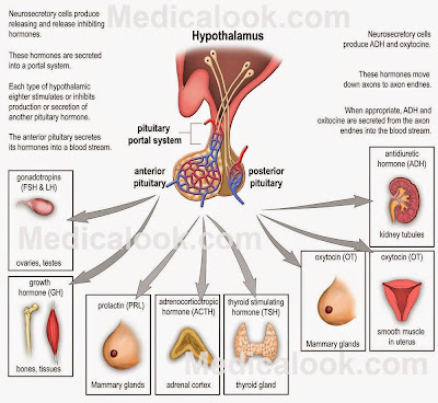
The anterior pituitary isn’t part of the brain at all. The tissue for the adenohypophysis(anterior pituitary, adeno = secreting) comes from the roof of the embryonic mouth. It is epithelial in origin and has cells that produce hormones in response to signals from the hypothalamus (also hormones). Here’s how it happens.


The pituitary gland, or hypophysis, has two small
portal systems that carry blood from one place to
another instead of to a particular tissue and then
back to the heart. The anterior system carries
releasing hormones from the hypothalamus to the
ant. pituitary and then carries stimulating hormones
from the ant. pituitary to the rest of the body. The
posterior system moves hormones from the
hypothalamus directly to the rest of the body.
Image credit to Medicalook.
Some portions of the hypothalamus sense changes from the body and produce hormones called releasing hormones. These stimulate the epithelial cells of the adenohypophysis to produce hormones that then act on the endocrine glands throughout the body. Confusing, yes?

Well, it gets worse. There are six releasing hormones from the hypothalamus that stimulate production and release of seven hormones from the adenohypophysis that then act on at least 20 endocrine glands and neuroendocrine cell types. Hypothalamus to adenohypohysis to endocrine gland – it’s called an axis, and there are several of them.

On the other hand, the posterior pituitary (neurohypohysis) is derived from brain tissue. Instead of the neurons of the hypothalamus producing releasing hormones that then act on the neurohypophysis, the hypothalamic neurons project right into the posterior pituitary where they deposit their hormones (oxytocin and antidiuretic hormone).  The neurohypophysis doesn’t make any hormones itself, it just stores what the hypothalamic neurons produce and then releases them to the circulatory system.

That’s a heck of an exception- part of your brain isn’t actually part of your brain. Cells from your mouth control most functions in your body! Some people I know have mouths that completely ignore their brains! Next week, let’s talk more about the neuroendocrine system. Your thyroid gland size correlates to which hand you use to write – say what?


Gu, X., Karp, P., Brody, S., Pierce, R., Welsh, M., Holtzman, M., & Ben-Shahar, Y. (2014). Chemosensory Functions for Pulmonary Neuroendocrine Cells American Journal of Respiratory Cell and Molecular Biology, 50 (3), 637-646 DOI: 10.1165/rcmb.2013-0199OC

Song, H., Yao, E., Lin, C., Gacayan, R., Chen, M., & Chuang, P. (2012). Functional characterization of pulmonary neuroendocrine cells in lung development, injury, and tumorigenesis Proceedings of the National Academy of Sciences, 109 (43), 17531-17536 DOI: 10.1073/pnas.1207238109

Porzionato A, Macchi V, Parenti A, Matturri L, & De Caro R (2008). Peripheral chemoreceptors: postnatal development and cytochemical findings in Sudden Infant Death Syndrome. Histology and histopathology, 23 (3), 351-65 PMID: 18072092


For more information or classroom activities, see:

Homeostasis –

Neuroendocrine system –

Hypothalamus –

Pituitary –


Thinking Asymmetrically About Hormones

$
0
0
Biology concepts – neuroendocrine system, bilateral asymmetry, internal asymmetry, hormones, endocrine glands


Jack Nicholson did some try asymmetric thinking
in The Shining. The fact that he was driven insane
by the ghost of a horrid past shouldn’t think less of
his accomplishments. Predicting that Shelly Duvall
would go for the radio – brilliant. Sneaking up on
Scatman Crothers – inspired. Following the boy
into the maze –oops.
Independent thinking; thinking outside the box; free thinking; lateral thinking; these are all terms for trying to come up with answers to problems through rejection of established logical methodologies. In today’s business, military and political worlds, they like to call it asymmetric thinking.

An asymmetric military engagement might be one where a traditional force is thwarted and confused by a nontraditional force of computer geeks hacking their communications and selling all their weapons on Etsy. Asymmetric thinking approaches what might be a traditional question at a creative angle, rejecting the tools and assumptions that are normally found “inside the box.”

Asymmetric thinking asks why outrageous solutions aren’t being considered. Many job interviews now include asymmetric thinking questions. A man goes into a restaurant and asks for a glass of water. The waiter points a gun at him and the man thanks the waiter and leaves. What’s your explanation for that? (see end of post)

Today we'll talk about asymmetric thinking in a different way – a lateral thinking approach to asymmetrical thinking, as it were. We have talked about the asymmetry of the brain hemispheres and how they sit asymmetrically in your skull. We have also talked about the neuroendocrine system and how it is controlled by a part of your brain that isn’t really part of your brain. Now let’s talk about the asymmetry of the neuroendocrine system as it begins in your brain and ends in the endocrine glands.


Most views show the hypothalamus from the side
(see last week’s post), so you don’t appreciate that
there are two of each nucleus. For those that dump
hormones into the pituitary gland, both sides
participate, but our story today shows that they don’t
necessarily participate equally.
The hypothalamus, as well as the paired endocrine and neuroendocrine glands (see last week's post), demonstrate significant asymmetry. I suppose that’s not so unusual - all the structures we’ve talked about in the past few weeks have structural and functional asymmetries. The weird part about it here is that we are talking about hormones being moved into the blood.

The whole purpose of the hormone system is that it can be used to bathe the entire body in functional hormones at the precise levels, so that all cells that can respond will respond. How does that jibe with an asymmetry where one gland of a pair does more than the other?

The hypothalamus is a good example. You have two halves of your hypothalamus, one in each hemisphere (sort of, see above), but they both deposit releasing hormones into the same pituitary vein complex so they can stimulate your single pituitary gland. Yet, studies show that the right hypothalamus makes more gonadotropin-releasing hormone than the left hypothalamus.

On the other hand, thyrotropin-releasing hormone (stimulates the release of thyroid stimulating hormone, TSH, from the pituitary) is higher in the left hypothalamus. Together, the results of several studies shows that the right hypothalamus plays a bigger role in controlling reproduction, while the left hypothalamus works more in metabolic rate. You have a lateralization of structure and function in your two hypothalami, just like in your two cerebral hemispheres, even though both halves work on a single pituitary gland.

How about some of the other endocrine and neuroendocrine glands?

Thyroid –  You only have one thyroid gland, which lies over the front of your windpipe in your neck. There are two lobes, one on the right side and one on the left, connected by the isthmus across the windpipe.


The thyroid receives stimulation from the anterior
pituitary release of TSH (thyroid stimulating
hormone). If for some reason you get to much
stimulation (maybe autoantibodies), or the thyroid
start making too much thyroxin on its own (tumor),
then you have Grave’s disease. In some cases, you also
get an autoantibody during Graves that attacks the
fibroblasts around the orbit of the eye. The
inflammation makes the lid retract and pushes on the
eyeball, making it bulge out of the socket. Which of these
two has thyroid eye disease?
The thyroid releases thyroxine hormones T3 and T4 into the blood that function to control your metabolic rate (see this post). Yet the right lobe of the thyroid is more vascularized and is almost always larger than the left lobe. The thyroid also has a sexual dimorphism, as it is usually bigger in women than in men, and the asymmetry of right > left is even larger in women.

The size difference may not be innocuous. Many studies have shown that thyroid diseases and cancer affect the right lobe more often than the left. And it gets weirder. A 2009 research paper from China showed that handedness may also play a role. They found that the right > left size difference was larger in right-handed people. However, the left lobe was about the same size no matter which hand the person preferred. So, does the hand you use influence the size of the thyroid, or does your thyroid predict which hand you will use? Or, is it a correlation without significance?

Parathyroid – You have four parathyroid glands – maybe. These are located on the backside of your thyroid gland (hence the name para = by). The parathyroids are important in regulating calcium levels in the body. This may seem weird, having four glands to control the levels of one element. But consider that calcium plays some major roles, from controlling muscular contraction, to neuron transmission, to at least a dozen different second messenger systems in every cell.

The parathyroids are small, only about 33 mg each, so they are easy to lose when people have surgery on their thyroid gland. A 2011 studysought to find out where they sit normally so surgeons would be able to find them and preserve them. Unfortunately, they found that position and number are quite variable. Forty-three percent of people have at least five glands instead of four. And the positions of the four common ones can be variable, they aren’t always in the same place. The extra ones can be just about anywhere! Good hunting Mr. surgeon.


If you soak bone in vinegar (acetic acid), it will remove
the calcium and leave the protein matrix. Notice that it
must be the calcium that gives bone rigidity. This isn’t
how PTH works. PTH stimulates osteoclast activity,
which removes the calcium AND the protein matrix. PTH
also decreases the amount of calcium lost in the urine,
but for some reason, we work just fine without PTH or
without its balancer hormone, calcitonin.
The thyroid and parathyroid seem to do different jobs, but they’re linked by more than just anatomy. The parathyroid hormone (PTH) made and released by the parathyroids works to increase calcium availability, by increasing bone break down (osteoclast activity, see this post) and increasing the amount of calcium recovered from the urine.

But wouldn’t you need a balancing hormone to decrease calcium levels if they get too high, so a balance could be established? This is how many hormones work; there are hormone pairs that have opposite stimulatory functions. The balancing hormone for PTH is calcitonin, made by the neuroendocrine parafollicular cells (C cells) in the thyroid gland.

But here’s the exception. Calcitonin is important in fish and birds, but it seems people and many other mammals can get along fine without it. Remove someone’s thyroid and they have to take thyroid hormone for the rest of their life. But they get along just fine without calcitonin. This is one instance where the balancing hormone isn’t necessary. It seems an asymmetry of function in calcium regulation is just fine in people.

Adrenal glands -  These glands sit on top of each kidney. You think of them as sources of epinephrine in the fight or flight syndrome, but they do much more. Adrenal (ad = of or near, renal = kidney) glands have a cortex, which is toward the outside (not the core, this always confused me), and a medulla, which is in the middle (makes more sense).


cortex of three layers and a medulla. The cells of the two
regions are of different origin, the medulla is nerve like,
while the cortex is epithelial in origin.
The medulla is neuroendocrine, the cells look like neurons and are stimulated directly by sympathetic (autonomic) nerves (see this post). The medullary chromaffin (they take up lot of stain) cells release epinephrine and norepinephrine in response to a fight or flight situation. Epinephrine and norepinephrine are neurotransmitters in many parts of the brain, and in this case they work on cells to increase heart rate, glucose availability to muscle and the like.

The adrenal cortex is made up of three layers, each produces hormones to be released into the blood. The outside most is the zona glomerulosa, which makes aldosterone to help control osmolarity and blood pressure. The zona fasiculata is the biggest, and makes cortisol that controls the metabolic rate. The zona reticularisis inner most and makes sex hormones, androgens specifically.

You have two adrenal glands – and they each do the same things in response to the same signals, either hormonal or neural. So why is the left adrenal gland almost always bigger than the right? Is it because the venous drainage of the right and left adrenals is different? In the right, the veins dump into the inferior vena cava, while the left drains into the left renal vein. I really don’t know if that would make a difference.


This is just weird. A paper from 2005 states that since
the autonomic (sympathetic and parasympathetic)
nervous system controls the neuroendocrine system,
asymmetries in the ANS can have ramifications on
endocrine function. Asymmetry in behavior can
affect ANS, which then can affect endocrines. This study
showed that feeding cows from the left side (affects right
side ANS) improved reproductive ability and lactation; so
the right ANS must have more influence on reproductive
endocrine function. Could I have some left-hand milk please?
But here’s the kicker, a group in 2002 studied the size of the adrenal glands and their functional abilities in wild animals and their domesticated counterparts; several species of foxes, minks, etc. They found that domesticated animals had a larger size difference in adrenals than did the wild versions.

What is more, when they compared aggressiveness, no matter whether the animal was wild or domesticated, the most aggressive animals had the largest size differential – always left bigger than right. This makes it sound like the medulla was involved – aggression being involved, but it wasn’t.

The increase in left adrenal size in domesticated animals was due to an oversized zona fasiculata (cortisol and other glucocorticoids), while the left adrenal asymmetry in the aggressive animals was due to a larger zona reticularis (sex hormones). Ah… now that makes some sense. Doesn’t it always come back to sex?

Next week, let’s look more into the neuroendocrine system and gender. The testes and ovaries have the most spectacular asymmetries.

(The man had the hiccups)


Ying M, & Yung DM (2009). Asymmetry of thyroid lobe volume in normal Chinese subjects: association with handedness and position of esophagus. Anatomical record (Hoboken, N.J. : 2007), 292 (2), 169-74 PMID: 19051270

Trut LN, Prasolova LA, Kharlamova AV, & Plyusnina IZ (2002). Directional left-sided asymmetry of adrenals in experimentally domesticated animals. Bulletin of experimental biology and medicine, 133 (5), 506-9 PMID: 12420075

Rizhova, L., & Kokorina, E. (2005). Behavioural asymmetry is involved in regulation of autonomic processes: Left side presentation of food improves reproduction and lactation in cows Behavioural Brain Research, 161 (1), 75-81 DOI: 10.1016/j.bbr.2005.01.007

Hojaij, F., Vanderlei, F., Plopper, C., Rodrigues, C., Jácomo, A., Cernea, C., Oliveira, L., Marchi, L., & Brandão, L. (2011). Parathyroid gland anatomical distribution and relation to anthropometric and demographic parameters: a cadaveric study Anatomical Science International, 86 (4), 204-212 DOI: 10.1007/s12565-011-0111-0


For more information or classroom activities, see:

Hypthalamus – see last week’s post

Thyroid gland–

Parathyroid glands–

Adrenal glands-

What the Heck Are Those Doing There?

$
0
0
Biology concepts – internal asymmetry, neuroendocrine, reproduction, hormone, absence symmetry


This is the Decatur County Courthouse in Greensburg
Indiana. A mulberry tree has been growing out of the
steeple for over a hundred years. Nature has things out
of place, sometimes for a good reason, sometimes
through sheer tenacity.
“That really shouldn’t be there… “ but it is. You can probably think of many examples of things that seem out of place, but with further investigation and reasoning, you figure out that there’s a plausible explanation for that particular thing being in that particular place.

A couple of examples - in December of 2013, a University of Arizona team announced that they had found a planet orbiting a star about 300 light years from Earth. No problem, we’ve found many exoplanets. But to our current understanding, it shouldn’t be there.

Planet HD 106906b is 11x the size of Jupiter and travels in a path 650x larger than Earth’s orbit around the sun.  This makes it the largest planet we have noted orbiting such a long distance from its star. Our current hypotheses of planet formation can’t account for this planet.    

Usually a planet will accrete from the left over material in the debris disk that forms a star. But a planet this large would take too long to form in that manner. And the outer edge of a debris disk, where this planet is located, doesn’t have enough material to build a large planet. So why is it there?

Perhaps it was a failed star, formed at the same time as its star. Many binary star systems form by quick accretion in two areas of the same disk. But binary star systems usually have a mass ratio of 10:1 or less. HD106906 (the star) and the presumed “failed star” (HD 106906b) have a ratio of >140:1. There’s no good reason for it – yet there it is.

Now for a less universe-shaking example. My family and I live in Indianapolis, the largest city in the United States without a navigable waterway. Yet, there's a US Naval Station located on the near north side of the city. Built in 1936 as a WPA project, the Heslar Naval Armory housed sailors that trained on Lake Michigan (200 miles away) each summer.


This is the Naval Armory in Indianapolis. There is a river
behind it, but the last time anyone tried to navigate the
White River from the Ohio to Indianapolis they ran
aground and the entire boat had to be disassembled.
It turned out to be a good idea. In WWII, a landlocked naval station was just the place to train naval communications officers without worrying about being bombed or spied on. The naval armory was also the single most important site for developing US naval war strategy, including the D-Day invasion. Sometimes out of place things are there for a good reason. And here we meet our topic for the day.

In terms of reproduction and neuroendocrine function, there’s a true disconnect with the testes and their function. Animal testes do a couple of important jobs. They produce the male gamete cells for reproduction, but they also produce sex hormones that control reproduction and secondary sex characteristics.

If the testes are so important, why are they housed in harm's way, outside the body cavities? You think you know the answer, but really you just know a portion of the story. The testes are housed in the scrotum instead of inside the abdominal cavity. Think how silly it would be to suggest that a woman’s ovaries should be housed in a sac outside her body – yet there are the male testes, in a sac outside his body.

One weird theory is that large testicles and scrotums are a sexual dimorphism ornament; it costs energy to make large testicles, so the larger they are, they better health the animal must have to be able to afford to invest so much in ornament size. To take advantage of this as a way to draw mates, they have to be seen. Therefore, they're outside the body. It's an example of the handicapping hypothesis– an organism handicaps itself by investing so much energy in one thing, but it pays off in a reproductive advantage.


Testicle size varies according to species and breeding
strategy. The right whale has the largest testicles,
about 500 kg (1100 lb) each, 10x larger than those of the
blue whale.  In general, monogamous males tend to
have smaller testicles. A paper from 2013 showed that
testicle size in humans is negatively correlated to
parenting ability. Those with smaller testicles were more
likely to be nurturing fathers. 
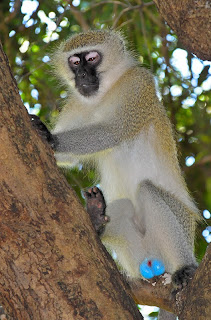
However, there isn’t any evidence to suggest that this is how it works in humans, or in most animals. If it were the case, then the genital size should get bigger and bigger, until it becomes a burden too great to overcome. And elephants and some other animals do have their testes packed into their abdominal cavity. In fact, the vervet monkey(Chlorocebus pygerythrus) is about the only example where the scrotum and testes are a sexual ornament, being neon blue (see picture).

This idea of scrotum as ornament is understandable, but if this isn’t the reason, then what is? Believe it or not, the answer explains several testicular asymmetries.

It’s all about temperature – male gamete cells survive only a short time at normal body temperature, so you save their active period for when they need to swim to the egg. By having them outside the abdominal cavity, they are cooler. The skin of the scrotum is thin, and the arteries of the scrotum and testicles lie right next to the veins; more opportunity to pass heat to blood that is moving away from the testicles. Everything is geared to lowering the temperature a few degrees.

Believe it or not, temperature is also the reason why they are positioned asymmetrically. In the majority of men, the right testicle doesn’t hang as low as the left. A 1997 paper actually studied this and showed that about 62% of right-handed men and 58% of left-handed men had a left testicle that hung lower. But the number of men with a lower right testicle was exactly the same in both right handers and left handers (around 21%). What changed was the percentage of men that had testicles that were positioned horizontally in the scrotum.


The vervet monkey (Cercopithecus aethiops) is an old
world monkey that lives Acacia woodlands of the
mountains of southern Africa. There are five subspecies,
all of which are threatened. They are used in research,
because they have hypertension, anxiety and alcohol
dependence as humans do. You can see why someone
might get the idea that testes and scrotum might be a
sexual ornament for mate selection.
An earlier paper had attributed this to better and larger muscular development on the dominant side. In abdominal muscles, this would mean more tension on the testicular cord from abdomen to testicle (a remnant of the descending testicle in fetal development). More tension means that it can’t hang as low. But this isn’t the reason.

Again we go back to temperature. There's a muscle called the cremaster. It responds to several signals, including temperature and adrenaline and testosterone. This muscle can raise or lower the testicles independently of one another. In a dangerous situation, the testicles are drawn up closer to the body to reduce chance of injury. In cold temperatures they are drawn up in an effort to keep them warm.

As we said above, the scrotum holds the testicles outside the body to minimize gamete activation until needed. But they don’t want to be too cold either. There is a small range of temperature in which function is optimal, and each testicle needs to be at that temperature. So, the body senses the temperature and the cremaster moves the testicle closer to and then farther away from the body to modulate temperature. Believe it or not, they both are constantly moving in independent orbits!


As with this statue of Hercules, the Greeks got it wrong.
Very accurate in their sculptures, they most always had
the left testicle hanging lower than the right in their
male nudes. But, they also had the left testicle as larger.
They fell prey to the idea that bigger would hang lower.
My thoughts on this – someone went around examining
and measuring the testicles on Greek sculptures?
According to a 2008 paper, if the testicles hung side by side, they would warm each other; the maximum surface area of the scrotum for temperature dissipation would not be utilized. Therefore, one hangs below the other for maximum independent temperature regulation. Apparently it isn’t a deal breaker, because 20% of men have testicles at equal heights; perhaps they have a slightly lower reproduction rate.

This leads to another asymmetry in testicles – one is usually bigger than the other. It varies amongst the species of the world, but in most mammals, the left is usually slightly larger than the right. But we humans are exceptions, in men, the right is usually larger than the left. It’s larger, but hangs higher – an example of antigravity?

The working hypothesis is that one testicle is dominant, usually the right in humans. This testicle contributes more to reproduction and plasma testosterone levels, and is affected more by changing levels of luteinizing hormone. But the other one is just as important. It is evolution’s back up plan. If the dominant testicle is injured, the other one takes over. By reducing it’s role in normal function, it reduces the risk of use problems developing; when it is needed, it is fresh and ready to go.


The leatherback turtle has a skylight in its skull (2014).
The bone is so thin that sunlight can reach the pineal of
the epithalamus. It has light sensing receptors, just like
the eye. In fact, most vertebrates have this ability. We
don’t since our cerebral expansion has buried the
epithalamus and hypothalamus deep on our brain.
In non-mammals, the testicles are usually similar in size, but there are exceptions in size asymmetry and even in number. Sharks usually have a much larger right testicle. In birds, one testicle may be larger, but it switches back and forth amongst the different families and species. The bufflehead duck has a left testicle that is four times the size of the right!

In animals that have a distinct breeding system, the sizes of the testicles can change. Just before the breeding season, the testicles may swell to double or triple their normal size. This occurs in the many bird species, and amazingly, their brain senses the day length directly. A 2000 paper looked at mallards, but other papers have shown in other species as well that the hypothalamus of many non-mammal vertebrates has light sensitive cells and can detect lengthening days right through the skull!

In some species, including a couple of birds, only one testis develops. The black coucal cuckoo has only a right testis. Jawless fish have a single testis; it forms in the midline and is probably a fusion of the two testes. The worm, C. elegans, has just a right testis. Its bowel is on the left side, so this is probably a space-saving mechanism.


This ground beetle is one of thousands of carabid
beetles. They are predators, fast and have nasty
secretions as a defense. And most them only have one
testicle. Interestingly, it’s always the right one.
Likewise, a 2005 paper showed that 174 species of carabid beetles are monorchid (one testis). This is also to save room in the exoskeleton. They hypothesize that the testes are the only of the paired organs not to be linked to the same mechanisms of development, so they are more likely to go off body plan. This is called absence asymmetry. The authors suggest that the trade off comes for maximizing some organs within the limited space.

So now you can ask not only, "why are they located there," but also, "does there really need to be two of them?" Next week, we shouldn’t leave out the girls. Ovaries have just as many exceptions as testes.


Davenport, J., Jones, T., Work, T., & Balazs, G. (2014). Pink spot, white spot: The pineal skylight of the leatherback turtle (Dermochelys coriacea Vandelli 1761) skull and its possible role in the phenology of feeding migrations Journal of Experimental Marine Biology and Ecology, 461, 1-6 DOI: 10.1016/j.jembe.2014.07.008

Kumar, A., & Kumar, C. (2008). Swinging high and low: Why do the testes hang at different levels? A theory on surface area and thermoregulation Medical Hypotheses, 70 (3) DOI: 10.1016/j.mehy.2007.06.023

Bogaert, A. (1997). Genital asymmetry in men Human Reproduction, 12 (1), 68-72 DOI: 10.1093/humrep/12.1.68

Will, K., Liebherr, J., Maddison, D., & Gali�n, J. (2005). Absence asymmetry: The evolution of monorchid beetles (Insecta: Coleoptera: Carabidae) Journal of Morphology, 264 (1), 75-93 DOI: 10.1002/jmor.10319

Mascaro, J., Hackett, P., & Rilling, J. (2013). Testicular volume is inversely correlated with nurturing-related brain activity in human fathers Proceedings of the National Academy of Sciences, 110 (39), 15746-15751 DOI: 10.1073/pnas.1305579110


For more information or classroom activities, see

Not many classroom activities involving testicles. Sorry.


Ovaries March To A Different Drummer

$
0
0
Biology concepts – neuroendocrine, ovary, bilateral asymmetry, internal asymmetry, absence asymmetry, hormones, ovulation


Many things we are taught in school just aren’t so.
The Salem witch trials, for example, did not result in
women being burned at the stake. Sure, some were
imprisoned and a couple dozen were hanged, but
none burned at the stake.
A lot of the things we think we know just aren’t so. I’ll give you a few examples. Vincent van Gogh cut off his ear in an insane rage, right? Nope, his “friend” Paul Gauguin cut it off as he drew his sword in a drunken fight with van Gogh. They made up the story so Gauguin could avoid jail. Not nearly so tragic, but it has the ring of truth if you knew Gauguin.

Engineering professors and even physicists at university will teach you that glass is an amorphous liquid. The reason that windows in extremely old buildings are thicker at the bottom is because the glass has had time to flow. Nope.

Glass doesn’t have a crystalline lattice when solid, but it doesn’t flow. The reason old windows are thicker at the bottom is because they used to make glass panes by pouring molten glass on a wheel and spinning it. The force would spread it out, but it would be thicker on the outside edges. When the panels were cut for panes, they installed the thick side at the bottom for stability. So there.

Finally, there were 13 original American colonies… or maybe not. Delaware was swapped back and forth between Maryland and Pennsylvania. Delaware didn’t come into existence as its own colony until the Revolutionary War. It was known as the ”Three Lower Counties” from 1664 until 1776 and shared a governor with Pennsylvania for the last 75 years of its existence.

Speaking of things that just aren't so, there are couple of things in the neuroendocrine system that most people think they know. There are asymmetries in every part of the endocrine system, ant the others are no exception to having exceptions.


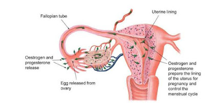
The ovary produces hormones and releases ova. Notice
that the oviduct (fallopian tube) doesn’t connect directly
to the ovary. When the egg is ready to be released, the
estrogen and progesterone cause the fimbria at the near end
of the oviduct to swell and come closer to the ovary. When
the egg is released, the cilia on the fimbriae cells sweep
it into the oviduct.
The ovaries are the source of eggs to be fertilized; those eggs might become small people with wrinkly skin.  But they are so much more. As part of the neuroendocrine system, they are stimulated by hormones and neural impulses and respond by releasing hormones of their own. Depending on the time in a woman’s fertility cycle, they release various amount of estrogen, progesterone and even testosterone.

The ovaries are paired organs like the testes of males, but not all animals have two functioning ovaries. In the Greater Horseshoe Bat (Rhinolophus ferrumequinum), there is only one functioning ovary, the right one, and it's 3-4x the size of the non-functional left ovary. On the other hand, the Natal Clinging Bat bat (Miniopterus natalensis) has only a left functioning ovary and it is several times the size of the non-functional right ovary.

Single ovary examples also exist in the primitive fishes. Lampreys have only one ovary as result of fusion of the two gonadal primordial into a single functioning gland. Hagfishes have a single ovary simply because the other one doesn’t develop.


The hagfish is a primitive fish. The females only have one
ovary, but that isn’t the weirdest part. They produce
proteins and mucins that mix with water and form a slime
when they are disturbed. The filaments are 100x thinner
than hairs, but 10x stronger than nylon, so they are a
subject of much research.
Interestingly, sometimes it’s the right that doesn’t develop and sometimes it’s the left. On the other hand, sharks start out with two ovaries, but the left one atrophies over time, leaving one ovary but two oviducts.

Many birds have one ovary – almost always the left one. A study from 2013 made use of very rare early bird fossils that preserved the ovary tissue; preservation of soft tissue elements is indeed rare. They found that these early fliers had already donated one ovary to the cause of flying.  The hypothesis is that dinosaurs laid many eggs because they had two ovaries, but early birds sacrificed an ovary to reduce weight and make it easier to fly.
The survival advantage afforded by flight offset the disadvantage of fewer eggs, so it was basically a reproductive no harm, no foul. These basal birds had already moved away from the reproductive mechanisms of dinosaurs and present crocodilians toward more bird-like strategies. What this doesn’t explain is why many raptors – like hawks and eagles, have two functioning ovaries. A 2014 paper showed that the right ovaries were functional and capable of responding to, and producing, estrogen and progesterone.

Some birds of prey have two ovaries and some have one. In some, the two are both functional and in others the right is vestigial. This makes me wonder about the evolution of birds. Did the loss of an ovary occur independently several times in different lineages? Or did it occur once in the progenitor of all birds, but some of the descendants evolved the second on again?


The mountain viscacha is a rodent, but looks like a cross
between a rabbit and a chinchilla. They have short forelegs
and long fluffy tail to go along with the rabbit-like ears.
They live in dry places, so they almost never drink. They get
all their water from the plants they eat.
There are even a couple of mammals with a single functioning ovary. The waterbuck (Kobus ellipsiprymnus) lives in sub-Saharan Africa. It is related to antelopes, but differs in that the females of waterbucks have only a left functional ovary.

The mountain viscacha (Lagidium viscacia) is a rodent that lives in the rocky, high altitude, mountainous regions of South America. It starts out with two functional ovaries, but about the time of their first breeding season, the right ovary overgrows, the left shrinks a bit; only the right becomes functional. The exception in this exceptional animal is that if the right ovary is injured or diseased, the left will grow and take over its functions. This doesn’t occur in our other examples.

Similar to testes, there is also a functional asymmetry in ovaries, and this is where we get into the major fallacy that people are taught about the female reproductive system. We are taught that ovaries are good sharers, they take turns ovulating, right-left-right-left, one each month. No……it just ain’t so.

They can take turns, but they usually don’t. And women who want to have children should be glad that they don’t split the load equally. For humans (women mostly), there is slightly less than 50% chance that the opposite ovary will release an egg in the next cycle, according to a 2000 study. This means that side of ovulation is basically random for any given month, but this doesn’t mean that every ovulation has an equal chance of producing an embryo.


Notice that after ovulation, the fertilization of the egg takes
 place in the oviduct, not the uterus. The embryo already
has 32-128 cells by the time it hits the uterine wall. This is
why it is important for the ovary to be producing estrogen
and progesterone the whole time. The uterus must be made
ready for the incoming embryo.
Another 2000 study showed just how unequal ovulations can be. In thousands of ovulations tracked in fertile and infertile women, 64% of pregnancies occurred after right ovary ovulations. In infertile women treated with intrauterine insemination or in vitro fertilization, pregnancy rates were low, as they always are. But if coordinated with right-sided ovulation, they were twice as likely to produce pregnancies as when compared to left-sided ovulations.

However, it isn’t just a right-sided ovulation that produces the best odds of pregnancy; the series of previous ovulations matters as well. If you were to monitor which ovary ovulated over three cycles, there would be eight possible sequences: left-left-left, left-left-right, etc. all the way to right-right-right. This is exactly what a 2011 study tracked, along with pregnancy rates.

The researchers found that the pattern most likely to produce a pregnancy was left-left-right. These results would need to be repeated several times, especially since significant results are difficult to assess when there are eight variables, but their numbers were very convincing. So asymmetry in the function of the ovaries can have a very real affect on hormone levels and pregnancy rates.

Ovulation of a single follicle might be a 50/50 shot each month, but over time the right side of the reproductive system in women seems to be dominant. If the follicles were counted in each ovary (sort of a permanent record of ovulations) of a woman late in her reproductive years, about 62% of them will be on the right side.

Likewise, progesterone and estrogen blood levels are higher during a right-sided ovulation cycle. This data, along with the pregnancy data, indicate the female reproductive system is really right-side dominated. Why is the right side dominant?


The top image shows the corpus luteum that develops from
the follicle that released the egg. This is a huge source of
hormones. It also shows the follicle atresia, where primary
follicles degenerate before releasing eggs. The bottom image
is the timetable of the happenings in the ovary. Notice that the
degenerating corpus luteum is still bigger than the primary
follicle, so over time, the ovary does get bigger. Then as she
gets close to menopause, they get smaller.
A good explanation comes from the fact that the drainage of blood for each ovary is different. The left ovary is drained by the left renal vein, but the right drains in to the inferior vena cava (like the adrenals, see this post).  There tends to be higher venous pressure in the left renal vein and so this side drains slower.

If the blood moves out slower, then the corpus luteum (the leftover follicle of ovulated egg) stays around longer, and this makes it less likely that the left ovary will be ready to ovulate again the following month. Over time, the right will have more follicles from ovulations. The hormone levels would also be higher if carried out of the ovary faster, so this is probably why plasma hormone levels are higher after a right-sided ovulation.

The right side dominance is likely to switch to the left side later in a woman’s reproductive years because of the relatively lower numbers of ova left on the right side. This may be why it is less likely that a woman will become pregnant in her later years.

In most mammals, right and left ovaries are about the same size (given the exceptions of mountain viscacha and waterbuck we talked about above). But in humans, this is merely how they start out. Later in the reproductive years, there is often an acquired size asymmetry.


A dichotic listening test is for attention and picking out one
noise precisely. Most people have a right ear advantage (REA)
for speech because the speech centers are on the left side of the
brain. Research shows that women have a reduced REA compared
 to men and it is affected strongly by estrogen and progesterone
(2011). This may be so that they will focus in less on one sound
and might then be able to pick up on distress from their baby.
After ovulation, the follicle expands and becomes the corpus luteum. This structure produces hormones that would stabilize the system to prolong the pregnancy. If the egg is not fertilized or the pregnancy is not carried for a long time, the corpus luteum reduces in size and hormone production falls. However, the leftover follicle is larger than the pre-ovulatory situation. 

Over time, the number of follicles increases and the size of the ovary increases. More right-sided ovulations (since the right side drains faster and therefore means it can be ready to ovulate again the next month) means that it enlarges more than the left, and a size asymmetry develops.

Hormones, number of ovulations, pregnancy rate – there isn’t anything about the ovaries that isn’t asymmetric – and yet people are taught that they have a symmetric size and function. Right-left-right-left.... yeah sure.

Next week, asymmetries abound in the human body, but they're usually a little here a little there. But what if every organ in you body turned out to be on the wrong side?


Cowell, P., Ledger, W., Wadnerkar, M., Skilling, F., & Whiteside, S. (2011). Hormones and dichotic listening: Evidence from the study of menstrual cycle effects Brain and Cognition, 76 (2), 256-262 DOI: 10.1016/j.bandc.2011.03.010

Rodler D, Stein K, & Korbel R (2015). Observations on the right ovary of birds of prey: a histological and immunohistochemical study. Anatomia, histologia, embryologia, 44 (3), 168-77 PMID: 24895012

Zheng, X., O’Connor, J., Huchzermeyer, F., Wang, X., Wang, Y., Wang, M., & Zhou, Z. (2013). Preservation of ovarian follicles reveals early evolution of avian reproductive behaviour Nature, 495 (7442), 507-511 DOI: 10.1038/nature11985

Fukuda, M. (2000). Right-sided ovulation favours pregnancy more than left-sided ovulation Human Reproduction, 15 (9), 1921-1926 DOI: 10.1093/humrep/15.9.1921

Ecochard, R. (2000). Side of ovulation and cycle characteristics in normally fertile women Human Reproduction, 15 (4), 752-755 DOI: 10.1093/humrep/15.4.752

Fukuda, M., Fukuda, K., Tatsumi, K., Shimizu, T., Nobunaga, M., Byskov, A., & Yding Andersen, C. (2011). The ovulation pattern during three consecutive menstrual cycles has a significant impact on pregnancy rate and sex of the offspring Fertility and Sterility, 95 (8), 2545-2547 DOI: 10.1016/j.fertnstert.2011.02.010


For more information or classroom activities, see

Not too many classroom activities for ovaries and hormones. Sorry. Although I am impressed by the idea of using a pomegranate as a model for the human ovary.


Organs Don’t Always Follow The Plan

$
0
0
Biology concepts – internal anatomy, asymmetry, symmetry breaking, primary ciliary dyskinesia, situs solitus, situs inversus


You hold your right hand over your heart for the national
anthem, correct? So what happened here?  Photoshop - they
even reversed the buttons on his jacket and the lapel pin to
sell it.  But notice the medals on the marine behind him; the
photoshopper didn’t switch them back to the left side.
Michelle came much closer to covering her heart. He missed
by a mile. He must be honoring the pack of cigarettes in his
breast pocket.
The national anthem starts. You rise from your seat and place your hand over your heart. But where’s your heart? Most people place their right hand over their left pectoralis major, halfway between the sternum and the armpit. Don’t put your hand in your armpit; nobody wants that.

But the heart is actually located in the middle of your chest, protected by the bony sternum. True, it’s tilted so some of it sticks out to the left side. This is why your left lung is smaller and has the cardiac notch (see this post). The reality is that some of the heart is located right along your midline and some of it is just barely in the left half of your thoracic cavity.

So if you want to cover your heart when the national anthem is played, put your hand over the center of your chest – but your going to look silly next to all those people who don’t really know their own anatomy. Or maybe they do; the heart isn’t always where the anatomy textbooks say it is supposed to be. And neither are the rest of the internal organs.

We’ve talked a lot in the previous weeks about internal and external asymmetries. Despite the fact that most animals are mostly bilaterally symmetric, there are parts of animals that just can’t be symmetric; we only have one of them. Unless they position themselves right along our midline and each half looks exactly like the other, internal asymmetry just isn’t possible.

Even for single organs that are on the midline, they're often not completely symmetric – see our posts on the brain (here and here). The heart itself isn’t symmetric, with the left heart ventricle being much thicker and a stronger pump than the right ventricle. We talked about the thyroid being larger on the right side; the pancreas crosses the midline but the head of the pancreas is much larger than the tail.


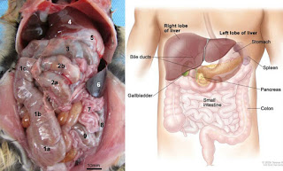
The abdomens of most mammals look pretty similar. The rat
on the left has a slightly more centrally located liver, and the
uterus is very different, but you can’t see it here. The spleen
(#6 in rat) is larger and wraps around to the front in the rat
as compared to the human.
The organs of the thoracic (chest) and abdominal (GI) cavities are examples of the asymmetry inherent in most animals. In your abdomen you have one stomach, liver, pancreas, gall bladder, spleen, and one set of small and large bowels. Each is located in a very particular place; like packing for vacation, you have to make the best use of the room you have.

Your thoracic organs are indispensible – you might be able to live without one lung, but every try getting along without both of them? And forget about going without a heart; although the Grinch did fine for a while with one that was three sizes too small.

Paired organs aren’t necessarily symmetric either, but they are most often located symmetrically within their cavity. Each kidney is located pretty much the same distance from the midline. And even though your lungs are asymmetric, their location on each side of the thoracic cavity is fairly symmetric. Part of the advantage of paired organs (kidneys, lungs, adrenal glands, testes, ovaries) is that you have a back up if something goes wrong with one of them.

For single organs, there is no back up. If they go bad, you’re in trouble. You don’t have another source of insulin outside your pancreas, no other organ makes bile besides your gall bladder, and your spleen is crucial for removal of old blood cells and immune complexes. Yet, you can get along without some of your abdominal organs.


If Hannibal Lecter had eaten just part of the census man’s liver and
left him living, it is very possible that his liver would have grown
regeneration. The hepatocytes start to split and regrow the
mass. But they don’t have the signals to produce the original
shape; they just form a functional mass of tissue.
A bunch of people are walking around without a spleen. Some lose it surgically after an injury. The spleen is located to the left of and behind your stomach.  It isn’t protected by bones as your ribs protect your lungs, and it has a thin, brittle covering that is vulnerable to damage. An accident that involves sudden sideways forces can tear the spleen and lead to substantial bleeding.

The injury often involves an automobile, but football players (American football) and rugby players have also lost spleens after tackles. One clinical case detailed a splenic rupture that didn’t occur until 70 days after an abdominal injury. You can get along without your gall bladder too, but you’ll have to read about that yourself - and don't eat potato chips while you read about it.

So how are our pieces fitted together – your heart points to the left of center, the liver and gallbladder are on the right. The stomach is on the left, and so is the spleen. The pancreas is about in the middle. For the digestive tract, the situation makes sense and is driven by the position of the stomach. Food exits the stomach to the right side, so all the things that help digest the food should be on that side – liver for glucose storage from small bowel, gall bladder for fats, head of pancreas for proteins and other things.

The normal arrangement of internal thoracic and abdominal organs (stomach, spleen and heart on left; liver pancreas, gallbladder on right) is called situs solitus. This is the picture you see in your anatomy textbooks and in bodies ripped open by movie werewolves and zombies. But sometimes the pictures aren’t right…. or left.

Some people have mirror image internal organs; what is usually on the right is found on the left, and what usually points to the left now points to the right. This situation is called situs inversus totalis. Luckily, the inversion of organs doesn’t really present a medical problem on its own. Since everything is reversed, the connections from organ to organ and from blood supply to organ are maintained.  Sometimes there is a reason for the inversion, but other times it’s random.

A study from Norway indicated that the chance of situs inversus increases with maternal age, and it is also more common in cultures with small gene pools and higher incidence of inbreeding. However, it is not more common in twins than in multiple single births of a family.

In the cases of situs inversus that are not sporadic (random), what controls the differentiation of right and left and how does it go wrong?


The nodal cilia rotate rather than beat. The third image shows
how they tilt backwards while positioned in the posterior of
each cell.
The identification of how left and right are determined in the embryo has been a long and arduous road, starting in the 1700’s. Different pieces of evidence, usually from mutant animals or from people with diseases, have been added together to form a picture of left-right determination.

The system goes back to the cilia that we talked about last Fall and Spring. The node cells that run along the midline of the embryo have monocilia(one cilium/cell). These are specialized cilia that don’t have the central pair of microtubules, instead they have a 9 + 0 configuration.  They gyrate instead of beating.


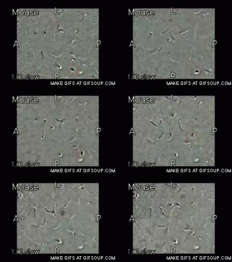
Follow the debris to see the leftward flow 
of the fluid.
Nodal cilia gyrate with a long, curved stroke to the left and then a slightly bent return stroke. This movement generates a strong leftward flow of the thick liquid that surrounds the embryo. Normally, gyrating cilia would just create a circular vortex, but these cilia are positioned posteriorly on the nodal cells and tilted posteriorly. Flow studies show that the position and tip are responsible for the leftward flow instead of a whirlpool.

The leftward flow is somehow responsible for turning on different genes in the embryonic cells that will be on the right and left sides, but people and other animals with a defect in nodal cilia are just as likely to exhibit situs inversus as situs solitus. It isn’t that non-functioning nodal cilia cause situs inversus, they just don’t promote situs solitus as they normally would.


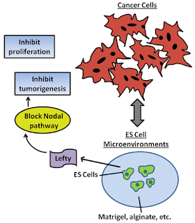
Nodal is important for the growth of some cancer cells. If you
harvest lefty from embryonic stem (ES) cells, you can use it to
inhibit nodal and stop the cancers cells from proliferating. This
could be one of the new therapies for cancer in the next few years.
The initial thought was that the leftward flow generated by nodal cilia created a concentration of some morphogen (a shape [morpho] generating [gen] molecule). The morphogen would have a high concentration on the left side and lower on the right, and this would signal for left side genes to be expressed on the left side.

It’s true that there is strong asymmetric gene expression on the left and right sides. The controllers are a couple of proteins related to an immune system protein called TGF-beta (reviewed here and here). The nodal protein is expressed only on the left side, while lefty protein is expressed on the right side. Nodal drives development of a left side, while lefty is a feedback inhibitor of nodal and therefore prevents a left side development on the right side. The asymmetry of nodal and lefty expression was supposedly driven by the concentration gradient of some morphogen generated by the cilia.

However, no one could find the morphogen. Meanwhile, it was discovered that mutations in another type of cilium also resulted in situs inversus. These cilia are immotile; they’re the sensory cilia we also talked about last fall. This led to the two cilia model to compete with the morphogen concentration hypothesis.


In some people with primary ciliary dyskinesia, or immotile
cilia syndrome, the mucociliary elevator isn’t operable, so they
are subject to recurrent respiratory infections.
The two cilia model supposes that the leftward flow generated by the nodal cilia bends the immotile sensory cilia and this sets off a signaling cascade in the cell. The signaling is different on the left and right sides because the left side receives a much stronger current that bends the immotile cilia more, while the right side cilia don’t bend much less or not all.

All this was discovered by accident; different researchers were looking several different diseases. Primary ciliary dyskinesia (PCD) is a rare genetic disease where the motile cilia don’t move. About half the people with PCD have situs inversus, if they do then this PCD is called Kartagener’s Syndrome. Other researchers were looking at polycystic kidney disease (PKD). These people had a defect in immotile cilia in the kidneys, but also had a high incidence of situs inversus.


In polycystic kidney disease (PKD), the defective cilia don’t 
sense flow and they trigger overgrowth of epithelium to 
form cysts. A normal kidney is on the right.
While situs inversus totalis isn’t a problem on its own, patients with PCD and PKD often have other problems. These problems are related to the cilia; it isn’t just the nodal cilia or sensory cilia that are defective, it will be other cilia as well, as well as the organs in which they work. Next week we’ll look the other side of the coin; sometimes organs in the wrong place cancause very big problems.


Babu, D., & Roy, S. (2013). Left-right asymmetry: cilia stir up new surprises in the node Open Biology, 3 (5), 130052-130052 DOI: 10.1098/rsob.130052

Nonaka S, Yoshiba S, Watanabe D, Ikeuchi S, Goto T, Marshall WF, & Hamada H (2005). De novo formation of left-right asymmetry by posterior tilt of nodal cilia. PLoS biology, 3 (8) PMID: 16035921

Shiratori H, & Hamada H (2014). TGFβ signaling in establishing left-right asymmetry. Seminars in cell & developmental biology, 32, 80-4 PMID: 24704359

Resteghini, N., Nielsen, J., Hoimes, M., & Karam, A. (2014). Delayed splenic rupture presenting 70 days following blunt abdominal trauma Clinical Imaging, 38 (1), 73-74 DOI: 10.1016/j.clinimag.2013.09.003


For more information or classroom activities, see:

Situs inversus –

Liver regeneration –

Primary ciliary dyskinesia –

Polycystic kidney disease –


It’s 11 PM, Do You Know Where Your Organs Are?

$
0
0
Biology concepts – situs inversus, situs ambiguous, dextrocardia, dextroposition, isomerism, canalization


Catherine O’Hara has situs inversus, but she doesn’t
have any strange stories to tell about it as do Dr. No
and Donny Osmond. On the other hand, a weird
movie like Beetlejuice deserves a star with a
strange anatomical quirk.
Being an arch criminal has its good points and its bad points. You have a lot of disposable income, but then again a lot of people are trying to kill you. Criminality is sort of a mirror image of polite society, so perhaps it fits into our discussion of situs inversus (mirror image organs) last week.

Dr. No of the James Bond stories was the quintessential bad guy, but he still had a congenial relationship with 007. During a discussion over tea, or perhaps as Bond hung over a pool of sharks, Dr. No explained to James that he once survived an assassination attempt when a his would be murderer tried to stab him in the heart.

Little did the assassin realize that Dr. No had situs inversus. Even though he was stabbed on the left side, right where his heart should have been, Dr. No laughed it off and then cut up the poor guy with a laser. It helps to have an edge that people don’t know about.

Then again, sometimes situs inversus can be a pain. Donny Osmond, of the Osmond Brothers and then later of Donny and Marie, had situs inversus. As a child he had a bout of lower left abdominal pain. Nothing special is located in the lower left abdomen, so his family ignored it - so did the doctors. When they arrived in England, he had to have emergency surgery because of appendicitis. His situs inversus meant that his appendix was on the opposite side.

In the cases mentioned above, the condition was situs inversus totalis, but it isn’t always totalis. This is rare, but it happens, and most often it involves the heart. Even when it comes to location, the heart wants what the heart wants.


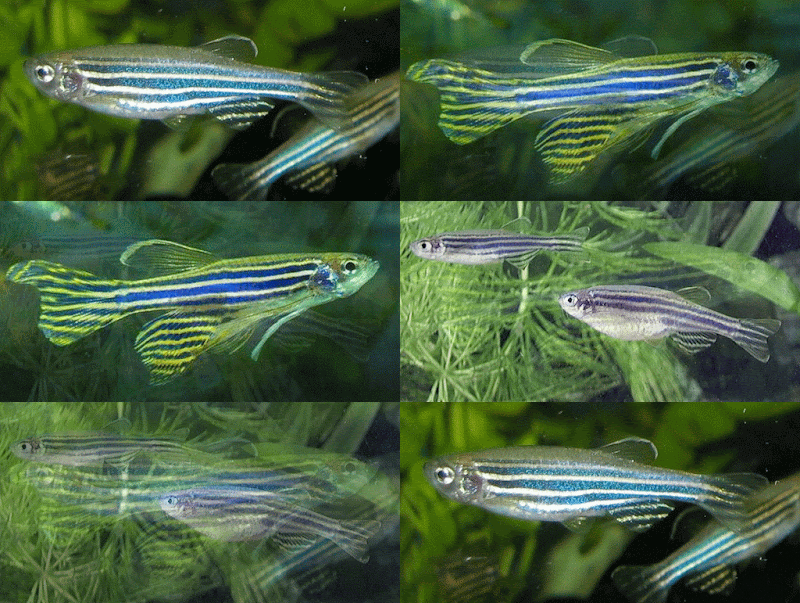
We’re talking about looking for horses instead of zebras
and we’re talking about hearts, so why not zebrafish
hearts? It turns out that zebra fish can regenerate their
heart after injury, as can many animals. A new paper is
showing how the cells can change into the cell type they
need and then proliferate.
One of the tenets of medical training is that when you hear hoof beats behind you, think horses not zebras. Another way of saying the same thing – common things occur commonly. So if a patient presents in your clinic, you should expect that their organs are where they most commonly are. However, they could be situs inversus totalis; this would be a Shetland pony instead of a horse.

But zebras do exist, and you have to know that they exist and what they look like. One zebra is called isolated levocardia (levo= left, and cardio = heart). If situs inversus is rare (1in about 10,000 live births), then isolated levocardia is very rare (in about 25,000 live births). Levocardia means that the apex (the bottom of the heart comes to a blunt point) is directed to the left – this is normal. But in isolated levocardia, the rest of the organs are situs inversus, only the heart is in the normal orientation.

Remember that the lungs are designed to fit around the heart. The left lung has a cardiac notch for the apex of the organ. If the lungs are switched, then the cardiac notch is on the right lung; this is fine if the heart is mirrored as well. But in isolated levocardia, the right lung has room for the heart apex, but the heart is point to the left.


The top left image is the normal arrangement. B shows
something called dextroposition. This happens with
congenital malformations of the chest or sometimes in
injury. The heart points left but is moved to the right side
of the chest. C is dextrocardia without situs inversus, there
are often congenital heart defects in this condition. D is situs
inversus totalis. The heart works fine because
everything is switched.
This in itself is a problem, but not a huge one. The problems really come because there are often congenital heart defects that accompany the change of orientation. The valves of the heart may be misshapen so they don’t close completely (called a heart murmur), the muscle fibers may not align for efficient pumping. None of these problems allow for maximal blood flow through the body.

The prognosis of patients with isolated levocardia is not good, only 5-13% live beyond the age of five years. This is usually due to severe cardiac defects. The problems with the vessels may be a part of it, but these patients often have defects of the heart organ itself. The chambers may be transposed or the septa between the chambers could have holes or other defects.

Situs inversus with a normal heart position is one exception, but how about the opposite exception? Can all the organs be in the normal orientation except the heart? Yes – it’s called dextrocardia. If you want to be picky, dextrocardia refers to any situation where the apex of the heart points to the right instead of to the left, but the vast majority of these cases occur in situs inversus. What we are talking about is an isolated dextrocardia, although the terms in dextrocardia get somewhat muddled. I prefer dextrocardia without situs inversus or dextrocardia with situs solitus (situs solitus is the normal organ arrangement).

Dextrocardia without situs inversus does occur on its own (sporadic), but it seems that it occurs more rarely in mammals as compared to lower forms (fish, amphibians). This may be an incidence of canalization, the tendency for a conserved trait to become more resistant to mutation or environmentally induced perturbation over evolutionary time.


The normal heart s on the left. The aorta is connected to the
left ventricle (the connection is hidden behind the
pulmonary artery. The right image transposition of the
arteries. The aorta empties the right ventricle and the
pulmonary artery empties the left ventricle. This sets up two
different circulatory systems: heart-body-heart,
and heart-lungs-heart.
It’s no wonder heart position is canalized in humans; individuals with dextrocardia without situs inversus also have a very high incidence of severe cardiac malformations and defects. Most patients with isolated levocardia or dextrocardia without situs inversus die before they can reproduce, so any mutations that lead to it are not passed on; therefore most cases are in fact sporadic.

One of the most common congenital heart defects seen in dextrocardia with situs solitus is called transposition of the great arteries. There are five great vessels that carry blood to and from the heart. The superior (1) and inferior (2) vena cava bring blood from the body back to the heart. The pulmonary artery (3) is an exception, being one of the few arteries that carries deoxygenated blood, takes the blood from the right ventricle to the lungs to pick up oxygen. Soon after the pulmonary artery (PA) leaves the heart, it splits in two, one for each lung.

The pulmonary veins (4) bring blood back to the heart from the lungs. Yet another exception – it’s a vein but it carries oxygenated blood. There are actually four pulmonary veins, two from each lung, but they all join together as the get to the left atrium of the heart.

Finally there is the aorta (5). This vessel carries oxygenated blood from the left ventricle to the entire body. This is why the left ventricle has to be so thick and strong. It has a long way to pump the blood. Of course, our descriptions above are of the way the vessels are supposed to fit together.

In transposition of the great arteries (TGA), the aorta and the pulmonary artery have switched connections. The aorta is attached to the right ventricle instead of the left, and the reverse is true for the pulmonary artery. Do you see the problem? In TGA, the aorta receives deoxygenated blood from the SVC and IVC via the right atrium and pumps it right back out to the body. The pulmonary artery receives oxygenated blood from the PVs via the left atrium and pumps it right back to the lungs!


These are the holes in the heart. The left image is the patent
ductus arteriosus from the aorta to the pulmonary artery.
The middle image is the atrial septal defect with a hole
between the two atria, while the right image shows the hole
between the two ventricles in the ventricular septal defect.
There are in fact two small circulatory systems in TGA: 1) heart – lungs – heart, and 2) body – heart – body. This isn’t a recipe for long-term survival. The only blessing is that we can often see another heart defect in these babies, a patent ductus arteriosus. In utero, a fetus doesn’t need to breathe air, the oxygen for the blood is supplied by mom via the umbilical artery. There is a connection between the pulmonary artery and the aorta called the ductus arteriosus. The blood can travel from the aorta to the pulmonary artery or vice versa through the ductus depending on which side of the heart just contracted.

Usually the ductus arteriosus closes on its own a couple days after birth. In the rare case, the ductus will remain open (patent). These are some of the babies you hear about with a “hole in their heart” (the hole could also be from a patent septal defect). The PDA is the only thing that keeps TGA babies alive – some oxygenated blood from the heart – lung – heart system can mix with the deoxygenated blood of the body – heart – body system.
Patent ductus arteriosus is more common in babies with other congenital heart defects, but doctors often give the infants prostaglandins to ensure that the ductus will stay open until they can get in there and switch the vessel connections surgically. The key is to find out there is a problem before the baby is born.


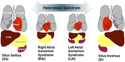
The two middle images show situs ambiguous. The heart is
in the center and has defects, the liver is all across the
abdomen, and the spleen is absent in one and too
numerous in the other. 
One last exception for the day, something even more rare than isolated levocardia or dextrocardia. In situs ambiguus (sometimes called heterotaxy, where hetero= other, and taxis = arrangement), there is a bad arrangement of the organs. The condition comes in two forms, right and left isomerism.

As with all sciences, terminology is important, and there is a discussion now as to whether isomerism or even heterotaxy are appropriate terms to uses for situs ambiguous (reviewed here and here).  Isomerism (iso = same and mer = part) means that parts are duplicated, like two right halves of the heart, but it isn’t all the organs or even the whole heart, just the atria. So is this an appropriate term?

Right isomerism usually comes with more heart defects than left isomerism, and there is no spleen in right isomerism as opposed to several spleens in left isomerism. So if I had to have one or the other, I would pick left isomerism for my heterotaxy. There are also malrotation problems in the intestines and the stomach is sometimes on the wrong side. If you’re ever given the choice, just take situs solitus; it’s boring, but it’s safe.

Next week - how similar are twins? Sometimes they are so similar because they didn't completely separate.


Kikuchi, K. (2015). Dedifferentiation, Transdifferentiation, and Proliferation: Mechanisms Underlying Cardiac Muscle Regeneration in Zebrafish Current Pathobiology Reports, 3 (1), 81-88 DOI: 10.1007/s40139-015-0063-5

Yousif, M., Elhassan, N., Ali, S., & Ahmed, Y. (2013). Isolated subpulmonic fibrous ring, mirror-image dextrocardia and situs solitus in a young lady unreported and a near miss Interactive CardioVascular and Thoracic Surgery, 17 (6), 1043-1044 DOI: 10.1093/icvts/ivt278

Anderson, R., Brown, N., Meno, C., & Spicer, D. (2015). The importance of being isomeric Clinical Anatomy, 28 (4), 477-486 DOI: 10.1002/ca.22517

Loomba RS, Hlavacek AM, Spicer DE, & Anderson RH (2015). Isomerism or heterotaxy: which term leads to better understanding? Cardiology in the young, 1-7 PMID: 26088959


For more information or classroom activities, see:

Situs inversus –
situs ambiguous –

transposition of the great vessel

One Egg, Two People, A Bunch of Reasons

$
0
0
Biology concepts – twinning, placenta, amnion, monozygotic, conjoined twins, in vitrofertilization


Twins are always a subject of interest. Quicksilver and
Scarlet Witch are twins in the Avenger and X-Men series.
Would they be fraternal (dizygotic) twins or identical
(monozygotic) twins? The answer – either. Stick around
for a few posts and find out why.
You may not know it, but mankind has achieved human cloning. We can produce two or more individuals from a single organism, each with exactly the same genetic material. What's more amazing, the people who first achieved this feat had no scientific training what so ever.

How can this be? Cloning humans is a highly technical affair, with all kinds of molecular biology and developmental biology knowledge required. It would require a team of scientists with a handful of PhD degrees ….. or …. a woman could just had monozygotic twins.

Twins come in two flavors – monozygotic and dizygotic. You may know them by other names. A zygote is a fertilized egg, so monozygotic twins come from one (mono) egg and dizygotic twins come from two separate fertilized eggs.

Monozygotic twins (MZ) are commonly called identical - they aren’t - but more on that later. Dizygotic twins are known as fraternal, but fraternal means brother, so this name is bad too.

You see a set of twins on the street. Do you think you would know if they are monozygotic or dizygotic? I bet you can’t.

For many years scientists were sure they could tell the difference by just looking; sure enough that they used the sight test to enter twins into studies on genetic traits. When DNA testing became more common, they found that many of the twins they studied were actually dizygotic. Science had to throw out forty years of twin research.

We’re going to talk about many exceptions amongst twins, but perhaps we should start with just what MZ twins are and aren’t, and how they might come about. First, there is an egg that is fertilized by a male gamete cell – a zygote is formed. The zygote splits two to three times over the first couple of days.


The twinning process isn’t as simple as monozygotic or
dizygotic. There is also how many placenta (chorions)
and amniotic sacs to deal with. If they have one placenta,
they mix blood, so genetic testing is going to be harder.
Which type they are depends on when the zygote split
or if there were two fertilized eggs.
The egg eventually implants into the wall of the uterus and develops an amniotic sac and a placenta. The amniotic sac contains fluid that makes the developing baby buoyant. This cushions the baby and allows it to move freely in the uterus. The placenta (chorion) connects the developing individual to the uterine wall and supplies the baby with oxygen and nutrients via the umbilicus (see this post). Inside the amnion inside the placenta is where the embryo develops into a fetus.

This how things normally happen, but about 3.5 times in 1000 live births, the zygote or embryo splits and becomes two embryos. This is possible because the cells of the early embryo are totipotent stem cells; they have the potential to become any type of human cell. Later on, the cells at different parts of the embryo are merely pluripotent– they can become several different types of cells. Even later, the cells begin to differentiate into the tissues that the fetus will need.

Therefore, those 3.5 embryos that split and become MZ twins have to split early in development. The placenta (part of the chorion) develops around day three, and most zygotes don’t split that early; therefore, 75% monozygotic twins are monochorionic(they share one placenta and chorion).

To become a dichorionicset of twins, the split must occur within those first two days, and this only occurs about 25% of the time. On the other hand, the amniotic sac develops around day nine. If the split occurs later than day nine, then the MZ twins will be monochorionic and monoamniotic(MoMo).

This is also rare because most splits occur before day nine. The result? – most MZ twins are monochorionic, diamniotic (MoDi). Can you explain why there can’t be a dichorionic monamniotic set of MZ twins? Of course you can.


The top image shows a time lapse of the blastocyst
emerging from the zona pellucida – hatching. The bottom
left image shows the components of the blastocyst before
it hatches. The right bottom image shows how the zona
can be nicked with a laser to assist in hatching.
Did you know that human eggs hatch? There is a zone of extracellular matrix proteins and “stuff” that surrounds the egg as it matures in the ovary. This zona pellucida continues to surround the egg even after fertilization. About eight days after fertilization, the egg “hatches” out of the zona pellucida so that it can implant into the wall of the uterus.

By this time (8-14 days), the embryo is more than 15-30 cells total, called a blastocyst. It is hollow and the cells are just starting to assume roles, meaning they are no longer totipotent. So the split needs to come before the end of that second week. One theory is that the blastocyst collapses and the cells on each side of the pinch becomes an individual.

This is the traditional view of when MZ twins form and how to account for the placenta and anmniotic sac number. But there are other views. A newer theory contends that the split occurs much closer to fertilization, maybe in the first couple of divisions after fertilization. Under this hypothesis, the chorionicity and amnionicity have more to do with random fusion of separate structures rather than their development based on time of split. By the way – there’s no evidence for this yet, just some twins that may not fit the traditional pattern - if, in fact, the recorded timing was correct.

The question you shouldbe asking about MZ twins is just why the egg splits into two (and very rarely, three) in the first place. It’s rare, but it has happened throughout history, so there is a cause. Or causes.

The short answer is science doesn’t know yet, but it has many hypotheses. The rate of MZ twinning has been stable over the years and doesn’t show a predilection for a race or area of the world. These all point to MZ twinning having no genetic basis. So what might be causing it?


Which twins are monozygotic and which are dizygotic?
Sometimes it isn’t easy to tell. This is why 40 years of
twins research before genetic testing had to be thrown
away. By the way, Mary Kate and Ashley Olsen on the top
are fraternal twins. The two women below are identical.
One smoked most of her adult life, the other didn’t.
An egg might be more inclined to split if it is older, meaning more time before it is fertilized. There might also be hormonal imbalances. Hormones play a big role in fertilization and continuation of an embryo’s development. Older women and younger women have more hormonal imbalances, and these are the women who are more likely to have MZ twins. A clue may come from reproductive medicine.

The increase in in vitro fertilization (IVF) therapies in the last decades has paralleled a slight increase in MZ twins. This isn’t because of more than one embryo implanted in IVF - those would be dizygotic twins, we’re talking an increase in MZ twins only here.

IVF therapies involve more than just implanting embryos. The reproductive system has to be made ready for the zygotes, and this means giving the lady lots of hormones. Might this throw off the balance and promote zygote splitting? Could be, but there are other possibilities.

The age of the embryo when implanted makes a difference. Five-six day embryos have a higher splitting rate than those that are implanted when they are only 2-3 days old. However, both age group embryos had a higher rate of MZ twinning than did control embryos from natural fertilizations. So perhaps we need to look further.


For in vitro fertilization. The eggs are removed from the
ovary, and each in turn is held in place by a slight suction
on a pipet (bottom right). The egg is pierced and one male
gamete cell is injected through the zona pellucida and into
the cytoplasm. Then, several are allowed to divide a few
times and then they are deposited into the uterus.
Maybe the tendency to split comes from manipulating the zona pellucida. Sometimes the clinicians will weaken the zona by laser (called laser assisted thinning) to promote hatching and implantation, and this may promote splitting, while those that don’t have the zona manipulated may have a delayed implantation. Each situation is accompanied by increased embryo splitting (weakened zona and delayed implantation in uterine wall) so perhaps its all zona regulated.

Then again, the idea that MZ aren’t linked to genetics may be wrong. There have been observations of MZ twins running through the males in some families, perhaps through a y-linked gene. Another study has documented seven families with higher than normal rates of MZ twinning.

It is through these mechanisms, or others we have no idea about yet, that an embryo might split to become MZ twins. But we have been assuming that the split embryo becomes two individuals. What if that doesn’t happen? What if the split embryos remain attached to one another? You get conjoined twins.


Chang and Eng were the most famous of the Siamese twins.
In fact, they are why we call conjoined twins Siamese. They
became rich plantation owners in the American South, but
lost all their money when the North won the Civil War. They
were each married and had 21 children total. There are
nor Eng had twin children themselves.
The incidence of conjoined twins is very low, only 1/45,000 to 1/200,000 births, and 40-60% of them are still born. It is a hard thing to carry or deliver conjoined twins.

There have been famous examples of conjoined twins, sometimes called Siamese twins because of the most famous pair – Chang and Eng of Siam in the 1800’s.

Conjoined twins can also be called imperfect twins, because the embryo split imperfectly. If the split occurs very late, perhaps after day 12 post-fertilization, the rate of conjoined twins might be increased. At this point the cells are already past the point of starting to differentiate, so they are becoming tissues and organs. The ones that split become organs on their own, but the cells that are shared will become organs and structures that the twins will share. In all, we don’t know the risk factors for creating conjoined twins, but it is interesting to note that Latin American women have a much higher rate of conjoined twins than anywhere else in the world. I can't imagine why.

Conjoined twins are described based on where they are joined. The majority are joined at the chest (called thoracopagus, pagos is from Greek = something joined) or the buttocks (pyopagus). The number and extent of organ and structure sharing can vary greatly, but thoracopagus twins often share a heart, while pyopagus twins might share a lower GI tract.


Dr. Ben Carson was the lead surgeon in the successful 1987
operation to separate occipital craniopagus conjoined twins.
He has also been lauded for his hemispherectomy method to
stop seizures and craniofacial reconstruction in
achondroplastic dwarfs. Now he is running for president.
What one has to do with the other, I have no idea.
Omphalopagustwins are joined near the belly button, rachipagustwins share a majority of their spine and ischiopagustwins are connected at the pelvis. In the later case, there may be two, three or four legs between the two individuals. Lastly, there are craniopagus twins. These twins are joined at the skull, and some brain tissue connection may also occur. Quite literally, these twins might know what each other is thinking.

Needless to say, there are many more papers describing them and their problems than there are solutions and treatments. Surgery is getting better, but it depends on what they share. What we have described are the equally conjoined twins. But sometimes, one twin dominates over the other. One will be smaller, weaker, and less able to survive. This is called a parasitic twin. Taken to the extreme ……. Well, that will have to wait for next week.


Herranz, G. (2013). The timing of monozygotic twinning: a criticism of the common model Zygote, 23 (01), 27-40 DOI: 10.1017/S0967199413000257

Kanter JR, Boulet SL, Kawwass JF, Jamieson DJ, & Kissin DM (2015). Trends and correlates of monozygotic twinning after single embryo transfer. Obstetrics and gynecology, 125 (1), 111-7 PMID: 25560112

Knopman JM, Krey LC, Oh C, Lee J, McCaffrey C, & Noyes N (2014). What makes them split? Identifying risk factors that lead to monozygotic twins after in vitro fertilization. Fertility and sterility, 102 (1), 82-9 PMID: 24794318

Machin G (2009). Familial monozygotic twinning: a report of seven pedigrees. American journal of medical genetics. Part C, Seminars in medical genetics, 151C (2), 152-4 PMID: 19363801


For more information or classroom activities, see:

Monozygotic twins –

Zona pellucida –

Chorion and placenta –

Amnion –

Conjoined twins -


When A Twin Vanishes

$
0
0
Biology Concepts – conjoined twins, resorbed twin, monozygotic twin, spina bifida, fetus in fetu, vanishing twin


Abby and Brittany Hensel are the ultimate in teamwork.
They drive a car, walk, go swimming, even use a computer
keyboard with each twin controlling one arm and one leg.
They each had to pass the driver’s test to get their license.
This picture is from their 22ndbirthday, but they are now
25 and teach elementary math.
Brittany and Abigail Hensel are a couple of miracles. They were born in 1990 as conjoined twins of the parapagus type (joined side to side with a shared pelvis, see last week’s post). Now 25 years of age, they inspire millions to expand their definition of what’s important and how to live a good life.

The girls represent a form of conjoined twins called symmetrical or equal. Each twin is developed to the same degree as the other. Even for symmetric conjoined twins, Abby and Brittany are unusually symmetric. Many of their internal organs are doubled; each has their own heart, spleen, stomach and spinal column.

Unfortunately, this isn’t always the case. All conjoined twins are termed imperfect, since the single embryo did not split completely, but some are more imperfect than others. Remember that 40-60% of conjoined twins are stillborn; we only see the miracles where they can be separated or where they can function and grown up to be strong, independent people.

Asymmetrical, or unequal conjoined twins lie along a continuum. In some instances, one twin is slightly larger and stronger, even in symmetrical cases like Brittany and Abby, Abby’s leg is two inches taller than Brit’s, so Brittany has a larger calf muscle from standing on her tip toes all the time. In other cases, one twin is unrecognizable as a small tumor in the body of the other twin, but more on that later.

If one asymmetrically conjoined twin is much larger and healthier than the other, it can have it’s health put at risk by the parasitic twin(ie. the smaller, less developed twin is terms the parasite). In many cases, the parasitic twin is dependent on the autosite (larger twin) for survival because he/she does not have a complete cardiovascular system of his/her own.

Despite it’s weaker state, the parasitic twin does survive to delivery because it gains energy and oxygen from the larger twin. Remember that these are conjoined twins, so they will share a blood system. This is why the otherwise doomed parasitic twin can live before and after delivery. This is one characteristic that distinguishes parasitic twinning from conditions like absorbed twin or vanishing twin (see below).


The first Total Recall movie had Arnold Schwarzenegger,
but the real star was a mutant, parasitic twin named Kuato.
His mutation allowed him to read the thoughts of other,
but you had to hold his hands. Kuato wasn’t far from the
real thing in some people – except for the being alive and
having mutant powers, of course. There are pictures of
parasitic twins on the web, but I wouldn’t recommend
eating before you look at them.
Several cases of living parasitic twins are in the literature. A 1783 case had the stronger twin surviving delivery and carrying his parasitic sibling to the age four. Then he was killed by a snakebite. Talk about irony.

In a newly described case, one set of rachipagus (joined at the spine) conjoined twins consisted of an individual whose only indication that they had a twin was a single leg extending from his back.

And sometimes it isn’t even that simple to see the twin. Spina bifida is an all too common congenital defect in which the spinal column and backbone do not close completely. There are varying degrees of spina bifida; including a mild form (occulta) in which has perhaps a dimple or some hair and no symptoms and a severe form (myelomengiocele) where the spinal column could be open to the environment. One reported case of parasitic twins consisted of a conjoined twin hidden within the defect of a spina bifida patient (2012).

Then there was a 2005 case in Egypt where the young boy had a parasitic twin that consisted of only a head. The head was located on the surface of the larger twin’s body. It could blink, cry, and even smile. The parasite didn’t cause the death of the larger twin, but he did unfortunately die from an infection obtained from the surgery undertaken to remove his sibling.

In other cases, the parasitic twin is completed enveloped in the larger twin’s body. The tissue is still alive, but invisible to the naked eye. This is sometimes called fetus in fetu (fetus within a fetus). But the definition is a bit more involved. Most fetus in fetu cases have a specific location in the body, the retroperitoneal space.


Kind of silly, but this is how fetus in fetu got its name. A baby
is born and it has a small fetus inside its abdomen. It looks
like the baby is pregnant; ie. fetus in a fetus. In real life, some
aphid insects are born pregnant (see this post).
The parasitic twin in fetus in fetu is also fairly well developed, if not large. There is usually a spine that can define right and left sides, and the organ tissues that are there are located on the correct side of the axis. There is usually a top and bottom distinguishable as well.  In some cases, the twin might look a lot like a human, but in most the features are distorted or incomplete.

However, if the axes are maintained and the tissues are organized, the site of the mass can be given a bit of leeway and still term the case as fetus in fetu. Amazingly, there is a case described in which the parasitic twin was found in the cranial cavity of the surviving twin, and even one where the twin mass was found in the scrotum of the survivor!

Even weirder, a case reported in India described two fetuses within the abdominal cavity of their male sibling. Talk about a bully, he ate his siblings. Notice that most of these cases involve boys as the surviving individual.  The known cases of fetus in fetu have a strong male predilection. Is there something genetic there, or is it the difference in how parents treat boys and girls? Maybe not.

When tissue is not viable, many things can happen, including calcification. Calcium can get added to the tissues so that the body kind of walls it off. This happens to some fetus in fetu tissues if they lose their blood supply as they grow. It can also happen in cases where the twin passes away while still in the uterus and is then absorbed by the surviving fetus.

Appropriately enough, these are called absorbed twins or resorbed twins. Like fetus in fetu, absorbed twins end up inside the other twin. Unlike fetus in fetu or parasitic twins, absorbed twins don’t have to be conjoined twins and the absorbed twin is not alive. They don’t even have to be MZ twins; they usually are, but they could be any type of multiples.


Dwight Schrute bragged that he grew strong by
reabsorbing his twin in the womb. If his twin is still inside
him, he resorbed it. If there’s no evidence, then it’s
a vanished twin.
Absorbed twin syndrome is less often brought to the attention of the medical community because there is rarely any symptomology. When discovered, it is usually because some other condition has brought a doctor to examine that part of the body. It is estimated that up to 25% of multifetus pregnancies could include an absorbed twin or a vanished twin (below).

Absorbed twins can cause some problems that other types of twins can’t. For instance, there is the potential to misdiagnose a trisomy or other nondisjunction problem (see this post) because there is extra DNA floating around in the amniotic fluid.

The mass of tissue that was a twin can be found just about anywhere in the body - but it doesn’t have to be a mass. In some cases, the twin tissues can be flattened against growing tissues of the other fetus and ends up paper thin and spread out – fetus papyraceous. This condition occurs about once in every 200 twins pregnancies, so it is not too uncommon in multiple gestations.

In vitrofertilization (IVF) techniques are increasing the rates of MZ twinning, dizygotic twinning, parasitic twinning (including fetus in fetu) and absorbed twins. As we said above, as many as 25% of pregnancies with more than one embryo could end up losing an individual to one of these phenomena.

One of these possible outcomes is closely related to absorbed twin. If the reabsorption comes early in the pregnancy with little or no evidence of the absorbed twin found in the survivor, then it can be called a vanished twin.

The vanishing twin becomes non-vital in utero, but it usually occurs earlier in the pregnancy and is diagnosed by ultrasound. The mother will have an image taken at, say, four weeks gestation, and there will be X number of small embryos implanted and developing placentas and amniotic sacs. Two weeks later she will be imaged again, and there will be one or two fewer embryos and amnions. The other(s) vanished; hence the name.


Gross picture, but you can see the different kinds of tissues
in the teratoma. You can even see some teeth (arrow). Some
clinicians consider parasitic or resorbed twins to be highly
differentiated teratomas, but it just ain’t so.
Because there is no real evidence of vanished twin if you don’t catch it with imaging during the early gestation, few studies have been done as to the mechanism and reason for vanished twins. It may come from poor attachment of the placenta to the uterine wall, but most causes remain unknown.

You may have heard of a teratoma. Many people use the term teratoma when fetus in fetu/absorbed twin/vanishing twin would be more appropriate. While they may appear similar, they have different origins. A teratoma is a tumor of stem cells, not the result of changes in an embryo. As they grow and propagate, some teratoma cells will start to differentiate into specific cell types – they are pluripotent after all. Teratomas may include fully or partially formed teeth, hair, and other recognizable structures, but there is no organization to the growth as one would see in fetus in fetu or an absorbed twin.

Today’s subjects have been interesting, but kind of a downer. Next week let’s talk about some happier kinds of MZ twins. There are several types, and they’re definitely not identical.


Navaei AA, Habibi Z, Moradi E, & Nejat F (2015). Parasitic rachipagus twins; report of two cases. Child's nervous system : ChNS : official journal of the International Society for Pediatric Neurosurgery, 31 (6), 1001-3 PMID: 25715843

Daga, B., Chaudhary, V., Ingle, A., Dhamangaokar, V., Jadhav, D., & Kulkarni, P. (2009). Double fetus-in-fetu: CT scan diagnosis in an adult Indian Journal of Radiology and Imaging, 19 (3) DOI: 10.4103/0971-3026.54890

Zahed, L., Oreibi, G., Darwiche, N., & Mitri, F. (2004). Potential trisomy 21 misdiagnosis by amniocentesis due to a resorbed twin Prenatal Diagnosis, 24 (12), 1013-1013 DOI: 10.1002/pd.918

Lakhoo, K., Ringo, Y., Sillo, T., & Drake, D. (2012). Parasitic twin within spina bifida African Journal of Paediatric Surgery, 9 (3) DOI: 10.4103/0189-6725.104728


For more information or classroom activities, see:

No real classroom activities for lost twins, thank goodness.


Epigenetics And The Evil Twin

$
0
0
Biology concepts – epigenetics, monozygotic twins, mirror image twins


The armadillo is a fascinating animal, and shares a couple
of characteristics only with humans. People and
armadillos are the only animals susceptible to leprosy, so
the armadillos are used for Hansen’s disease research.
Also, nine banded armadillos (the above is a six banded
armadillo) is the only animal other than humans that can
split an embryo twice, allowing for identical quadruplets.
Last week’s topic on parasitic twins was a bit depressing for me. This week, let’s focus on some amazing kinds of monozygotic (MZ) twins. Yes, there are many kinds of monozygotic twins, some we have talked about (conjoined, parasitic, absorbed, vanished) and some we’ll talk about today. Just because they’re monozygotic doesn’t mean that they only come in one type.

The popular idea is that MZ twins are “identical,” but nothing could be further from the truth. It may not even be the case that they share the same genes, but more about that later. When an embryo splits, each new embryo usually gets the same chromosomes. But a lot can happen after that.

We talked recently about the determination of right and left sides in the embryo by a flow of fluid from left to right (see this post). Well, that same flow can make MZ twins look different. One subtle difference can be their fingerprints. MZ twins don’t have the same fingerprints.

Fingerprints do have a genetic component, so the fingerprints of MZ twins will resemble each other more than non-twin siblings’ will. But the environment of the amniotic sac during gestation, especially during the first trimester, will help to determine the details of the fingerprint. Nutrition level, stress, movement within the sac, even a slight difference in umbilical cord length; these will all result in differences between the prints of MZ twins.

Talk about stress, wartime babies have different fingerprinting patterns than those born in peace, at least the seasonal variations in ridge counts disappear if the gestation is during war. All these things we have been talking about go beyond the foundation of genetics. Traits are established and influenced by genetics, but how they turn out (their phenotype, where pheno = visual form) can influenced by the environment. This is epigenetics (epi = beyond).


The top image shows the three general types of fingerprints
that humans display. Identical twins will have similar types
of prints about 80% of the time, because they are partly
controlled by genetics. The bottoms images A and C are
monozygotic twins. You can see they are of the same type,
but have different details. 
Purely environmental factors influencing phenotype might be considered a rather broad interpretation of the term epigenetics. The word does mean beyond genetics, but the strict definition involves factors that an individual encounters that will turn genes on or off. The changing expression then influences the phenotype.

The control of gene expression via chemical reactions is the example most often given. The chemical reaction affects the function of a given stretch of DNA, but doesn’t change the sequence of that DNA. The genetics remains intact to be passed on, but which genes are expressed is what changes.

There are two common examples of chemical reactions that affect DNA are methylation and histone acetylation. Let’s look at an example in nature that uses both systems. And it uses a multiple births in the example too – how convenient.

A honey bee colony has a strict structure. There is one queen (usually - of course there are exceptions), a few hundred male drones, and many thousands of female workers. The queen flies out to mate (with 12-24 drones of another colony) during first first two weeks of her life, but then settles down to lay eggs.


The top cartoon shows methylation of cytosine bases as a
way to control gene expression. The enzyme DNMT adds the
methyl group from SAM to the C base. CpG motifs (a C
followed by a G) are common targets for methylation to
silence genes. The bottom image is for histone acetylation.
Acetylated histones (yellow added) open DNA to be
transcribed (see orange lines in acetylated and deacetylated).
The workers build the honeycomb with different size chambers, small for workers, medium for drones and large for a new queen. It's the size of the chamber that tells the queen whether or not to fertilize the egg she lays in it - she has the choice since she stores the male gametes separate from the eggs.

If she doesn’t fertilize the egg, it will be a worker, a sterile female clone of herself. If she does fertilize the egg, it will become a male drone. But if she lays a fertilized egg in the large chamber, it becomes a female queen, not a male drone. Why the difference? Their diet.

There is a substance the bees produce called royal jelly. All the larvae are fed royal jelly for the first three days, but after that, only the larva that will become the new queen is fed royal jelly. In fact, that is all the queen will eat her entire life.

The royal jelly is a secretion that comes from the hypopharnyx of the worker bees. It's mostly water, with some protein and amino acids. The active ingredient is called royalactin. It ages and becomes less active over time, so the workers keep making it all the time. Get the subtle point here, the queen lays the eggs, but the workers make the queens.


Bees are highly organized and social. Workers are smaller
than drones or queens. These workers are older, because they
are leaving to forage. Younger workers make more royal jelly
and tend the queen and the larvae. Being older might account
for the bad eyesight and flying into each other. 
What does royal jelly have to do with DNA methylation and histone acetylation? I’m glad you asked. More methyl groups added to C’s of DNA keep the workers from becoming queens. There are enzymes that control methylation and de-methylation of DNA. What happens in the case of queen development is that more de-methylation activity occurs and less methylation. The methyl groups on DNA control whether the gene can be expressed or not; more methylation - less transcription.

We also know that queen development is promoted by more histone acetylation. Acetyl groups added to the histone proteins that help coil DNA make it loose and available for transcription (reading the genes that are there). If the histones are deacetylated, the chromatin becomes tight and the machinery can’t get access the genes.

It turns out that royal jelly has a histone deactylase (HDAC) inhibitor. Therefore, more DNA stays acetylated and open to be transcribed. The part that is transcribed might include the demethylase enzyme genes. This leads to less methylation and activation of genes that turn the larva into a queen. So her diet doesn’t change her genes, it just determines which ones will be expressed. And that makes all the difference.

Ask your friends if monozygotic twins are identical and you’ll many more yeses than maybes or nos. But given our discussion above of epigenetics and the power of environment to alter gene expression, do you have any doubt that there are changes that occur afterfertilization and after splitting of the embryo into twins? Epigenetic factors can produce monozygotic twins with different malformations, different lateral asymmetries and even different sexes! (reviewed here)


These are pairs of chromosome 3 from a three year (top) and
50 year (bottom) monozygotic twins. They used red tags for
one twin's epigenetic tags (methyls or histone acetylations) and
green tags for the other twin. If they are at the same place in
both twins in the overlaid image, the color will be yellow. If
they don’t overlap (tagged on different genes), you see the red
and green. Notice how many more differences there are in the
older twins. Image credit University of Utah.
In MZ twins, the environment in one amniotic sac will be slightly different than in the other. Flow of fluid will be different and even the position of the fetus can make a difference. This is why even twins inside one amniotic sac will still have differences. The differences grow after delivery, so more expression will be different at an older age than in utero (see picture on right). And this is just epigenetic, we aren’t even discussing the genetic changes that can take place via mutation once the embryo splits to become MZ twins.

We talked a few weeks ago about how the embryo distinguishes right from left so that the organs develop in their normal locations. Sometimes they don’t, and this is when we get situs inversus or situs ambiguous. If the split of the embryo that forms MZ twins occurs after the decision has been made as to right-left, then mirror image twins could be a result.

Mirror twins may have moles on their cheeks – one on the right and the other in the same spot, but on the left cheek. They may have mirror hairlines or defects like cleft lip and/or cleft palate. They might even manifest equal but opposite sleep patterns. In most cases, the twins will have some internal organs in mirror locations, but rarely will there be a situs inversus twin and a situs solitus twin. Here’s why.


Every television show gets to an evil twin episode sooner or
later. The point is that twins studies can look at physical
traits, but also at how genetics and epigenetics affects
personality…. and facial hair choice.  Top images are Spock
from Star Trek: The Original Series. The bottom image is
Michael Knight from the 1980’s version of Knight Rider.
The discrimination between right and left comes about after the blastocyst implants in the wall of the uterus and after the totipotent stem cells of the embryo start to differentiate into layers. The node cells have the monocilia that gyrate to develop the leftward current and the lateral cells have the immotile cilia that detect the current and respond by differentiating differently.

Most embryonic splits for MZ twins occur in the first 2-10 days, but if the split occurs after day 12 or 13, then some degree of mirror imaging is possible. This is the time when right-left decisions have already been made. Of course, the plane in which the split occurs will matter too, it would have to be directly along the node for a true right-left mirror to occur -there's epigenetics again. This is extremely rare, so most mirror image twins, have some mirror traits, not total mirror bodies.

Even in situs ambiguous (see this post) of MZ twins, there can be mirror imaging of some external and/or internal phenotypes in twins with heterotaxy. Yes, phenotypes – remember that these differences are affected by many things, but not directly genetics. They change the features of the twins while they still have identical genes. It’s epigenetic.


Twins studies are good for teasing out the influences of nature
and nurture in physical form and behaviors.  There are many
different ways to run the studies to find out different things –
type of twins, raised together or apart, degree of relatedness,
nature of environment, raised by birth parents or adopted.
The studies can involve social testing and/or physical testing.
The problem – how to control for unknown variables. These
are people lives, not laboratory mice.
This can be useful in medicine and science. It would be good to know which phenotypes are under strict genetic control and which can be influenced by environment. So while early twins studies concentrated on what was the same between MZ twins, new research is concentrating on what’s discordant (different) between them.

This research will go a long way to showing just how much of our health and life is actually passed down from parents and is inescapable. Even the differences between mirror twins can help to tease out the mechanics of how twinning occurs and the paths and patterns of normal embryology.

Now that we have a handle on epigenetics and its effects on MZ twins, let's talk next week about a couple of twin types that result from genetic differences. Yes, you read that right, some MZ twins are genetically different.


Spannhoff, A., Kim, Y., Raynal, N., Gharibyan, V., Su, M., Zhou, Y., Li, J., Castellano, S., Sbardella, G., Issa, J., & Bedford, M. (2011). Histone deacetylase inhibitor activity in royal jelly might facilitate caste switching in bees EMBO reports, 12 (3), 238-243 DOI: 10.1038/embor.2011.9

Kahn, H., Graff, M., Stein, A., Zybert, P., McKeague, I., & Lumey, L. (2008). A fingerprint characteristic associated with the early prenatal environment American Journal of Human Biology, 20 (1), 59-65 DOI: 10.1002/ajhb.20672

Zwijnenburg, P., Meijers-Heijboer, H., & Boomsma, D. (2010). Identical but not the same: The value of discordant monozygotic twins in genetic research American Journal of Medical Genetics Part B: Neuropsychiatric Genetics DOI: 10.1002/ajmg.b.31091

Thacker, D., Gruber, P., Weinberg, P., & Cohen, M. (2009). Heterotaxy Syndrome with Mirror Image Anomalies in Identical Twins Congenital Heart Disease, 4 (1), 50-53 DOI: 10.1111/j.1747-0803.2008.00229.x


For more information or classroom activities, see:

Epigenetics -

Looking Sideways In The Mirror

$
0
0
Biology Concepts – platyhelminthes, asymmetry, bilateral symmetry, evolution, cephalization, natural selection, fish, lepidophagy

What is the largest living structure on Earth? No, it’s not the 2200 acre Armillaria ostoyae fungus in Oregon that we talked about previously. That is the largest single organism, but there is something bigger.

The Great Barrier Reef houses more species of
coral than any other place on earth, more than 600
species call the reef home. You see how many shapes
they can take. Does this mean they are
asymmetric animals?
The Great Barrier Reef off the northeast coast of Australia is alive. Reefs are made of the exoskeletons of coral polyps, with the new corals growing next to and on top of the older ones. With all the nooks and crannies available, coral reefs are some of the most diverse ecosystems on Earth, with thousands of species per square mile.

This can add up quickly, because the Great Barrier Reef is more than 132,973 sq. miles (344,400 sq. km) in area. And if you still don’t believe it is a living structure, get this; it’s moving south! Climate change is warming the waters off the coast, and corals and tropical fish are moving south with the warmer temperature. Meanwhile, the northern edge recedes as the waters get too hot for corals.

Corals take all sorts of shapes (see picture above), and despite what we talked about last week, they don’t seem to be either bilaterally or radial symmetric. Sure, brain corals look radial, but most corals don’t have a repetitive shape. Are these asymmetric animals?

Nope - remember that the coral you see is the exoskeleton of the polyp, not the animal itself. Just because the apartment building isn’t symmetric, it doesn’t mean the animal is as well. Coral polyps are definitely radially symmetric, so our discussion of last week still holds.

Corals and other radial animals are exceptions, since 99% of animals are actually bilaterally symmetric. But there are exceptions with the bilateral animals as well. Some species that have been bilateral for millions of years then evolved a tweak to the system. Some part them became asymmetric in order to give them an advantage. Their stories are exceptional and we should explore some of them.

Coral polyps are cnidarian animals. They live inside
the calcium shells they produce, and it is the shells
that seem asymmetric in many cases. But the polyps
show radial symmetry. Each stalk and white “flower”
is an individual polyp.
But we begin with a challenge –as we discuss the different animals that break symmetry in the next few posts, see if you can find something else that is common to many of them.

Let’s start with the first animals that became bilaterally symmetric– the platyhelminthes, or flatworms. Most flatworms are small, just barely visible with the human eye, and most swim in the water. There are free-living versions and parasitic species and it is in the parasites that we find our first animal that has decided that completely bilateral isn’t necessary.

Polyopsithocotylea monogenea is a group of flatworms that live on and feed on fish gills. About 0.05-1 mm long, these platyhelminthes attach themselves to one of the gill ridges and take up residence there for life. The attachment they use is called a haptor, and they come in different shapes and sizes.

The oncomiracidium stage of the worm is directly after the egg stage and before it becomes an adult. This stage is completely bilaterally symmetric. But when the adult stage is reached and it’s time to settle down and starting feeding on some fish’s gills, they become asymmetric via their haptor attachment.

Opisthaptors, or just haptors, are the attachment
organs for many parasites, including parasitic
flatworms. They can use suckers or clamps and hooks
in order to anchor the worm in its preferred habitat.
You can see that these haptors maintain animal
symmetry, but not all do.
Different host species fish have different gill anatomy, so the attachment point and position will be different. The haptor(s) have to be located where attachment is possible. This means that they may be on one side of the body or the other, or two on one side and one on the other, etc.

The initial haptor is usually located on the posterior end, on one side only. So much for bilateral symmetry. In some species this haptor has suckers, in others it has hard clamps, and yet other species have both.

As the adult grows, more haptors may develop, just where the animal touches the gill. Some may have 50 or more haptors arranged around their posterior, becoming more and more asymmetric. But there is a plan, they only grow where attachment is possible; some signal is generated by contact and this stimulates growth of more attachments.

Here we have a family of parasitic worms that aren’t symmetric living within a phylum of worms that were the first to be bilaterally symmetric - exceptional. But, take one step up the chain and you see the fish they live on. It just so happens that at least one group of fish parasitized by P. monogean worms are asymmetric themselves.

These are the profiles of some monogean flatworms. The
haptors of these parasites grown in odd places and
destroy the bilateral symmetry of the animal. But it is
necessary for the worm to attach to the gills of their
prey fish.
Cichlid fish are one of the most diverse family of animals known, with more than 1700 known species. They are found in the Old World and the New. They exhibit some amazing adaptations, especially when many are found in one place, Lake Tanganyika on the border of Tanzania and the Democratic Republic of the Congo.

The cichlids are successful because different species have developed different feeding niches, and this is where we meet our asymmetric cichlids – they eat the scales off other fish! One genus, Perissodus, has at least six species that eat scales, all endemic to Lake Tangayiki (although there are also other scale eaters in other locations).

Scale ripping and eating is called lepidophagy (lepido = scale, and phagy = eat). Scales are an unexpectedly good source of nutrition. They chock full of protein and calcium phosphate, and their attachments are both cartilaginous, fatty, and come with some carbohydrate. Remember that the next time you order fish in a fancy restaurant. Tell the chef not to scale it – you’ll be quite the topic of conversation in the kitchen.

Fish scales have many uses. Some, especially from
herrings, are used to make the pearlescent cosmetics
that are sold today. Gals, your putting fish scales on your
eyes and lips. Fish scales can also be turned into artificial
bone, or they can be food for lepidophageous fish.
Mind you, this isn’t eating the scales of dead fish, or eating the scales that drop off live fish. Lepidophagy means eating the scales that are still attached to live fish. It’s a fish smorgasbord. This is both good and bad. Scales on live fish grow back fast, so there is always a ready supply of food.

But, you can imagine that the fish being unfrocked don’t appreciate it very much and fight back or swim away quickly. That doesn’t even take into account how hard it is to bite the scales off a swimming fish. Therefore, lepidophages must evolve anatomies and behaviors that give them a chance to succeed. Or perhaps it would be better to say, they acquired characteristics that made being a lepidophage an advantage.

Here are two P. microlepis scale eating fish. One is right
mouthed and the other is left mouthed. You can see how
the way their mouth develops breaks their bilateral
symmetry. The right-mouthed version (on the left) will
only eat scales from the left side of fish. How is it that both
versions can be maintained in a population?
The species P. microlepis has developed one particularly amazing way to help it eat the scales off of neighbor fish. It’s mouth and jaws have evolved so that they bend sideways. There are right-mouthed P. microlepis and left-mouthed individuals. The difference is obvious, right-mouthed individuals will feed only from the left side of their prey and vice versa. They have an asymmetry, or lateralization, of both anatomy and behavior

A 2012 video study showed that right-mouthed individuals almost always attack prey from the left, and their strikes are more powerful and successful when coming from the preferred side. One could ask, why are their both types? How did right- and left-mouthed individuals come to evolve and why are there still both types?

A different 2012 study shows that juveniles prefer one side or the other, even before their mouth bend has become pronounced, so it is a deeply penetrating characteristic, both heritable and perhaps partially acquired. There’s evolution and genetics at work here.

The prevailing model is that at any one time, right or left-mouthed individuals will predominate in a population. Let’s say that right now, right-mouthed feeders are the majority. The prey fish will learn to pay more attention to their left side, as this side is more vulnerable.

This makes it harder for right-mouthed individuals to feed, but easier for left-mouthed fish, because the prey fish ignore their right side relative to the left. In time, the right-mouthed individuals breed less well and the numbers in the population will shift. The left-mouthed feeders will become the majority. This is an example of negative frequency-dependent selection, where as a trait becomes more common it becomes less advantageous, and there is a balancing selection.

The flu virus comes with one of many hemagglutin proteins
and one of many neuraminidase proteins. If one genetic
version is too successful, most people will develop
immunity to it and it becomes less fit in the population of
hosts. Its success is its downfall and a more rare version
will rise up. This is negative frequency-dependent selection.
The cycle will begin again after left-mouthed individuals come to dominant in the population. Back and forth the population will go. If the population stayed 50/50, nobody would gain an advantage, and overall, they would all suffer. The system only works if a small number develop opposite to the majority. The gene regulatory complex that controls if an individual will be right- or left-mouthed must be very complex if it can take into account that a few need to be lateralized the other way.

However, a 2012 study throws this into question. They found equal distributions of right-and left mouthed individuals in five populations they studied. Also, left-mouthed and right-mouthed individuals mated with each other just as often as they mate with same-sided individuals (called disassortative mating). Perhaps the mouth bend is not a true dimorphism (di= two, and morph = shape).

Or, as a 2008 study suggests, negative frequency-dependent selection works best when there is disassortative mating. This may be necessary since a 2007 study showed that lefty:lefty matings give 2:1 lefty offspring, right:lefty mates give equal righty and lefty offspring, but righty:righty pairs ONLY give righty kids. Figure out the genetics of that. The authors proposed two possibilities – mendelian genetics with lefty being dominant and dominant homozygous being lethal, orcross-incompatibility that is predominant in lefty:lefty homozygotes, (meaning lefty homozygotes can’t mate successfully mate with the other types).

Today we have seen a swimming flatworm that feeds on some fish, and some fish that feed on the scales of other fish. And both of them achieve this only because they have adapted their bilateral symmetry to become just a bit asymmetric.

Next week, there are other animals that break symmetry to survive. Flatfish lay on their sides at the bottom of lakes and oceans, yet they still use binocular vision. How can that be?


Takeuchi, Y., Hori, M., & Oda, Y. (2012). Lateralized Kinematics of Predation Behavior in a Lake Tanganyika Scale-Eating Cichlid Fish PLoS ONE, 7 (1) DOI: 10.1371/journal.pone.0029272

Lee, H., Kusche, H., & Meyer, A. (2012). Handed Foraging Behavior in Scale-Eating Cichlid Fish: Its Potential Role in Shaping Morphological Asymmetry PLoS ONE, 7 (9) DOI: 10.1371/journal.pone.0044670

Kusche, H., Lee, H., & Meyer, A. (2012). Mouth asymmetry in the textbook example of scale-eating cichlid fish is not a discrete dimorphism after all Proceedings of the Royal Society B: Biological Sciences, 279 (1748), 4715-4723 DOI: 10.1098/rspb.2012.2082

Takahashi, T., & Hori, M. (2008). Evidence of disassortative mating in a Tanganyikan cichlid fish and its role in the maintenance of intrapopulation dimorphism Biology Letters, 4 (5), 497-499 DOI: 10.1098/rsbl.2008.0244

Hori, M., Ochi, H., & Kohda, M. (2007). Inheritance Pattern of Lateral Dimorphism in Two Cichlids (a Scale Eater, Perissodus microlepis, and an Herbivore, Neolamprologus moorii) in Lake Tanganyika Zoological Science, 24 (5), 486-492 DOI: 10.2108/zsj.24.486


For more information or classroom activities, see:

Great Barrier Reef –

Coral polyps –

Cichlids –

The Eyes Have It

$
0
0
Biology concepts – asymmetry, lateral polymorphism, flatfish, evolution, copepod, ecology, niche


Ray Harryhausen was the most famous of the stop
motion artists in the movies. This version of the
Cyclops was his creation for the 1958 movie, The 7th
Voyage of Sinbad. I can’t see how the Cyclops could
catch anything with just the one eye – he had no
depth perception.
We have been talking about bilateral symmetry in the past few weeks, and this would include having two eyes, one on each half of your face. Two eyes must be a pretty important evolutionary adaptation; can you think of an animal that has just one eye – other than a cyclops, that is? (the answer is somewhere in post) Some protists have a single eyespot for sensing light – but they aren’t animals.

Predators need to catch their food, so they need depth perception. For this you need two overlapping images. You don’t do the math consciously, but your brain uses the differences in each image to tell you how far away the target prey is. To get two images simultaneously, you need both eyes to be on the front of your face.

Prey animals, usually herbivores, have to worry about being chased down by a predator. Prey animals are usually quick, but they need clues to get a good start before the predator gets too close. Their eyes are designed to pick up motion; it doesn’t matter how far away it is. If something moves in their line of sight, they assume it’s a predator and they bolt. By having their eyes on the sides of their head, they have a maximal range of vision which gives them the best chance to see that lioness coming.

Just to show you how important evolution thinks it is to have at least two eyes, let’s discuss a group of animals that have found a way to make two drastic changes work for them. Flatfishhave evolved to lie on their side, but they’re predators so they can’t afford to have one eye constantly seeing nothing but the sandy ocean bottom. Consequently, they have moved one eye to the other side of their head!


This is a winter flounder (Pseudopleuronectes
americanus). Notice how it blends in to the floor. It
does more by flapping and tossing sand on its back.
The mouth points up when the fish is vertical, so the
left most eye is the one that migrated. Makes sense,
you wouldn’t want to move an eye under your chin –
that would be silly.
Flatfish are all from the order Pleuronectiformes (pleuro = toward the side), sometimes called the Heterosomata (hetero = differently, and soma = bodied). There are some 715 species of flatfish in 11 families, and they include the turbots, sole, flounder, plaice, and halibut. They are all predators that lie in wait on the ocean-, lake-, sea-, or riverbed. But they don’t start out that way.

All the Pleuronectifromesstart out as fry that swim upright. Their top is at the top, and the bottom is toward the bottom. They live nearer the top of the water than the bottom, have pigment coloration on both sides of their body, and feed on phyto- and zooplankton. But the surface isn’t the safest place to live; bigger fish are always around to eat the small fry.

So, as they start to develop into adults, many changes take place. They swim down to the floor of whatever body of water they call home. One eye starts to move! It travels over the top of their head and onto the other side, like the Mr. Potato Head of some deranged child.

As you can imagine, moving an eye isn’t an easy thing to do. Their brain has to move, as do the cranial and facial bones. Their mouth has to make room for the eye coming its way. All in all, it’s a tough piece of work.


The top image is the visible side of a rock sole, the
bottom image is the lower side of the same fish. As the
fish matures and lies on its side, the coloring changes.
Pigments are energetically expensive to make, so why
waste them. The middle image shows the line where it
goes from pigmented to unpigmented. What control!
Moving an eye from one side of the head to the other seems illogical, why not just develop with both eyes on one side, or make do with one eye? Creationists have used the flatfishes as an argument against evolution. We have fish with eyes on each side of their head, and fish with both eyes on one side of their head. They argued that if natural selection was responsible for the change in flatfish, there should be fossils or fish that are intermediates.

Well, there are evolutionary intermediates – even some living examples. A paper in 2008 introduced us to two extinct species of flatfish. In each (Amphisitium and Heteronectes), one eye had migrated, making the fish asymmetric, but it had not crossed the crown of the skull and made it to the other side. These are definitely intermediate species, but we can go one better.

The Psettodesgenus of flatfish (the turbots - yummy by the way) have one eye that is located right at the crown of their skull. Perhaps technically you could say that is has migrated to the other side, but just barely. And this brings us to another question, which eye moves?

Some flatfish are right-eyed (dextral) that swim left side down. Others are left-eyed (sinistral) and swim with their right side down. Some species are strict and some are more likely to have reversants (individuals that lay on their other side). A 2005 paper stated that only about 7 of the 700 species of flatfish show lateral polymorphism (lateral = side, poly = more than one, and morph = shape), ie. some right-eyed and some left-eyed individuals.


The bottom image has on the left side depicts the two
extinct species of flatfish where the migrating eye hadn’t
quite made it to its destination. The middle cartoon is
the extant Psettodidae, like the Indian Halibut on the top.
The eye has just made it to the crest of the cranium. The
right cartoon on bottom shows a species where the eye
has completely migrated. Looks like great support
for evolution.
For example, a 2009 paper describes reversants for two right-eyed flounder species which are the first left-eyed individuals ever seen in these two species (Microstomus achne and Cleisthenes pinetorum). It goes the other way too – a 2013 study describes the first right-eyed individual ever seen in a megrim (sometimes called a whiff, Lepidorhombus whiffiagonis). But in the left-eyed California halibut (Paralicythys californicus), up to 40% of the individuals are right-eyed. Perhaps right-eyed species are stricter than left-eyed species.

The exception to that rule is the starry flounder (Platichthys stellatus). It’s a member of a right-eyed family of flounders, but in some cases half of the individuals are left-eyed. In this case, there seems to be more to the story - the lateral polymorphism occurs only in populations of specific geographic areas.

A paper from 2007 looked into the mystery that 7 species that show lateral polymorphism, but only two (starry flounder and P. fleusus) show a geographical distribution in their polymorphs. Off the coasts of Japan and Russian, 100% of the starry flounders are left-eyed, but near Alaska they are only 75% sinistral and from Washington state to central California the populations are about 50/50.

The researchers looked into several questions. Was there more to being left-eyed or right eyed than just which side the eye went to? Does the side make a difference that could account for the geographic distribution? They found out – yes, and yes.

They saw that the right and left-eyed individuals have more asymmetries between them than just the side of the body that the eye migrates to. They have differences in mouth size and angle, as well as tail size of all things. Right-eyed individuals had significantly longer and wider tails than did left-eyed individuals!

The research also shows that in areas where the two groups compete the most, the differences between the dextral and sinistral individuals are the greatest. This suggested to them that the differences allow them to compete for different prey – to fill different ecological niches. The hypothesis is that the polymorphic asymmetries give them different advantages which they then exploit and this is why there is a stable geographic distribution in populations.


A very complicated but informative chart from the paper
on pitx2 reactivation during metamorphosis. Note which
side is right and left and then follow what happens for
the two species, one right-eyed and one left-eyed. The
right side shows what can happen if you block pitx2. In
the reversed individual, the left habenula (dd. l) enlarges
instead of the right.
Well, that’s cool, but how is it controlled – what makes a fish right- or left-eyed? A 2009 paper started to investigate this. During development of the embryo, certain internal asymmetries develop (will talk more about this in a few weeks). In fish and other vertebrates, this is controlled by expression of certain genes in the habenulaof the brain. One of the gene products (proteins) in the habenula is called pitx2.

There are actually two habenulae, one in each hemisphere of the brain, as part of the thalamus. The researchers looked at the brains of right-eyed and left-eyed flounder species and detected some things that were the same and some things that were different.

After the pitx2 did its job in the embryo, it was turned off. But right before metamorphosis – when the eye migrates and the fish lies down on one side, pitx2 was turned back on – only in the left habenula, regardless of which way the fish’s eye was going to migrate and which way it was going to lie down.

Not only that, but the right habenula started to grow bigger than the left habenula in both dextral and sinistral species. The only thing that was different was the rotation of the brain, with the left habenula moving forward in the right-eyed species and the opposite occurring in the left-eyed species. The same changes occurred in fish no matter which eye migrated!


This copepod is about to swim into the top border of
this image. The reason for this is that he only has one eye
(the light spot between the antennae), so he has no depth
perception! By the way, he’s only 2 mm long, so he
probably will just bounce off the edge.
However, in those fish that they experimentally blocked the second round of pitx2 activity, you could get a normally turned fish or a reversant. The only difference - when you got a reversant, it was the left habenula that enlarged, not the right. I think we still have some more to learn.

By the way – at the beginning of today’s post I asked if there were any real animals with one eye. I made it sound like there aren’t but in fact there are exceptions. In some small crustaceans (arthropods) called copepods, the majority of species have a single eye right in the middle of their head! What’s more, a 1994 study showed that the holdfast of a particular copepod parasite is asymmetric (like we saw last week) and this particular copepod is a parasite of only flatfishes! How symmetric this tale of asymmetry turned out to be!

Next week - can a single tooth render an animal asymmetric? Well, that depends on the tooth, doesn't it.


MacDonald P (2013). A rare occurrence of reversal in the common megrim Lepidorhombus whiffiagonis (Pleuronectiformes: Scophthalmidae) in the northern North Sea. Journal of fish biology, 83 (3), 691-4 PMID: 23991885

Suzuki, T., Washio, Y., Aritaki, M., Fujinami, Y., Shimizu, D., Uji, S., & Hashimoto, H. (2009). Metamorphic pitx2 expression in the left habenula correlated with lateralization of eye-sidedness in flounder Development, Growth & Differentiation, 51 (9), 797-808 DOI: 10.1111/j.1440-169X.2009.01139.x

Goto T (2009). Reversals in two dextral flounder species, Microstomus achne and Cleisthenes pinetorum (Pleuronectida; Teleostei), from Japan. Journal of fish biology, 74 (3), 669-73 PMID: 20735586

BERGSTROM, C. (2007). Morphological evidence of correlational selection and ecological segregation between dextral and sinistral forms in a polymorphic flatfish, Platichthys stellatus Journal of Evolutionary Biology, 20 (3), 1104-1114 DOI: 10.1111/j.1420-9101.2006.01290.x

Friedman, M. (2008). The evolutionary origin of flatfish asymmetry Nature, 454 (7201), 209-212 DOI: 10.1038/nature07108


For more information or classroom activities, see:

Flatfish –

Copepods –

Habenula -


The Search For The Unicorn - Slightly Off Center

$
0
0
Biology concepts – teeth, narwhals, unicorns, bilateral symmetry, evolution, mechanosensing, asymmetry


The movie Legend starred Tom Cruise and Mia Sara,
as well as a bunch of little people – you know, actors
that were small, not small actors. The unicorn pair
represented light and goodness, and kept the devil
at bay. Until Mia got cocky and touched one. Then
Cruise had to save the day.
It’s no secret that some pretty odd and awful stories have come out of North Korea in the past few years. Kim Jung Un and his recent ancestors have done some amazing things….. supposedly. Un’s father, Kim Jung Il apparently invented the hamburger, and he shot 11 hole-in-ones in his first round of golf.

Not to be outdone, Kim Jung Un made an amazing announcement in 2012. He and his archeologists discovered a unicorn lair. Yep, North Korea’s twenty-something leader proved the existence of unicorns. The lair was supposedly the resting place of the unicorn ridden by the great King Dongmyeong, around the year 0 CE.

The earliest writings that describe unicorns were those of the Greek, Ctesias, in the late 5th century BCE. He described the Indian Ass, an animal with a white, strong body and perhaps a red head from which sprung a long single horn of red, white, and black. It was said that a cup made from the horn could neutralize any poison.


There are real animals with one horn, like the
unicorn leatherjacket fish in the top left, and the
Indian rhinoceros at the bottom left. The rhinoceros
beetle has one big horn and fairly large part of his
jaw below, so I don’t know if he counts. On the top
right is the Meller’s chameleon. They say he a has a
horn on his nose, but you have to look close and
want to see it.
Four hundred and fifty years later, Pliny the Elder, historian of Rome, also wrote about a very strong animal with a single horn protruding from its forehead. He described an oryx (an antelope with a single horn), an Indian Ox (probably a rhinoceros – rhino = nose and ceros = horn), and the same Indian Ass with a horse-like build and a single horn.

Pliny wrote, “The unicorn (uni = one, and ceros = horn) is the fiercest animal, and it is said that it is impossible to capture one alive. It has the body of a horse, the head of a stag, the feet of an elephant, the tail of a boar, and a single black horn three feet long in the middle of its forehead. Its cry is a deep bellow.” Uh-huh. That doesn’t sound much like an antelope or a rhino, so I guess he meant the Indian Ass.

Soon, Romans were trading long spiral tusks, but no one was telling where exactly they had come from. These “unicorn” horns were snow white with a tight spiral. As a result of these horns, the unicorn in the West settled down to be a pure white horse with a very long, pure white, spiraled horn. This is the image we generally see in tapestries and illustrations.


Kirin Beer from Japan uses a unicorn (kirin) as its
logo. Look closely and you can see the single
horn on its head.
In the Far East there were unicorns as well. Known as the qilin (pronounced chee-lin) in China, there was a version in Japan too, the kirin. This was a benevolent animal, with shiny scales like a dragon and one or perhaps two horns. It avoided fighting and walked so softly that it would not disturb or harm a blade of grass. An animal like this (perhaps the saola) is most likely the one referred to in the North Korea stories.

But what about real life? Most likely, those horns in the Roman markets were really narwhaltusks, as discussed in a 2011 paper. It is very likely that the narwhal played into the unicorn legend, as their tusks could be offered as concrete proof of unicorn existence.

The narwhal (Monodon monoceros) is an amazing animal, and fits into our recent theme of animals that abandon bilateral symmetry. Monodonmeans one tooth, and monoceros means one horn; a pretty accurate name, all in all.


Our post today uncovers many of the problems
with these cartoon narwhals. Yes, they love where
there is ice. But they don’t have all those teeth, the
tusk isn’t centered and doesn’t come out of their
forehead, and they don’t have a dorsal fin
to speak of.
Narwhals are a species of whale, meaning that they are mammals. They live way up north. From Baffin Bay, around Greenland, to the north of Russian, they swim in pods of 10-100, but you’ll rarely see them even if you live near there. There are perhaps 45,000-50,000 narwhals today.

This is a steady number because it’s so hard to get to where they live. Consequently, narwhals haven’t been hunted into extinction. They spend a lot of their time on deep dives under the ice floes, so they aren’t seen often. No narwhal has ever been seen feeding; we only know what they eat from examining stomach contents.

Their most distinctive feature is the long (up to 10 ft/3 m) tusk on the males. Just one tusk, mind you, like a unicorn horn. The narwhal tusk - like elephant, walrus or warthog tusks - is a tooth.

Very young narwhals have six maxillary (upper jaw) tooth buds and two pairs of tooth buds in the lower jaw (mandible). However, only one pair develops any further. A tooth bud is what you find on an X-ray of a child (see picture).


You can see the teeth developing from crown to
root in the darker tooth buds. The pulp is usually
dark, but the middle tooth has had a root canal
and a filling has been placed in the whole pulp
chamber. The large tooth to eh left is the first
molar. It doesn’t have a baby tooth to push out
of its way.
Teeth form in the jawbones as tooth buds. Most narwhal teeth never go past the tooth bud stage, but occasionally a tooth will erupt where one shouldn’t. These are often misshapen or caught between the bone and the palate, or in the wrong place. This is all good evidence that the teeth are vestigial; they serve no functional purpose for the normal narwhal.

Just one tooth, almost always the left cuspid (most people call it a canine), does develop. Hold on though, it isn’t that simple. Instead of developing in a vertically directed tooth bud and erupting down through the jaw, the left canine stays horizontal and erupt right through the front of the jaw and through the narwhals lip!

Since the tusk is derived from the left cuspid, it erupts left of center, making the narwhal bilaterally asymmetric! A 2012 study showed that the bony attachment and length proves that the narwhal tusk is a canine, not an incisor as so many people think. But, it’s not just the location that makes the narwhal tusk amazing, it’s how it’s made and what it can do.

A 1988 study suggests that the tight spiral as it grows keep the tusk from curving. A curved tusk would make it hard of the narwhal to swim in a straight line. Whatever the reason, the spiral is an iconic image for both narwhals and unicorns.


The top image shows how the narwhal tusk is off
center. The bottom image is my analogy. The tusk
is offset like a knight with his jousting lance. This
is Heath Ledger in A Knight’s Tale. Um….why isn’t
he wearing armor?
Despite being a tooth, the tusk is quite flexible. It can bend up to a foot (0.3 m) in any direction without breaking. It’s awfully long, we said 10 ft. above, but most are in the 8-9 foot range. This is huge when you think that most male narwhals are only about 15 foot long in the body.

Teeth are normally built with the hard enamel on the outside. Enamel is harder than bone and protects the teeth from breakage when chewing. The mouth is a rough environment and teeth have to put up with a lot of abuse.

Deep to the enamel is a material called dentin. This stuff has a lot of similarity to bone, although it isn’t quite as hard and doesn’t have living cells within it (like osteocytes – see this post). The dentin does contain millions of tubules that go from the enamel junction all the way to the pulp in the center. The pulp has a nerve and blood vessels.

The dentinal tubules have fluid and small processes of the neuron in them. When you eat something cold or have a cavity, the fluid in these tubules moves and changes the pressure in the pulp chamber. The single neuron in the tooth is a pain neuron, so any pressure change is interpreted by your brain as pain. It teaches you to take care of your teeth, but it ain’t the most pleasant of all evolutionary adaptations.


The cartoon on the left shows the enamel crown
covering the dentin and the dentinal tubules.
Inside the tubules are the odontoblasts that lay
down dentin all during the life of the tooth and the
nerves that go into the tubules. The right image is
an electron photomicrograph of the tubules.
The narwhal tusk is different. It is the only tooth known that has the dentin on the outside, although a 1987 study showed that it has no enamel, so it isn’t really an inside out tooth. The dentin is covered by a thin layer of cementum. This is what normally covers the roots of the teeth and helps attach them to bone. The dentin of the narwhal tusk has about 10 million of those tubules, but it is different from human dentin.

A 1990 study compared calcium content and hardness between human teeth and narwhals. The narwhal cementum was more mineralized than human, but the dentin of narwhals was less mineralized than human dentin and was softer. This may be why the narwhal tusk is so flexible.

The tubules of the narwhal tusk dentin connect to channels in the cementum, so there is a communication to the outside. A group in 2014 showed this and used the information to hypothesize that the tusk is a mechanosensor. Experiments showed that their heart rate changed when the water touching the tusk was switched from freshwater to salt water. They hypothesize that the tusk senses temperature, salinity, pressure, and perhaps touch to help in navigation and hunting.

But if that’s the case, why do only males have them? Females have to hunt too. The group from the 2014 paper offers that males and females have sexually dimorphic foraging techniques – they eat different things and hunt differently, so females don’t need horns. This is not well-supported. Many scientists believe the long tusk is a sign of health and genes and is therefore an ornament for mate selection.


The dorsal fin of the narwhal is greatly reduced. It
has notches that scientists hop to use to identify
individuals. The lack of a dorsal fin is believed to
be so they don’t injure it on the under side of the
ice floes when they surface, but it could also be so
they don’t run it into the ocean floor as they feed
upside down.
Occasionally, one will see females with a tusk, but like with many tusked females (elephants, etc), they are usually shorter. You can also find narwhal males with two tusks. But two tusks doesn’t mean that they are returned to bilateral symmetry. Both tusks spiral to the left! There must be some strong left-hand genes at work.

One last thing. The offset tusk lead to another weird narwhal behavior. A group in 2007 put cameras and positional monitors on some narwhals and found that they tend to swim upside down a lot. Almost 70% of their time on the ocean floor was spent in the supine position. Since the tusk points down just slightly, scientists believe they hunt upside down so that the tusk won’t get stuck in the ocean floor and break! The tusk must be pretty important - or they just like lounging on their backs.

Next week – another whale has become asymmetric, but in a completely different way. This time, it’s the nose that goes.


Christen AG, & Christen JA (2011). The unicorn and the narwhal: a tale of the tooth. Journal of the history of dentistry, 59 (3), 135-42 PMID: 22372187

Kingsley, M., & Ramsay, M. (1988). The Spiral in the Tusk of the Narwhal ARCTIC, 41 (3) DOI: 10.14430/arctic1723

Nweeia, M., Eichmiller, F., Hauschka, P., Donahue, G., Orr, J., Ferguson, S., Watt, C., Mead, J., Potter, C., Dietz, R., Giuseppetti, A., Black, S., Trachtenberg, A., & Kuo, W. (2014). Sensory ability in the narwhal tooth organ system The Anatomical Record, 297 (4), 599-617 DOI: 10.1002/ar.22886

Dietz, R., Shapiro, A., Bakhtiari, M., Orr, J., Tyack, P., Richard, P., Eskesen, I., & Marshall, G. (2007). Upside-down swimming behaviour of free-ranging narwhals BMC Ecology, 7 (1) DOI: 10.1186/1472-6785-7-14


For more information or classroom activities, see:

Narwhals –

Tooth structure –


This Nose Knows

$
0
0
Biology concepts – evolution, asymmetry, bilateral symmetry, phonic lips, whales, echolocation, encephalization quotient, density


This picture gives you a good idea of just how big
a spermaceti whale is. Captain Ahab wanted to take
this guy on mano y mano. He was nuts.
Captain Ahab had an obsession for the white whale in Moby Dick. It was a killer, but not a killer whale. It swamped boats, rammed ships, and generally made a nuisance of itself. But it seemed to be intelligent as well, the way it planned attacks and how it looked at him sometimes. Is that weird for a whale?

Not for Moby Dick; he was a spermaceti whale. The fact that he was white and revengeful is nothing compared to how evolution has fashioned the real-life spermaceti whales. They have a head that is so weird - I just can’t wrap my head around it.

First off, the spermaceti whale is often known by its shorter name, the one that sounds like the cells in the male reproductive fluid. I have learned that using that word gets my posts blocked from most schools, so lets use the scientific name, Physeter macrocephalus, where macro = large and cephalus = head.

P. macrocephalusis the largest of the toothed whales. It is the fifth largest whale in the world, behind the blue, right, fin, and bowhead whales – all baleen whales that eat krill and plankton. P. macrocephalus averages 25,000-55,000 kg (60 US tons) and can be 70 ft (21.5 m) in length.

Fully one third of that length is head - and it gets weirder. The spermaceti whale’s head is very much an exception in the animal world. As it just so happens, its head is asymmetric, which brings P. macrocephalus into our growing list of asymmetric animals – the flatworm parasites of fish gills, the scale-eating cichlid fish, the flatfish, and the narwhals. See a pattern here?

A big head suggests a big brain, and P. macrocephalus has the biggest brain on the planet, averaging over 18 pounds (8 kg). So maybe Ahab was right when he and the other sailors described Moby Dick as having “intelligent malignity.”


The model is of an 18 lb. spermaceti whale brain. I
can’t vouch for the color. Notice how it’s as big as
your head! Bigger! The model of the left is named
Frank and he is available for lingerie and catalog
shoots.
Nope, it takes more than a big brain to have big thoughts. A more telling statistic for mammals is brain size relative to the predicted brain mass based on body size, something called the encephalization quotient (EQ). Brain size does usually increase with body size, but the increase isn’t linear, so scientists include a cephalization factor (C).

To compare relative cognitive power between mammal species, EQ is the ratio of C over the expected value for C of an animal of given mass (S), EQ = CSr. Humans have the highest EQ (7.4-7.8) but dolphins are pretty hefty as well (4.14). Whales of different types have different EQs,; P. macrocephalus’ value is very high (~3.8). But if he wanted to have a brain like humans, it would have to weigh several hundred pounds! The blue whale’s EQ is much lower (~1.0); in general the toothed whales (Odontocetes) have much higher EQs than the baleen whales. In fact, the toothed whales and dolphins as a group are pretty much second only to the humans in EQ.

A 2012 study sort of links humans and toothed whales like P. macrocephalus when it comes to EQ. Their paper suggests that the greatest variance in EQ occurs in primates and toothed whales, and suggests that the evolutionary constraints have been relaxed in these two groups of animals.

A 2013 study suggests that during evolution, the toothed whales underwent a body mass decrease, while baleen whales underwent a mass increase – each without changing the brain size much. This led to toothed whales having higher EQs, closer to humans than even some primates (lemurs are often below a 1.0).

But EQ doesn’t mean everything. A new study comparing killer whale brains and P. macrocephalusbrains suggests that the much smaller killer whale has a brain about the same size as P. macrocephalus. While it gives them a bigger EQ in general, the main difference in the brains of these two cetaceans (whales and dolphins) is in their cerebellums.

In this case, the killer whale is the exception – in all other mammals, the cerebellum size scales directly with overall brain size. The results suggest to the authors that the differences relate to what they eat and how they dive – the killer whales have to be much more agile, and this is one thing in which the cerebellum functions.


A great illustration of the P. macrocephalus head. The skull is in tan, the spermaceti and junk in yellow, brain fits in 
the little triangle made by the jaw bone, the frontal sac, and where the nasal passages go down to the lungs. You
can see the two nasal passages and their different paths in the transverse cut. See how the blowhole is so far
front? I put an arrow where it exits in the transverse cut. The right nasal passage goes to the phonic lips.
All this talk of big brains in the spermaceti whale may give you the wrong idea. Look at the picture above and you get a better appreciation for the size of this animal. And again, the head is just so weird. The vast majority of P. macrocephalus’ head is outside his/her skull (the tan portion)!

The biggest part of the whale’s head is devoted to the spermaceti organ and the junk organ (or melon). The brain is in the little case toward the back and behind the jaw. The real question is what all those compartments and tissues above the skull are for.
           
P. macrocephalusis one of the whales that uses clicks and rolls as well as echolocation. Lots of research has been done on the vocalizations of whales so let me explain….. no, is too long, let me sum up.

Echolocation uses high frequency short clicks, and they’re loud - over 230 decibels. We’ve talked about these before. The lower frequency coda (long rolls) are for gabbing, and slow clicks can be heard for 60 km so they are for males keeping track of other females during breeding season, according to a 2013 paper. These clicks and codas can be highly directional and are very powerful. This is what all that equipment is for.

Here’s how it happens. A vibration is produced just south of the blowhole (more on this below). That vibration is projected backward, through the spermaceti organ. This organ is filled with a whitish, waxy, fatty material. Sailors thought it was the whale’s male reproductive fluid (it isn’t) – and that’s how the organ and the whale got their common name. It is about 1900 L (502 gal) of very useful spermaceti oil for lamps. This is why they were hunted almost to extinction.


The left image is the surface of the frontal air sac
where the clicks and codas are reflected back through
the melon. The right image is the phonic lips of a
spermaceti whale. Made form a nostril, they act much
like our vocal chords, but I know people who can make
a heck of a noise with their nostril and a Kleenex.
At the back of the spermaceti organ is a knobbed plate in the frontal air sac. This reflects, focuses, and amplifies the vibrations. They bounces back toward the front of the whale head, through the junk (melon). This organ is also filled with waxes and oils, but the sailors didn’t think it was worth any money, so they called it the junk. This organ is made of many vertical compartments of spermaceti. When it leaves the front of the whale, it is one powerful click.

When the echolocation returns from the target, it vibrates the lower jaw and a fat pad at the back of the jaw. This connects directly to the auditory part of the brain, so the return click is processed to give a distance and direction to the prey. The slow clicks and social communication are made about the same way, but some are so powerful that they can stun or even kill nearby prey so they can be eaten easily.

Now you know another way that P. macrocephalus is an exception, few other animals can echolocate, although dolphins do have a much smaller melon for the purpose. We still need to talk about how that vibration is created.

The upper respiratory portion of the spermaceti whale is a thing to behold. There are two nasal passages as you would expect, but they take very different paths. The left nasal passage travels to the left of the spermaceti organ, while the right flattens out and travels between the spermaceti organ and the junk.

Add to this that while the left nasal passage ends in the left nostril – the blowhole, the right nostril doesn’t communicate with the outside world! It ends at the phonic lips, the source of the vibration. As a result, the spermaceti whales have one nostril while all other whales have two, and the one they have is set way off to the left side of the head. This arrangement makes the whale asymmetric.


This is not P. macrocephalus; it’s a blue whale. You can
see the difference easily. The blowhole is way back on
the head, and there are two holes in the blowhole, one
for each nostril.
The position of the blowhole is way up front. All other whales have their blowholes behind the jaw, as the nasal passages go almost straight up. The blowhole being set way off to the left helps make spermaceti whales easy to identify when they surface.

To explain the phonic lips, think of the honk and rumble when some people blow their nose. That’s from vibration of their nostrils. Well, P. macrocephalusdoes the same thing, although the nostril is inside its head, only located on the right side, and has been modified to look more like our vocal folds.

On first examination, the phonic lips looked like the lips of a monkey, so the French name is museau de singe (see picture above). This makes the P. macrocephalus the only whale with one set of phonic lips, all others have two - and this exception leads to another. Since the two nasal passages are quite separate, a 2005 study found that the spermaceti whale is the only whale that can breath and click at the same time!


In late 2014, seven sperm whales beached themselves
in Australia. This presents a problem because they have
to be cleaned up. As they decay, gas builds up inside.
Somebody (least seniority) has to release that gas.
World’s – worst – job.
Lest all of this hasn’t been impressive enough, the spermaceti organ may have another amazing function. P. macrocephalus dives deeper than any other animal, 3000 m or more. To swim down that far is hard, and if you sink easily, then staying on the surface would be hard. Scientists think P. macrocephalus conserves muscular energy by changing the density of the spermaceti fluid.

When diving, the whale can suck water in through the blowhole. This cools the waxy fluid in the spermaceti organ. The density goes up and helps the whale dive. When it wants to surface, it can increase the blood flow around the organ. This brings more heat and melts the spermaceti. Its lower density makes the whale more buoyant and helps it to surface! Evolution is amazing.

Next week, let’s leave the water and check out some asymmetric flying animals.


Ridgway, S., & Hanson, A. (2014). Sperm Whales and Killer Whales with the Largest Brains of All Toothed Whales Show Extreme Differences in Cerebellum Brain, Behavior and Evolution, 83 (4), 266-274 DOI: 10.1159/000360519

Oliveira, C., Wahlberg, M., Johnson, M., Miller, P., & Madsen, P. (2013). The function of male sperm whale slow clicks in a high latitude habitat: Communication, echolocation, or prey debilitation? The Journal of the Acoustical Society of America, 133 (5) DOI: 10.1121/1.4795798

BODDY, A., McGOWEN, M., SHERWOOD, C., GROSSMAN, L., GOODMAN, M., & WILDMAN, D. (2012). Comparative analysis of encephalization in mammals reveals relaxed constraints on anthropoid primate and cetacean brain scaling Journal of Evolutionary Biology, 25 (5), 981-994 DOI: 10.1111/j.1420-9101.2012.02491.x

Montgomery, S., Geisler, J., McGowen, M., Fox, C., Marino, L., & Gatesy, J. (2013). THE EVOLUTIONARY HISTORY OF CETACEAN BRAIN AND BODY SIZE Evolution, 67 (11), 3339-3353 DOI: 10.1111/evo.12197

For more information or classroom activities, see:

Spermaceti whales –


Encephalization quotient –


Echolocation in whales –


The Bird Jaws of Life

$
0
0
Biology concepts – ecology, radiative speciation, talpid, tomia, temporomandibular joint, cranial kinesis, bilateral symmetry, asymmetry


You’ve heard the phrase, “As scarce as hen’s teeth?” Well, you’ve heard it if you’re as old as I. Hens don’t have teeth, so the phrase means something so rare as to be fictitious. Sort of like Will Ferrell’s chances of winning an Oscar.


Chicks don’t’ peck their way out of their shell. They
use an egg tooth to pip the shell, putting a small hole
in it. This destroys the integrity of the shell, and
they can push their way out.
Are there are any birds with teeth? They would be an exception, and that’s what this blog is all about. Maybe they just don’t have a lot to smile about. Birds are edentulous– without teeth.

Most chicks do have an egg tooth, but it’s not really a tooth. An egg tooth is a small growth on the tip of a bird’s beak just before it hatches. The chick’s talons and beak aren’t sharp enough to break through the egg, so the egg tooth does the job. They pipthe outer shell (just break through) and push themselves out.

Of course, that’s how it usually works. There are of course, exceptions. The kiwi bird of New Zealand doesn’t produce an egg tooth. It uses its strong legs – even in the shell they are strong – to kick it’s way out of the shell. A few of the other large birds of Australia do it as well. No teeth here.

There are a few species of birds that look like they have teeth as adults. Anyone who has crossed paths with a Canada Goose with goslings probably knows that they can be toothy trouble makers. Called tomia, geese teeth these look a little like snake teeth; not the fangs, just the teeth. They slope backward to help grasp food.

Tomia aren’t covered with enamel and they don’t have a pulp chamber, so they are definitely not teeth. These serrations (serrated beaks) are made from the same material as the beak itself, and the rubbing of the top and bottom bills keep them sharp. Tomia are good for cutting and grasping, but they are more like fingernails or bone, not teeth.


These are the tomia of a domestic goose. They look
formidable. Tomia are located on the lower and
upper jaws, but also on the roof of the moth and the
tongue. See how they are pointed backward to help
grip things. But they aren’t teeth!
Birds have a common ancestor with dinosaurs, and dinosaurs had teeth. T. Rex himself is a direct ancestor of today’s birds, and boy did he have teeth! The genes to make teeth are still there in birds, they’ve just been turned off.

The common ancestor of all living birds did have teeth, according to new work from UC Riverside. Birds don’t really fossilize well since their bones are so light, so the six genes of teeth production will have to do for study. Did all birds lose teeth in one fell swoop, or did different lines of birds lose teeth independently, at different times?

The researchers found that genes in all birds had the same mutations, so all birds lost their enamel, and therefore their teeth, about 116 million years ago.

Another way we know that the tooth making genes are still there – sometimes they come back! There are chickens that show a certain group of mutations – they grow true teeth. Don’t get too excited, the mutations also kill them before they hatch. The mutation is called talpid, and involves the beta-catenin gene.

A 2006 study showed that teeth of the mutant embryos were very alligator like teeth buds, and turning on beta-catenin in normal embryos brings the same tooth development. Yep, birds used to have teeth and still have them buried deep in their genes.


For some reason, anatomists call the two jaws of man
a mandible (bottom) and a maxilla (top), but the two
jaws of a bird just upper and lower mandibles. See
how much more complex the human jaw joint as
compared to the bird. Also, see how close the human
joint is to your ear, that’s why you hear yourself chew.
The toothiness of ancient birds is just the beginning. Birds have some bizarre mouths as well. In humans, the mandible (bottom jaw) houses the most complex joint in our body. The tempromandibular joint (TMJ, between the temporal bone of the skull and the mandible) can move front and back, hinge up and down, and move down and forward laterally. It’s the only joint in the body that can do all these moves. On the other hand, our maxilla (upper jaw) is completely fixed, it doesn’t move at all.

However, in most birds the upper mandible (our maxilla) can move! The attachment to the facial bones is more complicated in birds than in mammals, the upper mandible attaches to the facial bones via some cartilage, called a syndesmosis joint. There are several types of movement based on just where the upper mandible is attached to the facial bones.

Being able to move the upper jaw is call cranial kinesis (cranial = upper jaw makes up part of the cranium or skull, and kinesis =moving). Snakes are big on cranial kinesis, they need it to eat things bigger than their head. But mammals can’t do it.


Cute yes, but you should pay attention to the top
part of the beak. It moves independently of the lower
mandible. We can’t do that. It’s called cranial kinesis
when the upper jaw can hinge against the facial bones,
and parrots are the birds that do it best.
Mammals and some other animals have developed a secondary palate made of bone. This affixes the maxilla tightly to the skull; there’s no way to get any movement out of it. The secondary palate is a consequence of needing to breathe while eating (necessary because of our high metabolism) and as a result of needing to develop suction for drinking milk as babies.

As far as birds go, many species move their upper jaw a bit, but the parrots move it the most. This helps then to eat the large and oddly shaped nuts that make up their diet. But oddly enough, they don’t move their lower jaw much side to side. Herbivores do, they have flat back teeth for grinding (see picture below).

Since birds don’t have teeth to chew food, they don’t really need to move their lower jaw laterally very much. This is true for most birds, but there is one exceptional genus of birds that can move their jaws laterally quite a bit - and they have to. Their bills grow sideways and cross each other like scissors!


Herbivores use lots of lateral mandiblular movement
to help grind their food. Birds have no teeth, so they
don’t move their jaws lateral much at all.
There are about 5 species of crossbill finches, all of the genus Loxia (Greek for oblique). They are the only birds whose upper and lower bills cross one other. One grows to the right so it can pass the other (which moves to the left). In populations of each species, the dextral individuals (bottom jaw crosses to the right of the upper) are about 1:1 with sinistral individuals (bottom jaw is on the left).

This makes the crossbill bilaterally asymmetric, and the top bill is longer than the bottom, assuring this asymmetry. Why did this crossed bill evolve? It’s based on what they eat. The diet of crossbills consists only of conifer seeds, harvested straight from the maturing cones. Each species dines on the seeds of different conifers, so their bills are slightly different based on what they are digging out.

The top image is the Hispaniola crossbill. This picture
shows the crossing of the upper and lower jaws. This
individual is a dextral crossbill. The lower picture is a
red crossbill feeding. Look carefully at the beak as it is
pushing the scales of the cone apart. The crossbill
moves its lower jaw laterally more than any other bird.
The crossbill turns his head to the side and inserts the crossed bill between two scales on a cone. He moves the lower mandible laterally while turning the head a bit back to vertical. This pries the scales open while it opens the mouth. When the space is wide enough, the tongue shoots out and grabs the exposed seeds. See the video at the end of the post.

Each bird attacks the cone based on which type of crossed bill it has, dextral or sinistral. Therefore, each bird can only access about half the seeds of a cone. This is why populations are 1:1 dextral/sinistral – it allows any population to get at all the seeds. If one morph (dextral or sinistral) predominated, some would starve. Having 1:1 ratio allows both morphs to feed maximally.

Different conifers have differently shaped cones. Over evolutionary time, individual differences were maximized until different species resulted. This allows different populations to live in the same area, because they feed on different trees. Called adaptive radiation, Darwin’s finches did it in the Galapagos Islands because of isolation and different foods. Here the crossbill species do it with in the same area to fill different feeding niches.


Some portion of the instructions for the crossing bill
is genetic, but how much? Look at the chick’s bill. It
isn’t crossed yet. They don’t cross until they are ready
to feed on their own. Is it an acquired characteristic?
Species have different bills, but so do individuals within a population, they are just smaller differences. A study in 2009 showed for the first time that differences in feeding ability of crossbills, based on individual differences (fluctuating asymmetry, will talk more about it in a couple weeks) in bill shape, may be used in mate selection. Those that are able to forage fastest seem to draw the attention of more females, and for longer times.

A second study indicates that differently individuals have different contact calls, and those with the most similar bills would respond best to each other’s calls. This would reinforce mating choice by assortative flocking. Feeding and calling based on bill morphology are two reasons behind ecological speciation in crossbills.

The above evidence of mate selection and radiative adaption suggest that bill shape is genetic, but it isn’t totally genetic. Chicks are born with straight bills, but they bend and cross at some point before the chick is required to search out food for itself. And a 2005 study found no evidence for simple or gender-based inheritance when examining captive bred versus will crossbills. More research is obviously needed.


The wrybill of New Zealand is the only bird in the world
with a beak that bends sideways. It is bilaterally
asymmetric like the crossbill, but in a somewhat weirder
way. It’s supposed to help find food, but other birds
with straight bill find just as much food in the rocks
as they do.
There is one bird that is even more asymmetric than the crossbills. The wrybill (Anarhynchus frontalis; wry is Old English for contorted) lives on the islands of New Zealand. Many birds have bills that bend up or down (or even cross), but this is the only bird whose bill turns to the side – always the right.

A single species of plover, the wrybill (Ngutuapore in native Maori language) is rare; only 5000 live on the North Island and fly to the South Island to breed each year. The turned bill is supposed to be for turning stones over and retrieving crustaceans, worms, and insects from the crevices of shore rocks.

However, the wrybill has been studied very little, and other wading birds do just as well at turning stones over, so the reason for the bill turn isn't understood. We don’t even know just how or if it is an adaptive advantage. Even if we don't know why it exists, it must play some role – the turn is ALWAYS to the right, and even the unhatched chicks have the turned bill. We’ll have to wait for the next turn in their story.

Next week - if we want to continue talking about bilateral asymmetry, we first have to talk about how males and females look different - well some do. But in animals like spotted hyenas, even the experts can tell the guys from the gals.


Meredith, R., Zhang, G., Gilbert, M., Jarvis, E., & Springer, M. (2014). Evidence for a single loss of mineralized teeth in the common avian ancestor Science, 346 (6215), 1254390-1254390 DOI: 10.1126/science.1254390

Smith, J., Sjoberg, S., Mueller, M., & Benkman, C. (2012). Assortative flocking in crossbills and implications for ecological speciation Proceedings of the Royal Society B: Biological Sciences, 279 (1745), 4223-4229 DOI: 10.1098/rspb.2012.1500

Benkman, C., Parchman, T., & Mezquida, E. (2010). Patterns of coevolution in the adaptive radiation of crossbills Annals of the New York Academy of Sciences, 1206 (1), 1-16 DOI: 10.1111/j.1749-6632.2010.05702.x

SNOWBERG, L., & BENKMAN, C. (2009). Mate choice based on a key ecological performance trait Journal of Evolutionary Biology, 22 (4), 762-769 DOI: 10.1111/j.1420-9101.2009.01699.x


For more information or classroom activities, see:

Tomia –

Crossbill –

Wrybill –

Cranial kinesis –

Radiative adaptation -


Why Do Males And Females Look Different?

$
0
0
Biology concepts – sexual dimorphism, phenotype, evolution, sexual selection, secondary sex characteristics, reproductive success, natural selection


Elephants are an animal that we can picture easily in
our head. But is this a male or a female? Don’t answer
quickly, in African elephants both the males and
females have tusks, but in the Asian elephants, it’s only
the males (usually).
We all know what a hippo looks like, an elephant, a duck. In most cases, if you name a species, you can picture the animal in your head. But are you picturing a male or a female? Sometimes they look the same, we can only tell the males from the females if we get close enough and are socially rude enough.

But in some cases, it’s much easier to tell the guys from the gals, so much so that sometimes scientists misidentify them as different species. The differences between how males and females look and how they look is called sexual dimorphism (di = two, and morph = form) and it can range from the subtle to the fantastic.

We have been talking about bilateral asymmetry in the past few weeks, and our next examples of bilateral asymmetry require a discussion of sexual dimorphism – a subject full of its own exceptions.

The mildest form of sexual dimorphism is when the difference lies in just in reproductive organs.  This may or may not be visible to the naked eye. Take the American white pelican (Pelicanus erythrorhynochos). On average, the males are just slightly larger than the females, but you couldn’t tell this by looking at them. Only their reproductive organs tell them apart and the external portion of the cloaca of a male looks just like that of a female. Maybe you could separate them another way – I hear only guys like the Three Stooges.

A better example would be the spotted hyena (Crocuta crocuta). The females probably like the Stooges more than the males, because this species has females that are extremely masculinized. Many studies have been done on just how this species is unique among mammals in its lack of sexual dimorphism.


Is this a male or female spotted hyena? Even experts
can’t tell. The females are just as aggressive as the males
and they could easily chase off a cheetah. Males and
females look exactly alike, but I’m betting a female
wouldn’t let a meal get away so easily.
A 2014 review discusses how the female external reproductive tissues look just like the males. Scientists have studied hyena individuals for years assuming they were males until all of suddenly they give birth to a litter of pups! The review goes over the data that shows that much of the external genitalia are masculinized before the reproductive organs can even start producing hormones, so much of the similarities between males and females is genetically driven. But not all – certain aspects could be stopped with anti-androgen drugs.

A 2012 study showed that spotted hyenas have 5x lower levels of SHBG (sex hormone binding globulin). This protein binds up estrogens and androgens and regulates how available they are to the tissues. The spotted hyena has a slight mutation in the gene. The result is that lower overall levels of that gene product (protein) are made. With less regulating protein, the androgens are free to strongly masculinize both the tissues and the behaviors of the females. They are bigger, stronger, and more aggressive than the males. This, along with their external reproductive organs looking so similar to males makes them a complete exception in the mammals.

But it isn’t always so hard to tell boy from girl. There are several external body features that may help if you find yourself needing to tell, say, a boy wombat from a girl wombat.

Size (mass, length, height, muscularity) is a common sexually dimorphic trait. In mammals and birds, the males are most often larger than the females, but our talk of spotted hyenas from above tells you that isn’t always the case. The exceptions carry over to birds as well. When the gender that is normally smaller in most species of a phylum turns out to be bigger, this is called reversed sexual size dimorphism or just reversed size dimorphism (RSD).


These are southern elephants seas, a mating pair. No, he’s
not a cradle robber, the males are just that much bigger
than the females. The penguins let you know just how
far south we are. Does she look scared to you?
Hawks, owls, and falcons (all raptors) show this RSD, which was investigated in 2005. The study found that the small-male hypothesis was supported – that males got smaller to become better foragers, while the females remained large or got larger as prey for their chicks got larger. The study concluded that RSD was a results of natural selection for resource and niche management rather than a selection based on who to mate with (sexual selection).

Amongst the mammals that follow the rule of larger males, the biggest size dimorphism is seen in the southern elephant seal (Mirounga leonina). The males weigh 8-10x more than the females, and they have a huge proboscis that the females don’t have. When hanging out together, they are often mistaken for an adult and a juvenile....unless she’s a trophy wife and he’s 50 years older than her. Then it’s completely believable.

Outside of mammals and birds, phyla generally have females that are larger than males. That’s if there is a difference in size between the sexes at all - many species don’t have sexual size dimorphism. One that does is the golden silk spider (Nephila clavipes) has a female that 35-70x the mass of the male and is 7-8x longer than he is. Many spiders have larger females.


On the left is the golden silk spider that lives in North
America, from NC to TX. The intruder above is the male,
while the female is hogging most of the picture. On the
right is A. aquatica where the male is bigger and both
males and females live underwater their entire lives.
But even in spiders there is an exception. The water spider(Argyroneta aquatica) is one of the few spiders where the male is larger than the female, but that’s not the weird part. It spins a web under water that acts as a diving bell. The spider pulls down air and holds it under the bell of the web. A 2013 study showed that the web contains a biogel that holds the air in the web. It can pull oxygen out of the water and replenish the air in the bell, so the spider can live and hunt under water without ever coming to the surface again.

Often, male and female animals have differences in secondary sex characteristics– traits that distinguish the two genders but are not related directly to the reproductive organs. Colors or ornaments (like wattles, antlers, etc.) can be used to tell the differences between males and females. These are phenotypic (pheno = observed and type= characteristic) differences; they make the two animals look different, not just be of different size.

Color is a good example of a phenotypic sexual dimorphism (sexual dichromatism). Cardinals are red (male) or kind of grayish-brown (female), while male and female Eclectus parrots (Eclectus roratus) are both colorful, they just have completely different coloration patterns (see picture below). Mandrill (a type of primate) males have coloration on their face and bums, while the females are basically all one color.


The Eclectus parrots on the left are also a mating pair.
The male is green and the female is red and blue. Why
might this sexual dimorphism have developed. Both are
bright and could be spotted easily, although in a forest
the male is probably hidden better. The right image is the
triplewart seadevil female. I superimposed a male about
the right size and where he would attach (see arrow).
Secondary sex characteristics often work in combination with differences in size. Perhaps one of the most dramatic examples is the triplewart seadevil (Cryptopsaras couseii), a type of anglerfish. The female is huge, up to 10 kg, with a bioluminescent lure and a gaping mouth. But the male is 1/25th her size and only 150 g at most; he looks nothing like her. He exists only as a parasite that attaches to her side and gets nourishment from her body. He is there when it is time to mate because he’s always there, just hanging on.

Why would it be advantageous for species to show a sexual dimorphism – like size, phenotype, or even behavior? There are sexual dimorphic behaviors, like male penguins presenting pebbles to prospective mates or male manakin birds dancing for females. Some birds dance better than others – at least according to the females, so this is a selection criterion just like other sexual dimorphisms, but these are beyond our discussion today.

Sexual selection (mate selection) criteria are good reasons for sexual dimorphisms. If a male (or every once in a while a female) has enough energy to make ornaments (or even better, larger ornaments like horns, wattles, etc.), then they must be good at finding food or have good genes. This would give those with larger ornaments a reproductive advantage and would select for genes that promote larger ornaments. Over time, there would be greater and greater separation between males and females.

Likewise, larger tusks or antlers would allow a male to compete better against other males. This would again help separate those with supposedly stronger genes. Winning a battle might reflect bigger muscles, again a sign of better energy procurement or the ability to resist disease. All in all, he’d be a better mate for a female looking for physical survival traits. The more a species starts to control its environment (ie. humans), the less these survival or strength genes matter.


This is a form of sexual behavior dimorphism in the
manakin bird. The male dances for the female. Now we
know where Michael Jackson got the idea for the
moonwalk.
Then again, sexual dimorphism may be a survival advantage. If the two genders are put together somewhat differently, then perhaps they will exploit different food sources in the same area. This would allow both males and females to get enough energy and more of each gender would survive to reproduce because they aren’t competing with each other for resources. This is what happens with some hummingbird species where the males and females have different bill shapes and lengths that allow them to drink from different types of flowers.

A new paper shows that plumage color in birds is often related to survival advantage - not mate selection advantage. Plumage can be used for camouflage, when males live in slightly different environments than females. The alternative - if they don’t survive, they probably won’t mate. On the other hand, sexual size dimorphisms can promote stronger mate selection if the males are bigger (sexual selection), or may allow for the mothers to hunt better and find more food for offspring if it is they that are larger (natural selection).

In some arthropods, there is often a sexual size dimorphism where the female is larger. This would allow them to lay more eggs – more eggs means more potential offspring might survive to reproduce themselves. Likewise, female humans have a wider pelvis to allow for passage of the baby through the birth canal - a dimorphism not associated with mate selection. Males don’t need that – thank goodness.

We see here that the point of sexual dimorphisms can be for reproductive success or survival advantage. These are what keeps a species living generation after generation. However, evolution has deemed reproductive success even more important than individual life span. In pheasants, the females live much longer, so the males have to make themselves stand out so that they will mate as often as possible in their shorter lives. Therefore, they are colored much more brightly.

Next week, sexual dimorphism isn’t just an animal thing. There are genders in plants too. Sometimes they different sexes have very different characteristics so that they can mate as well, but do plants select mates?


Dunn, P., Armenta, J., & Whittingham, L. (2015). Natural and sexual selection act on different axes of variation in avian plumage color Science Advances, 1 (2) DOI: 10.1126/sciadv.1400155

Neumann, D., & Kureck, A. (2013). Composite structure of silken threads and a proteinaceous hydrogel which form the diving bell wall of the water spider Agyroneta aquatica SpringerPlus, 2 (1) DOI: 10.1186/2193-1801-2-223

Cunha, G., Risbridger, G., Wang, H., Place, N., Grumbach, M., Cunha, T., Weldele, M., Conley, A., Barcellos, D., Agarwal, S., Bhargava, A., Drea, C., Hammond, G., Siiteri, P., Coscia, E., McPhaul, M., Baskin, L., & Glickman, S. (2014). Development of the external genitalia: Perspectives from the spotted hyena (Crocuta crocuta) Differentiation, 87 (1-2), 4-22 DOI: 10.1016/j.diff.2013.12.003

Hammond, G., Miguel-Queralt, S., Yalcinkaya, T., Underhill, C., Place, N., Glickman, S., Drea, C., Wagner, A., & Siiteri, P. (2012). Phylogenetic Comparisons Implicate Sex Hormone-Binding Globulin in “Masculinization” of the Female Spotted Hyena Endocrinology, 153 (3), 1435-1443 DOI: 10.1210/en.2011-1837

Krüger, O. (2005). The Evolution of Reversed Sexual Size Dimorphism in Hawks, Falcons and Owls: A Comparative Study Evolutionary Ecology, 19 (5), 467-486 DOI: 10.1007/s10682-005-0293-9


For more information or classroom activities, see:

Sexual dimorphism –

sexual selection –


Boy Plants Are From Mars …..

$
0
0
Biology concepts – sexual dimorphism, plants, monoecious, dioecious, pistil, stamen, floral scent, ecology, ecological selection


Charles Darwin missed the boat on linking his sexual
and natural selection in animals to plants as well.
This is odd because he was quite the botanist and
spent many years studying grasses and such with his
son. The parallels between selection in plants and
animals might have strengthened his initial argument,
but there would still be dissent about the descent of man.
Charles Darwin was a smart guy. He had a lot to do with recognizing sexual dimorphism and natural selection and sexual dimorphism to come up with the idea of sexual selection (natural selection based on preference of one sex for certain characteristics in the other) in evolution. But he blew it when it to recognizing sexual dimorphism in plants.

Does it seem weird to talk gender differences in plants? Yes, they do have genders and sex chromosomes, but it's way more complicated in plants than in animals. More complicated probably means more exceptions, but now let’s focus on the main types of reproductive systems in plants and the types of sexual dimorphism they create.

We have talked about how plants come in many varieties-angiosperms, gymnosperms, bryophytes, etc. in previous posts. Many can reproduce both sexually and asexually, but for today, let’s limit ourselves to angiosperms (flowering plants) and sexual reproduction.

More than 90% of flowering plants have flowers with male reproductive organs (stamen) and female reproductive organs (pistil) on the same individual plant. The term in botany for this is monoecy– the plant is monoecious (mono = one and ecious = house).  

The anther produces pollen (the male microgametophyte, see this post) and the carpel produces the ovule with the egg cells (female microgametophyte). How could a plant have differences based on sex if both sexes are on the same flower? Can a flower be dimorphic with itself? Well…. no, but it isn’t always that simple.


Carnovali’s painting of Hermaphroditus and Salmacis
from 1856. The nymph became so smitten with him on
first sight that she prayed they would never be parted.
The wish was granted. Note the soft outlines, this was a
radical move in Italy in the 1800’s and got some of his
paintings rejected by the church.
If one flower has both the male and female reproductive structures on it, it is called a perfect flower - I wonder if they have inflated egos.  This makes them true hermaphrodites. The term comes from the Greek mythology; Hermaphroditus was the son of the Greek gods Hermes (dad) and Aphrodite (mom). Hermaphroditus (male) was fused with the nymph Salmacis (female) so that the result was a demigod with male and female characteristics.

In science we use the term more strictly; it means one individual that has both and female reproductive organs. This happens to rarely in animals. But in flowering plants it’s the rule rather than the exception.

But in some monoecious plants, the flowers aren’t perfect. Individual flowers will have either male or female reproductive parts. These are called…. you guessed it, imperfect flowers.

So, can an imperfect monoecious plant be sexually dimorphic? On one hand, it is a hermaphroditic individual plant, just like a plant with perfect flowers; both male and female reproductive organs are found in one individual. This would argue that it can’t be sexually dimorphic, just like the perfect plant.

But on the other hand, it has two different types of flowers, and they look different because one has staminate structures (anther + filament) and the other has pistillate structures (pistil = ovary + stigma + style). Because of this, the two types of flowers are different morphs (different shaped versions), and that makes them sexually dimorphic. Does it matter that they’re on the same individual plant? I leave that argument to you.


The top line drawing is a perfect flower, both male and
female. The bottom drawings are of a male (left) and
female flower. These can exist on the same plant –
monoecious imperfect, or on dioecious plants. It’s not
quite this simple, but wait until next week for that.
That takes care of the hermaphrodite majority, but about 6% of angiosperms are what is called dioecious (two houses). They have individuals with male flowers and individuals with female flowers. Now we’re talking sexually dimorphic for sure; different individuals of a species with different characteristics based on sex. Sounds just like the animals we talked about last week.

Sexual dimorphism in plants comes in two main flavors, just like in animals. One is obvious; differences in reproductive organs (primary sex characteristics) will make the flowers look different. The second type is more interesting. You can have difference in characteristics not directly related to reproductive organs (secondary sex characteristics). Dimorphisms could include the shape, color, number, or smell of the flowers, or even differences in the vegetation of the plants. Who knew that plant sexual characteristics could be so complicated?

In general, male flowers are smaller and more numerous than female flowers. Think about it. Males need to spread as much pollen as possible, whereas females spend much more energy to make fruits and seeds. Just like in animals, males make lots of reproductive cells, and females make fewer – so more, smaller flowers in males makes sense.


The male soapwort flowers are on the left. They have
smaller flowers and more variability in coloring. The
female flower is on the right. The tall structures are the
pistil. Compare them to the anthers in the male flowers.
But there can be differences in individual flowers too. A 2014 study showed that in Saponaria officinalis (soapwort), an insect pollinator can discriminate between male flowers and female flowers based on shape and color (males are a little pinker).  The most successful male flowers (whose pollen got to female flowers and fertilized them) were just a little different from females, so the insect works to keep the dimorphism low.

Scent can be another dimorphism. Most often, the scent of male and female flowers is very similar; this is so they can attract the same pollinators. But an exception is Phyllanthaceaeplantsas shown in a 2013 paper. A parasitic moth is the pollinator. It gathers pollen from males, offers it to the female flowers where it lays its eggs. The larvae then eat the seeds - so the females need to be fertilized for this system to work. This means that the beetle really needs to find male flowers first.

Mated female beetles prefer the smell of the male flower, so female beetles being mated drives them to collect pollen by attracting them to the male flowers more than the female ones. Then, when it visits the female flower to lay its eggs, it brings along the pollen to ensure a food source (seeds) for the larvae.

The differences between the sexes can be seen in the plants as a whole too. Longer living dioecious plants often have males that are larger. Male seeds will be heavier and germinate earlier than females. The extra endosperm gives them a chance at establishing themselves and growing larger, and the early germination also gives them a head start on the females. So no wonder they are often bigger.


The quaking Aspen is an exception. It is long lived, but
the females are usually bigger and have more clonal
propagation. And the exception to the exception – Pando,
one of the oldest and largest organisms on Earth is a
clonal population of quaking aspens in Utah –
but they’re male!
The investment females make in fruits and seeds also keeps them smaller; they have to conserve energy for building expensive reproductive structures. A 2010 study showed that in many serotinous species (plants that release seeds after a fire), the females branch less than the males and have fewer but bigger leaves. In studying which individuals release the most seeds after a fire, those that looked least like males did the best. Saving energy by making fewer branches and leaves really does pay off, especially because the females have to invest so much in keeping the cones alive until there’s a fire.

Another example of plant size dimorphism is the Rumex hastatulus from a paper from 2012. This is a wind pollinated and wind dispersed (for seeds) plant. The male grows taller than the females early, so it's taller when the pollen is dispersed (better distance). Then the female grows more and is taller than the males when the wind disperses the seeds (better distance again). Neat how that works out.

On the other hand, plants that live shorter lives usually have females that are bigger. In the perennial plant Silene latifolia, growth and survival are same in male and female until reproduction begins, the females grow bigger and live longer. There is a live fast and die young strategy for the males– their job is done first, a lot like female spiders that eat the males after mating. By providing their mate with a meal (himself), the male spider improves the chance she will lay healthy eggs.


On the left is the male L. xanthoconusplant with flowers.
These provide the Pria beetle in the middle with nectar.
The female plant and flowers on the right are shaped
very differently and provide the beetle with shelter. It
doesn’t mean to pollinate the female, it’s just trying to
get out of the rain.
Ecology plays a role in plant sexual dimorphism as well. The environment and the pollinators can bend plants to their will. The Leucadendron xanthoconus of South Africa is pollinated by a single beetle species (Pria cinerascens) according to a 2005 study. It gets nectar and lays it’s eggs in the male, but seeks shelter from the rain in the differently shaped female flower. It only gets food from the male, and only receives shelter from the female. Even though the female doesn’t offer any nectar – the system works and therefore evolution keeps the males and females from being similar.

The same plant demonstrates another ecologically driven dimorphism.  Males that maximize their number of flowers get visited by more beetles, but the investment makes them die sooner. Bond and Maze in 1999showed that the males spend more on non-photosynthetic flowers (because they aren't green) and that these flowers cover up more of the photosynthetic leaves. The dimorphism is that the female plants live longer.

In many dioecious plants, the sex ratio is nearly 1:1 male:female.  That makes sense, unless there are longevity issues induced by one sex or the other (like females dying younger because they put more into making fruits or the example immediately above). However, in some plants the ratio may be way off. Males may be bigger, and they may make less defense toxins, therefore, they may get eaten by herbivores more. This example not withstanding, male bias is more common than female bias.


Biologically, an interesting picture. Stephen Colbert is
about six feet tall, sexually dimorphic than most females
based on height and haircut. The bald eagle has reverse
sexual size dimorphism, the females are usually larger.
And the pistachio tree is dioecious and has a sexually
dimorphic ratio; there is about one male for every
10 females.
On the other end of the scale, dimorphism can end up affecting the environment as well.  Spatial segregation of the sexes (SSS) can occur if the resources are spaced differently in the environment and the different sexes need different resources (it happens). This could lead to areas that are mostly male and areas that are mostly female. It’s not a problem unless they get so far apart that the wind or the animal pollinators don’t make it from the males to the females.

A report in 2010 stated that SSS usually works out so the males end up in areas of less resources and females in areas of more. Females need the resources more, and while the less ideal areas mean fewer competitors for the males. That’s the general rule – males are limited by competition, and females are limited by resources – think about that.

Next week, the sexual dimorphism of plants seems strange to us because we don’t really see the difference between males and females (maybe in holly plants). But it does get weirder. Animals have males, females, and hermaphrodites. But plants take it to a whole other level.


Davis, S., Dudle, D., Nawrocki, J., Freestone, L., Konieczny, P., Tobin, M., & Britton, M. (2014). Sexual Dimorphism of Staminate- and Pistillate-Phase Flowers of Saponaria officinalis (Bouncing Bet) Affects Pollinator Behavior and Seed Set PLoS ONE, 9 (4) DOI: 10.1371/journal.pone.0093615

HEMBORG,., & BOND, W. (2005). Different rewards in female and male flowers can explain the evolution of sexual dimorphism in plants Biological Journal of the Linnean Society, 85 (1), 97-109 DOI: 10.1111/j.1095-8312.2005.00477.x

Bonduriansky, R., Maklakov, A., Zajitschek, F., & Brooks, R. (2008). Sexual selection, sexual conflict and the evolution of ageing and life span Functional Ecology, 22 (3), 443-453 DOI: 10.1111/j.1365-2435.2008.01417.x

Pickup, M., & Barrett, S. (2011). Reversal of height dimorphism promotes pollen and seed dispersal in a wind-pollinated dioecious plant Biology Letters, 8 (2), 245-248 DOI: 10.1098/rsbl.2011.0950

Okamoto, T., Kawakita, A., Goto, R., Svensson, G., & Kato, M. (2013). Active pollination favours sexual dimorphism in floral scent Proceedings of the Royal Society B: Biological Sciences, 280 (1772), 20132280-20132280 DOI: 10.1098/rspb.2013.2280


For more information or classroom activities, see:

Monoecious/dioecious –


Spatial segregation of sexes –

Serotiny –


Boys Will Be Boys… And Then Girls

$
0
0
Biology concepts – botany, monoecious, dichogamy, imperfect and perfect flowers, self-pollination, cross-pollination, self-incompatibility, heterostyly


This clip shows the mating of hermaphroditic
leopard slugs. Each may provide male gametes
for the other, or it may just go one way. They hang
from a branch to do this, and the male reproductive
organs spiral around one another. The trait has gone
mad – in some species, the male organ has reached
92 cm long!
There are a few ways for animals to make new animals. Asexual reproduction is possible in a few species, while sexual reproduction is much more common. In between, there are the hermaphrodites. These animals carry both sets of reproductive organs.

Some gastropod (snails and such) breeding is strictly sexual; they have male and female snails (marine), while most terrestrial snails are hermaphrodites. But even then, most need another gastropod to mate with. Each hermaphrodite fertilizes the eggs of the other. There are the rare cases where a hermaphroditic slug will self-fertilize and produce clones, but this is the exception, not the rule.

These examples show that weird reproduction does take place in animals, but the plants have us beat by a long shot. Even the simple types of flower breeding systems turn out to be not so simple. Sometimes being both sexes is the easy part.

If a single flowering plant (angiosperm) can produce both male and female reproductive cells (pollen and egg), the plant is called monoecious (one house). This represents a nice tight bundle, reproduction wise. One plant can make the male gamete cells (pollen) and the female gamete (egg) – it’s a hermaphrodite. Let’s look at the types of monoecious flowering plants, maybe it's not so simple:

Monoecious perfect - These plants have flowers that contain both the male reproductive structures and the females reproductive structures, so they are said to be perfect flowers. Because they occur on the same flower, any time the plant flowers, the blooms have both female and male structures and qualities. These are true hermaphrodites.


The king flower of the apple blossom is the key to
getting good apples. It has to be pollinated for the
cluster to all produce apples. The king flower opens
first, is pretty, and smells good.
Apple trees are monoecious perfect, although most are pollinated by other apple trees, not themselves (see below). Interestingly, in order to get the most and best fruit, the king flower (the largest and first bloom of a cluster) must be pollinated and be pollinated first.

Monoecious imperfect– In the simplest form, monoecious imperfectplants have some male flowers and some female flowers, but they both bloom on the same plant. The flowers themselves aren’t hermaphroditic, but the plant is, since it has both male and female structures.

A single American chestnut tree will have both male and female flowers at the same time. Some long catkins (an arrangement of small flowers on a single stem) have only male flowers while others have male flowers at the tips and female flowers at the base.

Male and female flowers on separate individuals at the same time like with the chestnut is one form of monoecious imperfect, but there are others as well.

Gynomonoecious or andromonoecious – These are a mixing of perfect and imperfect flowers on the same plant. Gyno- means female, so these plants have imperfect female flowers AND perfect flowers at the same time. Andro- means male so, you guessed it, they have male flowers and perfect flowers.

A 2003 study of four Solanum (a large genus that includes potatoes and tomatoes) species showed that the number of male flowers compared to the number of hermaphroditic flowers can vary greatly. Some species were about 7% male flowers (weakly andromonoecious), while others were 69% male flowers (strongly andromonoecious). Weak species would change the number of male flowers produced according to how much fruit was produced, but strong species made the same number of male flowers no matter what.


The American chestnut was a towering species until
the late 1800’s when a nursery owner imported some
Japanese chestnuts that had a fungal parasite. The
American version had no immunity, and we lost these
huge trees in short order.
The small Spanish flower Silene littorea was recently (2013) found to be mostly gynomonoecious. Before this, it was thought that this species had two populations of plants, but the seed numbers and variable numbers of female flowers show that being gynomonoecious helps significantly in producing more seed with less flower investment.

Problems with Monoecy- With either perfect flowers or imperfect flowers one a single plant, it’s possible for the pollen of an individual to fertilize an egg on the same individual – self-pollination. However, self-pollination isn’t always a good thing. Self-pollination produces clones of the parent that provides both the pollen and egg. Pollen from one plant fertilizing the egg of a second individual is called cross-pollination.

Cross-pollination promotes genetic diversity. Clones tend to build up genetic mistakes, while cross-pollination help to spread genes through the population and makes the species more likely to possess genes that might help them withstand changes in the environment. Therefore, many plants take steps to prevent self-pollination and promote cross-pollination.

Heterostyly (hetero = different, and style = part of the female reproductive organ) prevents “selfing” in many animal pollinated plants. In this case, a certain species will have two morphs (shapes) of flowers. One will have a long anther (pollen producer) and a short style (where the pollen is deposited and grows down to egg). The other will have short anthers and long styles (see picture and caption to below). 


Morph 1 and Morph 2 are the same species, but different
individual plants. The large insect pollinator can easily
get pollen from A anthers (right) and deliver it to A pistils (left), but
how would it get pollen to the B pistil (right)? The reverse is true
for the smaller pollinator. Therefore, Morph 1 can pollinate
Morph 2 and vice versa, but neither can pollinate their
own morph. This is heterostyly.
A pollinator well-designed to gather pollen from a long anther would be poorly designed to accidentally (it’s almost always an accident) deliver that pollen to a short style. So it is unlikely that self-pollination will take place.

However, that same pollinator would be well-designed to deliver its pollen load to a flower with a long style, the kind found on the other morph of individuals of the species. This would promote cross-pollination. The strategy is equally successful for those pollinators best prepared to gather pollen from short anthers.

It is not known whether the move to heterostyly in some plants has been driven by genetics to avoid inbreeding or by pollinators and the need for efficient fertilization. A 2006 study in Narcissus flowers looked at both genetics and pollinator efficiency in breeding and concluded that the pollinator driven evolution was supported to a greater degree. This agrees with the pollinator hypothesis that Darwin proposed almost 150 years ago. He was pretty smart.

In other cases, the position of the flowers may discourage self-pollination. For instance, some wind pollinated fir trees have female cones up high and male cones down low. The pollen from the male flowers might travel on the wind and gain altitude to fertilize female flowers on adjacent trees, but it is extremely unlikely that the pollen would be blown straight up to fertilize the female flowers of the same tree.

The most common mechanism to prevent selfing is self-incompatibility. There are two main mechanisms of self-incompatibility; they both work at the genetic level to make sure that the pollen of a particular individual will not successfully fertilize an egg of the same plant.  


The blue pollen has one rearrangement of the
compatibility genes (S3, S4), while the red pollen has a
different rearrangement (S1, S2). If the ovule genome has
S1, S2, then S1, S2 pollen landing on the stigma will be
destroyed. S3, S4 pollen won’t be recognized and can grow
pollen tubes to fertilize the S1, S2 egg. Self pollen is
incompatible with the same egg; this promotes
cross-pollination.
Both mechanisms involve genes that can rearrange to form many slightly different gene products. One individual will have the same rearrangement of the gene in its pollen and its egg. Pollen of one type will not work with an egg of the same type. It works in the exact opposite fashion as self-recognition proteins in humans. In that case, tissues with different HLA markers are attacked as foreign; in plants, pollen and egg of the same rearrangement will be shut down.

So these are ways to prevent self pollination in perfect and imperfect monoecious plants. But monoecy can get weird on its own in an attempt to prevent selfing:

Dichogamous monoecy– This breeding system probably evolved as a way to prevent self-pollination in monoecious plants. The pollen and ovule mature at different times. This is equivalent to having a flower (plant) that can change its sex in just a short period of time, and these count as additional monoecious breeding schemes. Some animals can do this, but the change takes place over the period of a lifetime. Here were talking about in the period of a few hours.

If an individual plant can change its sex over a short period of time within one growing season, then it is called dichogamous (dicho = in two, apart, and gamous= gametes), also called sequential hermaphroditism or temporal dioecy. But which comes first male or female? If the plant first produces male flowers, then it is termed protandrous (proto = first). Protogynous is name for those that are female first. This is a great way to prevent self-fertilization and there are a couple of ways plants can employ dichogamy.

This chart will help explain the different monoecious
breeding systems. Each large circle is a population of
plants of the species. The circle with cross means female
flower, the circle with arrow means male flower, and the
circle with both means perfect flower. The line arrow
with a “t” means a change as time passes. For dichogamy,
the flowers may be perfect or imperfect, but they function
as male or female at each time point.
One system of dichogamy comes about if the flowers of the monoecious plant are perfect. In this case, the structures are all there, but the timing for maturity is different. The flowers of Scyphiphora hydrophyllacea, a mangrove shrub, are perfect and protandrous, whileCenchrus clandestinus, a Hawaiian grass, has perfect, protogynous flowers. The flowers are structurally perfect, but functionally imperfect.

In perfect protandrous plants, the pollen matures and is carried away (ind, insects, animals, etc) before the ovules mature on the same flower - so no selfing. In perfect protogynous, the opposite is true. The early maturing eggs must procure pollen from individuals who have already had mature eggs and have changed to produce pollen.

The other dichogamous possibility for monoecious plants is when they have imperfect flowers. This means that the plant would make flowers of one sex first, and then grow separate flowers of the other sex later on. The separate flowers are still on the same plant, but self-pollination isn’t possible because they aren’t there at the same time.

Corn is an imperfect, protandrous plant. This is why country kids detassel in the summer. The tassel is the male flower. If you remove it (de-tassel), it will prevent possible selfing when the female flowers come out. You can create hybrids by planting a few rows of a specific breed of male corn at the end of the rows.

It’s much harder to find an example of an imperfect, protogynous plant. In general, doesn’t it seem silly for a species to produce their female flowers first? They need pollen from the males in order to be fertilized, but if they’re all female, who provides the pollen? It doesn’t seem logical.


The left image is the female flower of corn, every silk is a
flower that can be pollinated. Each one that is will produce
a corn kernel on the ear. The right image is the male
flower, the tassel. This get pulled off by teenagers trying to
make money for that prom dress or new stereo.
The key is in the timing. Not all the individuals of a protogynous perfect population will flower exactly at the same time. So some will have moved on to being male while others are still female. This would then provide pollen for them without resorting to self-pollination.

But why no protogynous imperfectplants? The wasted energy in making purely female flowers very early when little pollen is present probably dooms this breeding system. At least with perfect protogynous, they get some benefit by dispersing pollen later from the same flower. No extra energy is consumed in producing an entirely different flower.

Duodichogamy - This system can help with the timing issue above; it’s dichogamy taken a bit further. Instead of being one sex then the other, they go back and forth and back and forth – like Mystique of the X-Men.In Bridelia retusa, a tropical tree from India, the switching between male and female occurs several times within a single week! To my mind, this is like sexual dimorphism in antlered males of some species - the antlers just keep getting bigger and bigger. Where will it all end - how many times will this plant change sex?

Next week - another mechanism for preventing self-pollination is to separate the reproductive parts to different plants. These are the dioecious plants.


Casimiro-Soriguer I, Buide ML, & Narbona E (2013). The roles of female and hermaphroditic flowers in the gynodioecious-gynomonoecious Silene littorea: insights into the phenology of sex expression. Plant biology (Stuttgart, Germany), 15 (6), 941-7 PMID: 23174011

Pérez-Barrales, R., Vargas, P., & Arroyo, J. (2006). New evidence for the Darwinian hypothesis of heterostyly: breeding systems and pollinators in Narcissus sect. Apodanthi New Phytologist, 171 (3), 553-567 DOI: 10.1111/j.1469-8137.2006.01819.x

Miller, J., & Diggle, P. (2003). Diversification of andromonoecy in Solanum section Lasiocarpa (Solanaceae): the roles of phenotypic plasticity and architecture American Journal of Botany, 90 (5), 707-715 DOI: 10.3732/ajb.90.5.707


For more information or classroom activities, see:

Monoecy –

self-pollination and cross pollination –


The Flower Child Must Be Confused

$
0
0
Biology concepts – dioecy, heterodichogamy, diphasy, plant breeding systems, evolution, botany


Bananas grow from cuttings of the previous banana
plant. Each plant produces just one bunch and is then
cut down. None of the bananas grown by Chiquita or
Dole are known to wear pajamas. Maybe the kids’
television program was a way of showing how
genetically similar we are.
Did you know that humans and bananas have about 25% of their genes in common? We share 50% of DNA sequence with this fruit, but a smaller percentage of genes. How can that be? The difference is one is sequence and the other is functional units.

DNA sequence can include of a lot of A’s, C’s, T’s, and G’s that are not in coding regions. The genes are sequences of DNA that code for a specific product, not just a run of nucleotides. The genes of the banana and man may be different in sequence (slightly to moderately), but they each code for a protein that does the same job. Overall, it’s easier to find DNA that matches outside of genes because there is so much of it (humans have about 2% of their DNA invested in genes).

Still, 25% similarity in genes is pretty amazing, though some people are truly bananas and should be better matches. In reality, we aren’t that different from any organism that descended from those first algae that moved on to land.

Our molecular biology is very similar to plants. Consider all the genes responsible for making DNA, proteins, modifying all the different biomolecules, and making energy in the form of ATP. In those ways, we are almost identical to plants. True, most plants do some things we can’t, like photosynthesis. And we do some things they can’t, like calculus and making videos of cats playing the piano, but our biochemistry is remarkably similar.

One way in which plants and animals are often different is that about 99% of animal species have two separate groups of individuals – guys and gals. Sure, there are the exceptions, and we have talked about some, but for the most part animal species have separate sexes.


These are some of the ways plants can be dioecious.
The heterodichogamy shown is the simplest form. It
can also occur with duodichogamy (two switches on
one plant) or can involve just one set of individuals
while the other stays a single sex all the time.
The flowering plant world on the other hand is made up of mostly hermaphroditic species. Over 93% of angiosperms assume one of the monoecious breeding systems we talked about in the previous post. Even though the rest may be exceptions, they’re not all the same exception. Just like how we described the several ways to be monoecious last week, there are several systems in which the sexes are divided amongst different individuals – called dioecy. Having separate male and female plants makes an angiosperm dioecious.

But before we get to the ways of being dioecious, we better look at one mysterious exception, called subdioecy. It’s hard to define, because it’s hard to tell if it is real or not. Subdioecy is really just what scientists call it when they can’t tell what they’re looking at. Most succinctly, subdioecy is when a dioecious plant has flowers that aren’t clearly male or female. And that may be because the flower hasn’t decided what it wants to be, speaking on an evolutionary scale, of course.

Flowers that are in the middle of becoming bisexual after being unisexual for a long time, or those hermaphrodites that are just starting to lose male or female structures will be hard to define as male, female, or bisexual. The structures might be there, but be small, they may not look like the typical structures, or they may be there but be non-functional.

All these would be reasons to classify a plant as subdioecious. However, if a species keeps functional hermaphrodites and its unisexual plants in a stable system (over time), then that’s another exception that we can talk about next week. Three sexes, you say?

But for now, let’s move on to the more definable exceptions, the first of which is the situation when there are clear-cut female and male plants.

Dioecy– About 4% of flowering plants have individual male and female plants, but the incidence is higher on island systems than on continents. Hmmm. We talked a couple of weeks ago about how areas that are relatively resource poor, ie. dirt with fewer nutrients or areas of less water or higher stress, seem to promote development of maleness. It turns out that they promote dioecy as well.


Tropical islands and palm trees; they go hand in
hand. Island conditions are most suited to dioecious
plants so there are more there than on continents.
All the date pam species are dioecious, but the
coconut is an exception, it’s monoecious.
Island soil is often poor in nutrients, maybe because so many islands come from lava rock and lava has fewer nutrients. Or maybe it's because islands have fewer species, and that means that more individuals are competing for the same nutrients, which makes those nutrients more scarce. Third, islands are more susceptible to nutrient leaching and erosion, so they can lose their nutrients to the sea.

Whatever the reason, it seems that dioecious plants compete better in nutrient poor or stressed soils. If this is true, then you would also expect to find a higher than normal percentage of dioecious plants in tropical rain forests and along sandy beaches near salt water (high salt is a stressful environment). Does it work out that way?

Yep, many studies have discussed a higher percentage of dioecious plants on island systems. A 2005 paper extended this to a portion of SE Brail that is resource poor because it is rain forest (most of the nutrients are tied up in large trees) and because it is sandy soil near the beaches. In this locale, fully 32% of species are dioecious – that’s a huge number compared with a normal range of 4-6%.

The reason may be that by dividing the duties of the plants - males producing pollen and females producing ovules and then seeds – the resource needs of any one plant is lower. Some of each will survive and keep the species going. Once again, it’s related to males doing better in stressed situations. 

Androdioecous– This breeding system is related to that situation above. In androdioecy, there are male plants and hermaphroditic plants. The unisexual males can colonize more territory and produce a lot of pollen to ensure that the bisexual flowers get pollinated. On the other hand, a single hermaphrodite can produce several plants on its own, so it is good for colonizing new territory.


Dwarf Ginseng (Panax trifolius) has two different
types of flowers, but three different plants. One plant
has no flowers, so I can’t show them two you. The
flowers on the male plant have one short style, while
those on the hermaphrodite plant have three or four
longer styles.
An example of an androdioecious plant is dwarf ginseng. This exceptional plant is even weirder than just being androdioecious. It comes in small, medium, and large sizes. The small plant is sterile, it doesn’t have male or female flowers. The medium sized plant is the male, and is the one Goldilocks likes best. The largest is the hermaphrodite and is the only one to produce seeds (only one with female flowers).

Gynodioecy– I’m guessing you can figure out what this system must be like. Female plants and hermaphroditic plants coexist in one population. This system is more advantaged when resources are plentiful, so in areas of fertile soil, these species will gain a better foothold and out compete gynodioecious species.

In fact, gynodioecy is much more common than androdioecy. This may result from a pollen saving strategy, especially in areas where nitrogen is limited (pollen uses a lot of nitrogen). More likely, it is preferred because it gives more chances for seed production (more females). But whatever the case, both gynodioecy and androdioecy are considered to be important systems because they probably represent a transition between monoecious and dioecious systems.

This begs the questions as to which was first, monoecy or dioecy. Several recent reviews talk about the evolution of dioecy from monoecious plants, with stop-overs in gynomonoecy, andromonoecy, gynodioecy or andromonoecy. A 2012 review shows that we have good evidence for the first step – monoecy to gynodioecy, but little evidence of the second step – gynodioecy to dioecious.


Z. jujubai is a tropical plant that is heterodichogamous.
It’s fruits are dried and eaten as treats. This is where the
name for jujube and jujyfruit candies came from – mostly
from the colors and texture, not that any of the plant is in
them. Go ahead, ask your parents, they’ll remember
jujubes and jujyfruits.
But the advantage must be there, since most research shows that dioecy usually develops from monoecy, not the other way around. Cross-fertilization with increased genetic diversity is advantageous, even if it is risky. The risk is that dioecy gives just half (or so) as many plants capable of producing seeds, but is does eliminate the need for self-incompatibility strategies (see this post).

Heterodichogamy– This breeding system is the dioecious version of dichogamay we talked about last week. Here, the males turn into females and the females plants become male, but the two populations are out of phase. The timing is crucial so that cross pollination is accomplished.

An example is the Zizyphus jujuba. This tree has individuals that are male in morning and female in afternoon. Another set of individuals in the same population are male in late afternoon and female in morning. So there is always a female to accept the pollen being made by males and the possibility of self-pollination is reduced almost entirely.

A more perplexing example of heterodichogamous breeding system is found in a 2014 study of Platycarya strobilacea (family Juglandaceae). Most plants in this family of walnut trees are wind pollinated, but in this one case, pollination occurs via a thrip insect.


Monty Python acted diphasic. Sometimes they were
male, sometimes they were female. It all depended on
what the script called for. Many species of maple tree
are diphasic. One year an individual may be male, the
next year maybe female – it all depends on the
environment. But as the picture shows above, when
happy, they usually opt for female.
One of the two populations P. strobilacea fluctuates from female to male over a short period of time. The other population starts out male and turns female just as you would suspect, but then turns back to male within the same growing season. This is a heterodichogamous plant with a duodichogamous (two switches) male! The first and second male flowers look different and the thrip insect pollinator prefers the second version of the male flower. This works out well, since it times out better with the appearance of female flowers on the other individuals.

It may be that the dichogamy in dioecious plants occurs in just one of the populations of individuals. Now the names start getting long. Heterodichogamous androdioecious– one population is male while the other is first male then female. Some maple trees were thought to exist in this system, but a 2007 study by Renner cast doubt on the earlier observations and reclassified many as dichogamous or duodichogamous monecious (see last weeks post). On the other hand, there are no recorded instances of heterodichogamous gynodioecious (females and females that turn male). Do you have any ideas why?

Next week, a final flower breeding exception. Not one, not two, but three stable sexes in one population of flowering plants. Trioecy is the reason we have dyed silk and tropical fruit salad.


Fukuhara, T., & Tokumaru, S. (2013). Inflorescence dimorphism, heterodichogamy and thrips pollination in Platycarya strobilacea (Juglandaceae) Annals of Botany, 113 (3), 467-476 DOI: 10.1093/aob/mct278

Spigler, R., & Ashman, T. (2011). Gynodioecy to dioecy: are we there yet? Annals of Botany, 109 (3), 531-543 DOI: 10.1093/aob/mcr170

Matallana, G., Wendt, T., Araujo, D., & Scarano, F. (2005). High abundance of dioecious plants in a tropical coastal vegetation American Journal of Botany, 92 (9), 1513-1519 DOI: 10.3732/ajb.92.9.1513

Renner SS, Beenken L, Grimm GW, Kocyan A, & Ricklefs RE (2007). The evolution of dioecy, heterodichogamy, and labile sex expression in Acer. Evolution; international journal of organic evolution, 61 (11), 2701-19 PMID: 17894810


For more information classroom activities, see:

Dioecy –
see last week’s list of links (here)

Evolution of dioecy –

Plants Aren’t Just Male Or Female

$
0
0
Biology concepts – trioecy, sea anemone, sex determination systems, hermaphroditism, ecology, polyploidy


We think of animals has having two sexes, male
and female. Of course there are a few
hermaphrodites too. But…. There are animals with
three sexes or even four. The wrasse, like this sling
jaw, is a fish with one female sex but three different
males. We are finding that plants can have even more
complicated breeding systems, including having
three different sexes.
In the past two weeks we have discovered that there are at least six different ways for a flowering plant to be monoecious (have male and female flowers on the same individual). We have also seen that there are at least five different ways for a plant to be dioecious, with male plants and female plants living separately.

So, 94% of flowering plants are monoecious and the rest are some version of dioecious, right? Nope. A few exist stably in three morphs: male, female, and hermaphrodite. This makes them trioecious (tri = three, and oecious = house).  The males and females can’t exist on their own, but could exist if it were just the two of them. The hermaphrodite could exist on its own, but it doesn’t. All three are present in a population and it stays that way. Stability of the three sex system is what separates trioecy from the transitional subdioecy we talked about last week.

It would be easy to mistake one of the dioecious breeding systems last week for trioecy depending on when you observed the population, and it has happened. In fact, most descriptions of trioecy have turned out to be dioecious populations in the midst of a switch of some kind (like heterodichogamous androdioecy), for example.

But some do exist. Atriplex canescens (the fourwinged saltbush) is a bush that lives in arid areas. Living in dry climates will be a recurring theme as we talk about trioecious plants. We have talked about this bush as an exception in the realm of polyploidy, and who knows, that may be involved in it being trioecious as well.

Saltbush has three different morphs of bushes, in terms of sex chromosomes and flowers; males, females and hermaphrodites. Trioecious it is; but this doesn’t mean that all the populations of saltbush are trioecious.

Add to this that fourwing saltbush can be diploid (2n) tetraploid (4n), or hexaploid (6n), and as we discussed previously, these different genotypes can be found in three different microenvironments. The hexaploid version is found in the driest areas, the 2n plants live closest to water and the 4n’s are in between.


The sex ratio in plants can change according to
environment (see text). We do it too. In war, more girls
are born than average, but after wars, more boys are
born. War brings death, so evolution has equated stress
as a need for animals that can have offspring – females.
In peace, better mates can be sought, so more males can
be born for more competition.
This makes one wonder: could ploidy, environment and sexual breeding system be interrelated? A paper from way back in the 80’s looked at this very question. They found that the male:female ratios did vary according to ploidy, with diploid being about 1:1 and the tetraploid and hexaploid populations being more likely to have higher numbers of female plants.

Location also made a difference in number of each morph. Populations in more northern parts of the range were likely to have more male plants and fewer hermaphrodites. Two things jumped out at me. One, the authors didn’t address whether the location with respect to dryness. I would love to know if populations in the dry areas were more or less likely to be stably trioecious, or if the stress of being thirsty affects the female:male ratio.

Second, the authors looked at the sex ratios in the different ploidies. Everything seemed to follow a pure X/Y determination, except for the hermaphrodite plants. Since the sex determining genes for dioecious plants haven’t been determined yet, who can say what effect an XXXXXY, XXXXYY, or XXXYYY hexaploid might have on sex determination. 

Papaya (Caricapapaya) is a trioecious plant and points out some the interesting aspects of studying plant breeding systems and sex determining genes. In fact, papaya may teach us about what makes males male and females female. The papaya females are XX, males are XY, but hermaphrodites have a slightly different Y, called Y(h).

Papaya grows as either dioecious or gynodioecious in cultivated fields because we plant them that way, but all three morphs exist. Why two ways to grow them? The fruit of the unisexual females is different from those of the hermaphrodite. Papain is an enzyme obtained only from papaya. It’s used as a meat tenderizer, to remove hair from hides before tanning, and to treat silk and wool before dyeing so that they will take up more color.


The papaya has sexual trimorphism (I made that up).
The male flower on on the left, the female is in the
middle. The hermaphrodite flower has a more bulbous
carpel than the female. The fruits are dimorphic. The
fruit from the female is rounder and has more papain.
We eat the fruit of the hermaphrodite.
The fruit produced by the female plants has more, and more active, papain than the fruit from the hermaphroditic plants, so this is one reason why some growers grow dioecious fields and some grow hermaphrodites only (they kill off the female plants once they identify their sex). The fruits of the hermaphrodites are usually longer and narrower, as opposed to rounder for the fruits from the unisexual female flowers. The papaya in the grocery store is from hermaphrodite plants.

The seeds of the hermaphrodite fruit may be hermaphroditic or female (do the Punnet square, YY males are lethal). This presents a problem to papaya growers who only want to plant hermaphroditic seeds. Consumers are used to a certain look for their papayas, both in shape and color, so fruit growers want as many hermaphroditic plants and as few females as possible. In their opinion, females are just a waste of resources and time.

Farmers plant several seeds in each hole, and wait until the plants are old enough to tell the sexes apart (they flower). Then they cut down all the females and keep the hermaphrodites. This added time and effort costs money, time, and reduces the initial output of the hermaphrodite plants. What the growers need is a way to produce only hermaphrodite seeds.

It would be helpful if they could engineer seeds to be hermaphrodite, but to do this they first have to identify the sex determining genes on the sex chromosomes. This is especially important because we haven’t identified the sex determining genes in any dioecious plant, so this work would be important on two counts.


We growers plant papaya for eating. They grow several
all together until they can tell the hermaphrodites from
the females. They chop out the females so they won’t
stunt the growth of the bisexuals. The trunk isn’t woody
like a tree, it is more like a stem. It’s hollow in the center.
Recent papers have started to identify the sex determining regions of the Y chromosome because they have the Y and Yh to compare against one another. Sequencing of Y and Yh show that they have lots of chloroplast DNA. One, this shows that the hermaphrodite form is stable because the chloroplast DNA is degrading from the Yh very slowly.

Two, this shows that they are young – Yh is only about 4000 years old and is derived from the Y chromosome. This is about the time that human domesticated the papaya, so hermaphrodite fruits of this type are probably a result of domestication.

And three, parts of the Y chromosome don’t recombine during mitosis or meiosis. This points to the areas that are important for sex determination; the MSY (male specific region of Y) and HSY (hermaphrodite specific region of Y). So now they can look at just these regions. It turns out that that MSY and HSY are 99.6% identical. Any differences would be good candidates genes for the suppression of the female reproductive structures that creates male flowers from hermaphrodite flowers.

In fact, they found one gene called SVP (short vegetative phase) that is different between Y and Yh. The SVP in the HSY is truncated (shortened) as compared to the MSY version. The hypothesis is that the SVP suppresses the female ovule formation in the male, but the truncated version has lost function and this is why the hermaphrodite version allows for the formation of the female structures. Find out what genes SVP controls and you could then make all the seeds from hermaphrodite plants be hermaphroditic. No more need to weed out the females.

How about one more example of trioecy in plants? This one shows how the environment is important in determining which breeding system will win out. In a species of Sonoran cactus (Pachycereus pringlei), the population can exist as trioecous or gynodioecious. It is pollinated by the nectar-eating bat, Leptonycteris curasoae, but not all populations of P. pringlei live near a bat cave.


We think of bats eating insects or fruit, or even drinking
blood. But many bats are nectarivorous – they drink the
sugary fluid from flowers. As such, they are important
pollinators. Here we see how their tongue is designed to
pull out the nectar from deep within the flower.
Those populations within 50 km of a L. curasoge cave are trioecious. But further away than 50 km from their pollinators, the plants are gynodioecious. Even weirder, the hermaphrodites that are fertile are self-compatible, they have no mechanisms to prevent self-pollination. The unisexual plants would need some competitive edge in this case, or the hermaphrodites would have such an advantage that soon all the plants would be their progeny, and therefore the entire population would be monoecious perfect.

The unisexual plants do have an advantage, they produce 1.6-3.0x more pollen or seeds than the hermaphrodite plants; this is the only way they can compete with the perfect flowering plants. That’s O.K. for females, because they can be fertilized by either the hermaphrodites or the males, but the only way to make more unisexual males is for their pollen to get to females. What might account for their ability to survive in the population – bats.

Living near the bats provides much more opportunity for the unisexual males to be visited. Since they make more pollen then the perfect flowers, the increased number of pollinators is enough to put them over the hump and keep them successful enough to remain in those populations. But if the pollinators are too far away to give them a required number of visits, the males slowly die out and you end up with the gynodioecious populations.


Nemo was one of about 30 species of clownfish,
Amphprion ocellaris, to be exact. The different species
are very specific with the sea anemones in which they
live. Now we know that the sea anemone A. diaphana is
trioecious. I am wondering if the clownfish prefer the
males, females, or hermaphrodites. Yet another area of
study to be carried out.
For zoologists, this discussion of plant breeding systems might seem trivial, but it’s not. A 2014 paper identified a trioecious animal, a sea anemone. Aiptasia diaphanais a cnidarian, as are all sea anemones. You would recognize them from Looking for Nemo; clownfish like Nemo and his dad live inside the tentacles of a sea anemone. They are immune to the venom in the anemone nematocysts, while other small fish would be stung, killed, and eaten.

Most animals have males and females. There are rare animal species that exist as only females (those that undergo parthenogenesis). Some animals exist as hermaphrodites, but A. diaphana is the first animal shown to exist as all three – males, females, and hermaphrodites in a single population.

The researcher in this study collected the propagules (asexual offspring) and allowed them to grow up. They then examined the sex chromosomes and sex steroid levels and determined that some were guys, some were girls, and some were hermaphrodites. Maybe studying plants can help us with animal research after all.

Next week, we've talked enough about the dimorphisms in plants. Back to the animal world with some of the most amazing exceptions to bilateral symmetry; their exactly half male and half female.


Armoza-Zvuloni, R., Kramarsky-Winter, E., Loya, Y., Schlesinger, A., & Rosenfeld, H. (2014). Trioecy, a Unique Breeding Strategy in the Sea Anemone Aiptasia diaphana and Its Association with Sex Steroids Biology of Reproduction, 90 (6), 122-122 DOI: 10.1095/biolreprod.113.114116

VanBuren, R., & Ming, R. (2013). Organelle DNA accumulation in the recently evolved papaya sex chromosomes Molecular Genetics and Genomics, 288 (5-6), 277-284 DOI: 10.1007/s00438-013-0747-7

VanBuren R, Zeng F, Chen C, Zhang J, Wai CM, Han J, Aryal R, Gschwend AR, Wang J, Na JK, Huang L, Zhang L, Miao W, Gou J, Arro J, Guyot R, Moore RC, Wang ML, Zee F, Charlesworth D, Moore PH, Yu Q, & Ming R (2015). Origin and domestication of papaya Yh chromosome. Genome research, 25 (4), 524-33 PMID: 25762551

Ueno, H., Urasaki, N., Natsume, S., Yoshida, K., Tarora, K., Shudo, A., Terauchi, R., & Matsumura, H. (2014). Genome sequence comparison reveals a candidate gene involved in male–hermaphrodite differentiation in papaya (Carica papaya) trees Molecular Genetics and Genomics, 290 (2), 661-670 DOI: 10.1007/s00438-014-0955-9


Sorry, I tried to find more information or classroom activities for trioecy, but there isn't much out there. 

Half Male, Half Female, Completely Weird

$
0
0
Biology concepts – sex determination system, gynandromorphs, non-disjunction, mitosis, bilateral symmetry, chimera, mosaicism


Ardhanarishvara is just form of the god Shiva.
As a male, he is considered the ultimate man,
James Garner mixed with a little Steve McQueen.
Parvati, his wife, wanted to share his experiences,
so he became half her. That’s one
progressive marriage.
In the Hindu faith, Shiva is the destroyer. Anything that has a beginning must have and end, so as Brahma made the world Shiva must destroy it so that it can be remade. On a more positive note, Shiva is also the god of change, making people better versions of themselves by destroying the ego and bad habits.

Shiva is male and celibate, although he has a female consort named Parvati (aka. Shakti, Devi, or Uma). There is also a deity called Ardhanarishvara, which is a half male/half female representation of Shiva + Parvati. The icon is found in most temples to Shiva, but this deity rarely has temples dedicated to him/herself.

Evolution chose to go the other way with nature. In more complex animals, the sexes are separated and join energies to reproduce. In biological terms, it’s a matter of increasing genetic diversity, the source of mutations and drift for natural selection.

However, like Ardhanarishvara, nature sometimes gives us a mixture; a normally two sex species will produce an individual that is part male and part female. And sometimes they're exactly half and half. This is going to take some explaining.

Every once in a while, some embryos have a mistake in mitosis. When the chromosomes line up for random assortment and portioning into the daughter cells, things can go wrong. Once in a while, two chromatids (the two copies of a replicated chromosome) may get pulled into the same daughter cell instead pulled apart with one going to each daughter (called a non-disjunction event).

This produces one cell with too many copies of that chromosome, and one cell with too few. Both outcomes can cause problems. Sometimes, the problem is just cosmetic; sometimes it’s deadly.


Gene loss can come from losing a part of one
chromosome, or you might lose the whole
chromosome (monosomy). It could occur from a
non-disjunction or from some toxic event. A 2015
studyshows that smoking can cause a loss of the Y
chromosome in some cells. This makes men more
at risk for some cancers due to smoking (those
outside the lung).  Still want a cigarette?
On the other hand, on very rare occasions a chromosome will be lost during mitosis (chromosome loss event). It ends up next to that sock you can’t find in the washer. Who knows where it is – it just ain’t where it ought to be. One daughter cell has the right number of chromosomes and the other has one too few. Again, the consequences can range from small to really big.

A third possibility exists, where a mutation occurs in one chromatid after replication, so that even if the mitosis is normal (which it almost always is) one daughter will have a mutation (one normal and one mutated gene on the two chromosomes of the same type) and the other won’t (two normal genes on two normal chromosomes).

From then on, every time the daughter cells divide they increase the number of mutated and normal cells. The animal, if it survives to be born, will be a chimera (a mixture of two genotypes). The original chimera was a Greek mythical figure made from the parts of many animals and which breathed fire. It was a half-brother to the Hydra and Cerberus, the three-headed dog. Here it means something less menacing, but just as interesting.

Special circumstances can bring special kinds of chimeras. Which type is formed depends on when the mutation, non-disjunction, or chromosome loss occurs. In some animals, the first cell division after fertilization establishes right and left halves of the animal. Every progeny cell from one of the first daughters will be one side of the body, while every cell coming from the other original daughter will be on the other half of the animal.


The lobster on the top is a mosaic, the mutation
which changed the pigment occurred at a point when
some mutated and some non-mutated cells were on
each bilateral half of the embryo, so there are patches
of each. The bottom version had a mutation that
occurred precisely as the embryo was determining
right and left sides.
If the chromosome change or gene mutation occurs at this point, then exactly one half of the animal will have the change and the other half won’t. This is a bilateral chimera. On the other hand, of the mutation/change occurs at some other point, the there will be patches of one type of cell and patches of the other. This is called a mosaic (see picture to the right).

A 2013 review talks about mutations in different populations of cells and the right left isolation of some mutations. The authors point out that in bilateral chimeras, it is easy to study subtle effects of the gene mutation – one half displays the mutation, and the other half doesn’t. A single animal (could be a person) can serve as the experimental model AND  the control.

For example, in fruit flies (Drosophila melanogaster) the males are XY and the females are XX. If there was chromosome loss early in development, with a single X lost in one daughter cell, there will be XX daughter cells and X (called X0) daughter cells. X0 cells are male because the primary sex determining is located on the X chromosome. In this case just described, the XX cells are female and the X0 cells are male, in the same animal!

This animal would be a gynandromorph chimera. The word is very telling, since gyno = female and andro = male. This is different from a hermaphrodite. The hermaphroditic animal has two sets of genitalia, one female and one male (whether they work or not is another question). In a gynandromorph, the two cell populations of the entire animal show different sex chromosomes.


The patterning on the thorax and abdomen is a bit
hard to see, but the eyes are easily picked out on the
gynandromorphic fruit fly. The pigment genes are
on the sex chromosome.
Gynandromorphs are extremely rare. In fact, they have been demonstrated in only two groups, but this is preliminary. Remember how we talked about animal sexual dimorphism a few weeks ago? Well, it’s only in sexual dimorphic animal species that you would actually notice gynandromorphs (of course, there are exceptions).  

Birds and arthropods are the two animal groups where we have seen gynandromorphs. We gave the example of fruit flies above. You can check out the picture of one to the left. This is specific example of gynandromorph, a bilateral gynandromorph. The left side is female and the right half is male.

In different systems of embryonic development, chimeras can develop side to side (bilateral), front to back (polar), or corner to corner (oblique). This is if the mutation or change in chromosome or gene number takes place at exactly the right mitotic event that divides an animal. If it is any of the other time, the animal will be a mosaic.

In the bilateral gynandromorphic fruit fly above, the color of the eyes is different on each side, as is the body coloring and some other characteristics. This is because the secondary sex characteristics that determine sexual dimorphism are linked to the sex chromosomes.


Spiders have funky sex-determination systems, but
they can still have gynandromorphs. The coloring is
different, but there’s more. It is hard to see, but only
the male side (purplish) has the palp organ growing
on the second appendage for the transfer of
reproductive cells. Image via: spider silk stockings
But wait – normal male and female fruit flies both have red eyes. Here one is red but the other is white. That’s because the gene for eye color and body color pattern in fruit flies is carried on the X chromosome too. If the loss of a chromosome leaves that side of the body with only one X (XO male) and the X it has carries the recessive white eye color gene, then that eye will be white. The other half (XX female) might have dominant red and recessive white eye genes on its two X chromosomes, so that eye would be red.

Is a bilateral difference in coloration enough to call an animal bilaterally asymmetric? They are phenotypically (how they look outwardly) asymmetric, but you cut them in half the silhouettes would be exactly the same (body plan is still symmetric). You can argue amongst yourselves as to what makes an animal bilaterally asymmetric.

Gynandromorphs in vertebrates are extremely rare. The reason for this is that sex characteristics aren’t only controlled by genes on the sex chromosomes. They are also under the control of hormones. But gynandromorphy does occur in birds – they’re vertebrates, but different somehow. No one is quite sure why gynandromorphs are possible for them.

It could be that the mistake comes when multiple male reproductive cells are successful in fertilizing one egg cell. When the fertilized egg cell divides, the daughters would probably be asymmetric with respect to sex chromosomes. Since the sex chromosomes control the production of the reproductive organs, and those organs then make the hormones, you can see how the two are linked.


The gynandromorphic chicken on the left is more a
mosaic than completely bilateral. The male side (your
right) has bigger breast muscle, leg spur, bigger wattle
and white feathers (see the sporadic darker feathers –
it’s a mosaic). A 2010 study showed that if you
transplanted male cells on to the female side, they
retained their secondary sex characteristics –hormones
ain’t everything. The cardinal on the right is striking –
the perfect Ball State University mascot.
The key is in determining which of the sexually dimorphic traits are under strictly genetic control and which are under hormonal control. A study in a gynandromorphic finch in 2003 showed that not everything is hormones. The brains of the males and females are different (in part this determines the song the bird sings) and gynandromorphic finches have brains that are half male and half female in structure. Even with hormones that circulate throughout the body, the brains are still different. The finch of the study sang a male song and mated with a female (no offspring). The male behaviors were controlled by the male part of the brain.

A very rare gynandromorphic cardinal was spotted and subsequently studied for 40 days from afar. The paper reporting this study stated that the bird never sang, never drew the attention of other birds, and never mated. It was a complete loner. But could he/she mate?

Most female birds have one horn of the uterus (left side) that is functional while the other is small and nonfunctional (makes them lighter for flight). Male birds usually have one long testis that is functional, the right one. Since neither male or female birds (most of them) have external reproductive organs, then a gynandromorph bird where the left half is female and the right half is male might actually have a shot at being fertile. It would all depend on how the hormone battle played out.

However, gynandromorphs in mammals don’t happen. The sex hormones control too much of the systems and flow throughout entire body, so you can’t really keep secondary sex characteristics limited to a geographically determined set of cells, even if the sex chromosomes are different in the cells.


Butterflies show sexual dichromatism (different colors
in males and females) and well as different morphologies
of wing (shapes), The gynandromorphs display both, so
they are truly bilaterally asymmetric. Butterflies have an
XX and XY (XO) sex determination system like fruitflies,
except here the XX’s are male.
But there are some characteristics that are hormone independent. Some sex characteristics are set BEFORE the sex determining genes on the sex chromosomes are turned on (reviewed here). The sex characteristic is a default, and therefore is seen both males and females despite later hormone differences. That’s why men have nipples. Nippled is the default state, no nipples isn’t possible. And three nipples is just weird.

The example I like to give for the bilateral gynandromorphy that shows true bilateral asymmetry is butterflies. The male and female often have different coloration and wing shape. This makes them sexually dichromatic within one animal but also bilaterally asymmetric.

Next week we’ll back to the butterflies and asymmetry. There’s one butterfly who is attractive to the girls precisely because he’s asymmetric.


Renfree, M., Chew, K., & Shaw, G. (2014). Hormone-Independent Pathways of Sexual Differentiation Sexual Development, 8 (5), 327-336 DOI: 10.1159/000358447

Dumanski, J., Rasi, C., Lonn, M., Davies, H., Ingelsson, M., Giedraitis, V., Lannfelt, L., Magnusson, P., Lindgren, C., Morris, A., Cesarini, D., Johannesson, M., Tiensuu Janson, E., Lind, L., Pedersen, N., Ingelsson, E., & Forsberg, L. (2014). Smoking is associated with mosaic loss of chromosome Y Science, 347 (6217), 81-83 DOI: 10.1126/science.1262092

Zhao, D., McBride, D., Nandi, S., McQueen, H., McGrew, M., Hocking, P., Lewis, P., Sang, H., & Clinton, M. (2010). Somatic sex identity is cell autonomous in the chicken Nature, 464 (7286), 237-242 DOI: 10.1038/nature08852

Peer, B., & Motz, R. (2014). Observations of a Bilateral Gynandromorph Northern Cardinal ( ) The Wilson Journal of Ornithology, 126 (4), 778-781 DOI: 10.1676/14-025.1

Ma, K. (2013). Embryonic left-right separation mechanism allows confinement of mutation-induced phenotypes to one lateral body half of bilaterians American Journal of Medical Genetics Part A, 161 (12), 3095-3114 DOI: 10.1002/ajmg.a.36188


For more information or classroom activities, see:

Sex-determination system –

Gynandromorphy -


Viewing all 288 articles
Browse latest View live