Quantcast
Channel: As Many Exceptions As Rules
Viewing all 288 articles
Browse latest View live

The Ugly Butterfly Gets The Girl

$
0
0
Biology concepts – fluctuating asymmetry, mate selection, honest signal, directional asymmetry
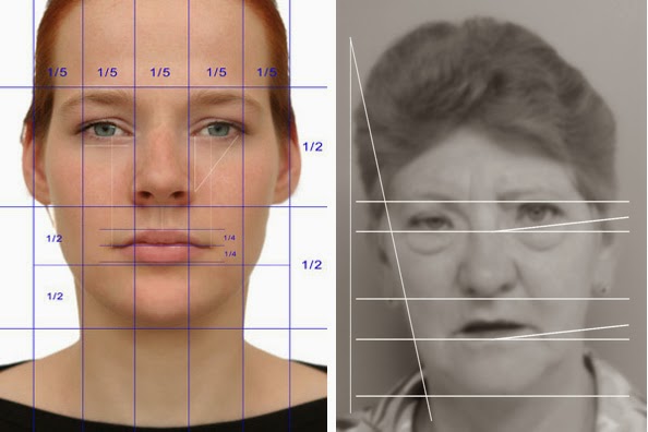


Facial symmetry supposedly plays a role
in physical beauty, but may represent
developmental stability, an ability to resist
disease, and of genetic strength.
The reasons that one human picks another as a mate are unique to the individual; personal history, social mores, and biology all play a role. No matter what e-Harmony or Match.com tell you, a complete understanding of how two people match for life is truly unknowable. Do opposites attract in a meeting of the minds? Is it all about chemistry and the way someone smells? Do big muscles and child-bearing hips play a central role?

Bilateral facial symmetry is thought to play a role in what people think is pretty, and pretty plays a role in mate selection. Why? Because symmetry implies a stable embryologic development and good genes – or maybe not. As Shakespeare wrote in A Midsummer Night’s Dream, “The course of true love never did run smooth;” it applies here.

Bilateral asymmetry takes three forms; fluctuating asymmetry, directional asymmetry, or antisymmetry. In terms of humans and pretty faces, it’s fluctuating asymmetry that we’re talking about.

Take a sharpie and draw a line from the top of your head down to the split in your legs – no, don’t really do it, this is just a thought demonstration. If you were to measure the distance from that midline to the same point on each of your eyes or ears or any body part, the length on the right will be close to the length on the left, but probably not exactly the same.

The same would be true if you measure the heights from the floor to the bottom of some body part like your eyes, or if you measure the length of fingers on each hand. The differences represent fluctuating asymmetry (FA), and your neighbor’s will be different from your own – we say that FA is what gives faces character.


President Lincoln had a defect called facial
micronesia. The left side of his face was much
smaller than the right. The middle image
above is his face. The left image is his right
side flipped and placed against his right side.
The right image is made from two left halves of
his face. Everyone’s right face and left face
look different; his is just a greater than average
difference.
Your body has a plan that determines where each eye or ear will go, and in a perfect world, it would be exactly the same spot on each side of the midline. But your genes don’t control the implementation of the body plan – what you end up with in terms of symmetry is based on the environment in the womb and how well your genetics can compensate for it.

In other words, the more instability there is in the developmental environment, the higher the FA could be. What might stop fluctuating asymmetry from becoming large? Some scientists believe it to be good genes. The instability in the womb could come from disease, toxins, parasites, drugs, stress… who know how many things might be involved.

But if your immune system is strong – partly a genetic trait, then perhaps you could control in utero diseases and FA could be kept to a minimum. For just about any stressor you can imagine, there could be a genetic response that, if healthy and strong, could minimize the damage from the stressor.

In terms of picking a mate, unconsciously sensing low levels of FA suggests to your primitive brain that the genes of this individual might be more adapted to the environment - good potential parent. And that’s all that really matters to an organism at the basal level – having strong offspring.


A 2005 study in Jamaican teenagers showed that
those teens that were rated as better dancers had
less FA. As always, cause and effect is elusive.
Did symmetry make them better dancers, or
 were they perceived as better dancers because
they were more symmetric? Regardless, good
dancers do have a reproductive advantage.
A 2014 study concluded that males with less body FA are stronger with respect to hand grip. They suggest that since strength is a quality used in mate selection and male competition, increased strength may be one reason why symmetric males are considered better mates – they win out in more intra- and intersexual competitions (they're more appealing and win more fights). Just how symmetry brings strength was not discussed. Together, symmetries are considered to reflect fitness, so in terms of non-verbal communication to potential mates, they are considered honest signals, traits that represent truth about the possessor.

A 2012 study in rhesus monkeys built upon the result of a previous experiment in that macaques stare at symmetric faces longer than faces with higher FA. This was supposed to mean that they preferred the symmetric faces. And there is evidence to suggest that our brain does find symmetry in objects, scenes, and art more pleasurable.

So the 2012 study measured both the FA and the health of female macaques. Using veterinary health standards, number of wounds and weight gain over first four years of life, the females with less FA had the best health. This supported the hypothesis that better genes result in better health.

To my mind, there is also the possibility that less FA leads to being treated better within the group, which results in better overall health. Since primates prefer symmetric images, a symmetric face (the seat of social communication) might result in more food offered, fewer fights, and/or less stress – and therefore a better overall health rating. This idea suggests that better health is a result of symmetry, not the other way around. In this case, wouldn’t facial symmetry be a dishonest signal? Is anyone studying this?

There is evidence that supports this notion, or at least lessens the strength of the symmetry and fitness hypothesis. A 2015 study in Senegal found no link between malaria rates and FA in teenagers. This suggests that FA doesn’t predispose to malaria (lack of fitness meaning higher susceptibility), and that malaria does not increase FA.


FA isn’t just a measure of “beauty.” A 2013 study
showed that urban lizards display more FA than
rural lizards. The hypothesis is that urban living
increases exposure to stress and pollutants, and
this manifests as increased FA. If true, tracking FA
could be used a measure of pollution and its effect
on wildlife.
Likewise, a 2011 paper showed that poverty was linked to increased FA. This is a touchy subject. The study found higher FA in young males from the poverty stricken sections of Ankara, Turkey. Does this mean that the stress of poverty destabilized the developmental environment of males, either in utero or afterward? Or does it mean that the stress of poverty increases FA? Does it mean that people with weaker genomes end up in poverty? Would people with better genes be more likely to resist the FA that might result from poverty-induced stress?  I leave it to you to argue that one out.

One could make a strong argument that humans have inventedtheir way out of needing to rely on finding mates with low FA. Our technology, medicine, and brains have helped us to overcome our environment (sometimes to our detriment) so that a different set of traits might be more telling as to the fitness of partners. Intelligence, puzzle solving, emotional quotient and even tendency to maintain monogamy might be more desirable today. But the instinct to look for low FA has been stuck in our brain by evolution and it doesn’t care about logic.

Why do I bring up emotional quotient and monogamy? Some studies reviewed in 2010 show that lower FA in human males and females correlates with more extramarital or extra-significant other affairs (called extra pair coupling, EPC). Males with more symmetrical bodies sought out more EPC and females who engaged in EPC were more likely to seek out men with low FA. Not a great commercial for symmetry.

It isn’t just humans that sense symmetry and use it for mate selection. The primate studies above show that it extends to macaques, but birds do it was well (as do probably animals in other phyla). Peahens look for symmetric tails in peacocks and barn swallow females look for makes with symmetric and long tail feathers.


The left butterfly is paler; it's a territorial male. The
right speckled wood butterfly male is non-territorial.
 He flits from area to area looking for the females not
in a sun spot. If he competes with a territorial male –
he always loses. He has more FA than a territorial
male, but no directional asymmetry.
Everything we have said above states that animals look for symmetry in mates, but there are exceptions. The speckled wood butterfly(Pararge aegeria)is a great example. The biggest exception of all is that the females prefer asymmetry – a bit.

This European and North African butterfly has two male morphs. One is territorial, it's a paler color and has fewer spots on it’s wings. The other morph is darker, has dark spots and is non-territorial. The territory they fight over is a sunny spot where the light comes through the forest canopy.

Females are more likely to be in the sun, so territorial males will fight over a sunny spot territory. The contest between males is aerial; combinations of acrobatics and duration determine the winner – except when the territory is being defended. Sound a bit confusing?

In 1978, a paper addressed this. The author found that the defender of a territory ALWAYS won the flying competition. It’s a weird way of determining who will probably mate more often if they already know the outcome. A non-territorial male will compete, but will always lose.

The only time the competition isn’t rigged for the defender is when the spot has no owner, or both males believe it to be their territory. Then the flight contest is much longer and more intricate. This is where the asymmetry comes in.


The territorial speckled wood butterfly males seek
out sun shafts in the forest. This is where the girls
will be, but it also makes them better at the flying
competition. Their time in the sun warms them up
according to a 1998 paper. Being ectotherms, warmth
means they will fly with more energy and win
more contests.
Males have some FA in their wings, but females have much more. What might be limiting the asymmetry in the males? The flying competition. A 1999 study looked at the asymmetry in the wings and noted that those males that had some asymmetry, but not too much, turned in the air faster and could stay aloft longer in a downward spiral maneuver – the main competitive move for males in an honest contest for a territory.

If asymmetry helps win the territory when it is up for grabs, then it will provide more contact with females. This means that this asymmetric (uglier?) male will have more reproductive success and his version of asymmetry will be passed on, if it's genetic.

FA is generally not considered genetic, but directional asymmetry is. The fluctuating asymmetry in the males is low, but the asymmetry that helps win competitions seems to be directional, so it could be genetic. The asymmetry that helps flying is slight, so it is a middling asymmetry that should be passed on.  The fact that females and non-territorial males have more asymmetry suggests that keeping asymmetry low is energetically costly (think about it).

Directional asymmetry in the wings has a functional advantage so it is worth the cost, whereas FA is allowed to get larger. It just so happens that directional asymmetry and antisymmetry are our subjects for next week.


Wade, T. (2010). The Relationships between Symmetry and Attractiveness and Mating Relevant Decisions and Behavior: A Review Symmetry, 2 (2), 1081-1098 DOI: 10.3390/sym2021081

Little, A., Paukner, A., Woodward, R., & Suomi, S. (2012). Facial asymmetry is negatively related to condition in female macaque monkeys Behavioral Ecology and Sociobiology, 66 (9), 1311-1318 DOI: 10.1007/s00265-012-1386-4

Fink, B., Weege, B., Manning, J., & Trivers, R. (2014). Body symmetry and physical strength in human males American Journal of Human Biology, 26 (5), 697-700 DOI: 10.1002/ajhb.22584

Thomas F, Doyon J, Elguero E, Dujardin JP, Brodeur J, Roucher C, Robert V, Missé D, Raymond M, & Trape JF (2015). Plasmodium infections and fluctuating asymmetry among children and teenagers from Senegal. Infection, genetics and evolution : journal of molecular epidemiology and evolutionary genetics in infectious diseases, 32, 97-101 PMID: 25725158

Lazić, M., Kaliontzopoulou, A., Carretero, M., & Crnobrnja-Isailović, J. (2013). Lizards from Urban Areas Are More Asymmetric: Using Fluctuating Asymmetry to Evaluate Environmental Disturbance PLoS ONE, 8 (12) DOI: 10.1371/journal.pone.0084190

Ozener B (2011). Does urban poverty increase body fluctuating asymmetry? Collegium antropologicum, 35 (4), 1001-5 PMID: 22397230


For more information or classroom activities, see:

Fluctuating asymmetry -


Hermit Houses And Fiddler Claws

$
0
0
Biology concepts – bilateral asymmetry, directional asymmetry, antiasymmetry, crabs, evolution, mate choice, sexual selection, sexual dimorphism


Hermit crabs aren’t true crabs since they don’t
have ten legs (two go to form claws in true crabs)
and their antennae and eyes are different from
true crabs. Many don’t even live in the water, but
that is true for true crabs as well. King crabs,
horseshoe crabs, coconut crabs, they all have
crab in their name, but they aren’t true crabs.
Our first exception today concerns the hermit crab and the animal that determines whether hermit crabs live or die. Hermits don’t eat snails, but snails are the most important things in the life of a hermit crab. And it all relates to our ongoing to tale of bilateral asymmetry.

Most snails live in shells of their own making. As they grow larger, they add to the aperture (opening) of the shell to make it bigger – they remodel instead of relocating. As they add on to their shell, most take on a curve, so that over time, they are spiraled.

The vast majority of snail shells spiral asymmetrically to the right. They are dextral, although the occasional sinistral(left-handed) shell is found, and some species are equally left and right shelled. Because the asymmetry of the shell is almost always to the right, this is called a directional asymmetry (see last week’s post).

The right handedness of the spiral isn’t a distinction without a difference – it matters. Since the snails reproduce with organs that are located in their heads, they have to be able to line up correctly for mating; this means they need right handed spirals that fit together well when they face each other.

How does this affect the hermit crab and its survival? Because hermit crabs use gastropod shells as protection for their soft abdomens, that's how. Hermit crabs have hard exoskeletons on the front of their body, but their abdomen is covered in a very soft shell that is easy prey for a predator.

Their abdomen is reducedas well. The legs are absent or very short, and this leaves a tapered back end that fits well into an abandoned gastropod shell. The furthest south point on the hermit crab abdomen has a specialized function for gripping the inside of the shell. So, he/she carries the shell around for protection – until he/she outgrows it.


Shells are at such a premium that even before
one is abandoned, there are others lining up to
use it. Notice how they line up from large to
small. When largest finds a new shell, each of the
others will be able to move up to new digs as well.
When the crab molts a few times and becomes larger, he has to find a new home. Therein lies the rub, because snails keep one shell their entire life. Consequently, a shell only becomes available when a snail dies. And even then, they have to die in a way that doesn’t destroy the shell.

Shell number is the main stressor in a hermit crab’s life. The availability of appropriate shells is the major limiting factor in keeping hermit crabs safe. If there are too few, hermits have to find alternate housing (maybe the neck of a coke bottle or some such thing). Snail shell numbers can and do limit the size of hermit crab populations.

We stated earlier that the vast majority of snail shells are dextrally asymmetric, so it isn’t surprising that hermit crabs greatly prefer dextral shells. This is a case of an animal born symmetric but grows asymmetric. The dextral spiral of the shell molds the abdomen as the crab grows, so that it becomes asymmetric.

When a hermit crab finds an appropriate new shell, it pulls its abdomen out and quickly enters the new shell. This is the time when the crab is most vulnerable to predators. When it comes out of the shell, you can see that the abdomen is soft and reduced, but you also see that it is straight. The asymmetry is an adaptation see when they coil, but under muscular control it will be straight.


You can see the asymmetric twist of the hermit
crab’s abdomen to the right side, but it isn’t
born that way. This happens after growing up
with a dextral shell for a home. They are born
with straight abdomens and can easily
straighten them out if they so choose, since
the shell is so soft on the abdomen.
So what happens if the hermit is presented with a sinistral shell? What if it's the only shell available? Well, they may use it, but they don’t necessarily like it. A 1994 experiment showed that when presented with dextral shells, the hermits turned them correctly to pour out any sand inside.

But with left spiraling shells, they only had a 50/50 chance of turning them correctly, based on which direction they were facing. The crabs don’t instinctively know how to make use of a sinsitral shell. Good thing their abdominal asymmetry is labile; they can use a sinistral shell if that’s all they have to choose from, or even an old pop can if need be.

The hermit crab’s story is one of directional asymmetry and acquired directional asymmetry, but another crab gives us a story of directional symmetry and antisymmetry. This crab is the arm wrestling champion of the crab world.

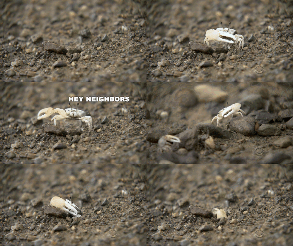
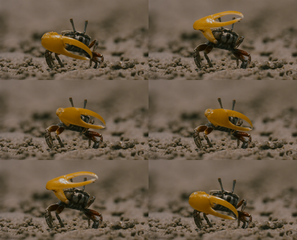
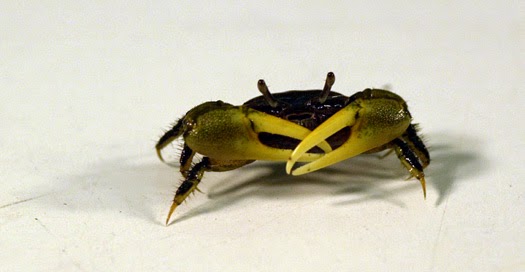
There are 92 species of fiddler crab (genus Uca). The males of this genus have one oversized cheliped, called the major claw. That makes this a sexually dimorphic trait (see this post). They wave the major claw (more on this below) and long ago it looked to someone as if they were playing a fiddle – so they're called fiddler crabs. One guy and his observation; he must have been some kind of big wig, because the name stuck.


This shows the major claw of one of the 92
species of fiddler crab. These are in fact true
crabs, as opposed to hermit crabs above. Not
that the burrow is out of the water, but will be
inundated by a high tide. That’s important
for mating.
The major claw has a few functions. One of the primary functions is as a sexual ornament for attracting females. But it’s an ornament with a reason for its size. The males prepare a burrow for the female to lay her eggs in, and the width of the burrow matches the width of the major claw.  A wider burrow leads to a lower temperature for egg development, so it takes longer for the eggs to mature.

A 2007 study showed that the tides are very important for reproduction by washing the larvae into the water. So, if the females are mating five days before the highest tides, they choose the burrows of the largest males to slow down the maturation of the eggs. If they mate late, they choose the smaller burrows of smaller-clawed males, so the temperature will be higher and the eggs will hatch faster and be ready for the coming high tide.


The male waves his major claw to attract
females, so it is a dimorphic sexual ornament.
But it also functional, as the length of the claw
determines the width of the burrow and is
used for fighting off other males. Some males
don’t wave, they just snatch females into their
burrow when they come close. How rude.
So how does a male fiddler promote his burrow size? He waves his claw in front of the females. This draws attention to his claw and therefore his burrow size. Interestingly, they play faster when in an orchestra – well sort of. A study in 2012 showed that in crowds, the males wave their claw faster. Waving for a long period is energetically costly, so they wave fast when competition is highest and slow down when fewer males are present.

In 90 of the 92 fiddler crab species, the major claw is antisymmetric. This means that roughly half of the individuals will have the major claw on their left side and the other half will be right-clawed. This suggests that the exaggerated growth of the major claw is not controlled genetically. The pressure on claw direction is either negative-frequency based (see this post), or there is no pressure to choose a side.

However, there are exceptions. In two species, the major claw is directionally asymmetric; 99% of the males will have their major claw on their right side. This does suggest a genetic component to the side on which the major claw occurs. It’s interesting that these two very different mechanisms occur in species that are so closely related. Did antisymmetry evolve from directional asymmetry or did only a couple of antisymmetric species become directionally asymmetric?


Here you see the exceptional fight; one right-
clawed and one-left clawed individual. If they
were both right-clawed, they could clench claws
sort of like a hand shake. This is how they were
designed to be used and work best for
crushing force.
The side the major claw is on has relevance for male fighting competitions. The fights between males are very different when they have claws on the opposite side as compared to when they are on the same side. The fight starts with the males facing each other and pushing against each other claw to claw. The fight might end right there if one is demonstrably stronger, but if not, they will interlock claws and try to move the other out of the way. Interlocking occurs one way if they are opposite and another if they are same handed. This could affect the outcome.

A left-clawed male in a right-clawed species is usually at a disadvantage because of the mechanics described above. A 2007 study showed that left-clawed males lost their burrows to right-clawed males more often and kept their burrows for shorter times than right-clawed males. The right-clawed males are also more likely to pick a fight and try to take another crab’s burrow.

Therefore, these researchers hypothesize that left clawed individuals in a right-clawed species are not advantaged in a negative-frequency dependent manner – in fact, they are significantly disadvantaged, and this may keep the percentage of left-clawed males low.

The idea of whether the antisymmetric claws are not heritable traits and the directional ones are is an interesting topic.  And it gets more interesting when you start talking about regeneration– fiddler crabs can regrow the claws or limbs they lose.


Every once in a while you may see a fiddler crab
with two major claws. This isn’t such a good
thing. It costs a lot of energy to build and maintain
one, let alone two, so they will be at a survival
disadvantage. More importantly, it’s hard to pick
things up and eat them using the major claw. The
small claw is important for feeding.
Sometimes the major claw is lost, but it can come back. In some species, the small claw will grow into a major claw, and the regenerated claw will become the small claw. This makes sense – regrowth is kind of slow because the growth is limited by the exoskeleton until there is a molt – or several. By making the small claw into the big one, a couple of molts are saved.

But in other species, handedness is set and can’t be broken – if the major claw is lost, the regenerating claw will be the one of the same size no matter what. Dr. Brook Swanson of Gonzaga University has observed these regenerations in a right-clawed species. He stated that the major claw will regenerate almost completely in one molting, but to accomplish this the overall growth and size of the crab will be reduced. They have to shrink to get the claw to grow fast. Remember, this claw is basic to their competition and mating.

This leads to an amazing feature of fiddlers who regenerate a major claw. The new one is often not as strong as the old one, but they aren’t going to tell their opponent that. The crab will behave as though it's as strong as ever; he will feign wanting to fight even though he is aware that he is not as strong. They fake it – like that bully in middle school that ran away if someone stood up to him. This is called a dishonest signal, as opposed to the fluctuating asymmetry and honest signal that we talked about last week.

Next week, let’s switch it up and talk about asymmetries that occur on the inside of the animal body.


Backwell, P., Matsumasa, M., Double, M., Roberts, A., Murai, M., Keogh, J., & Jennions, M. (2007). What are the consequences of being left-clawed in a predominantly right-clawed fiddler crab? Proceedings of the Royal Society B: Biological Sciences, 274 (1626), 2723-2729 DOI: 10.1098/rspb.2007.0666

Milner, R., Jennions, M., & Backwell, P. (2011). Keeping up appearances: male fiddler crabs wave faster in a crowd Biology Letters, 8 (2), 176-178 DOI: 10.1098/rsbl.2011.0926

Reaney, L., & Backwell, P. (2007). Temporal constraints and female preference for burrow width in the fiddler crab, Uca mjoebergi Behavioral Ecology and Sociobiology, 61 (10), 1515-1521 DOI: 10.1007/s00265-007-0383-5

Imafuku, M. (1994). Response of hermit crabs to sinistral shells Journal of Ethology, 12 (2), 107-114 DOI: 10.1007/BF02350055


For more information or classroom activities, see:

True and false crabs –


Left-Handers Have Prettier Brains

$
0
0
Biology concepts – evolution, internal bilateral asymmetry, lateralization of function, brain, neural plasticity


This MRI start at one ear and shows slices through
the head until it gets to the other ear. It looks as
though the loop reverses itself halfway through,
because we are bilaterally symmetric – supposedly.
But the brain has some very specific asymmetries.
Can you see the differences from side to side?
Brains are amazing things – and we know next to nothing about them. For instance, every once in a while a seemingly normal person will show up at a doctor’s office or hospital for a headache or perhaps a tingling in an extremity. When they have an X-ray or an MRI of their head, low and behold they’re missing some large part of their brain! Sometimes, an entire hemisphere just isn’t there.

In 2009, a 10 year old girl in Britain was found to have depth of vision and a full visual field even though she was completely missing her right cerebral hemisphere. As you might already know, some visual signals from each eye cross the midline and are processed by the alternate side of the brain, but her brain had reprogrammed itself to process all visual signals through the left hemisphere.

This cerebral hypoplasia often brings some functional defects, but not always. Defects might occur because the brain is functionally asymmetric; there are lateralization of functions (more on this below).

However, the brain is quite plastic in early life. Many possible defects can be worked around, especially if the problem is congenital or comes from a trauma that occurred early in life. Rerouting of functions is more likely if the brain is in the process of lateralizing function, rather than if the brain is set in its ways.

Take for example the case of an 88 year old man who came to the hospital in a bit of confusion with a tingling in one foot. A subsequent MRI showed that his brain was completely missing a structure called the corpus callosum (Latin = tough body). This is the main connection between the right and left hemispheres of the cerebrum.


The corpus callosum is just the largest connection
(commissure) between the different hemispheres,
but there are others. The anterior may be involved
in color perception while the posterior is for the
pupillary light reflex. The optic chiasm crosses some
visual signals to the opposite optic nerve, but the
functions for the middle and habenular are unknown.
His right half wasn’t speaking his to his left half (like many extended families), yet he functioned just perfectly. The reason is that he was most likely born that way, and his brain had ample time to build up the lesser communications between the hemispheres; this is plasticity. And yes, there are other ways for the two hemispheres to talk to one another, though most people believe it occurs only through the corpus callosum (see image to left).

We have filled several posts this year discussing the asymmetry of some animals and plants (see this, this, this, this, this, and this post). In each case, we have been talking about external asymmetries, but that isn’t the only kind. Our bodies, and those of many animals, also have internal asymmetries.

Most animals have significant internal asymmetry, but there’s no better place to start than the brain. It is both structural and functionally asymmetric. Here are a couple of stories of the brain structures/functions and the asymmetries that are built into them.

The human brain has many parts; each of which has varied functions, although most parts coordinate together. There are usually two cerebral hemispheres which make up the cerebrum (Latin for brain… well, duh). These hemispheres make up 80% of the volume of the human brain. It is here that our more advance thinking takes place – language, thought, attention, decision making, emotion, and consciousness just to name a few.

Each of the main parts of the brain can be broken down into many subparts, each with unique or coordinated functions. For example, the cerebrum can be broken down into lobes; frontal temporal, parietal, and occipital. Deep inside these are the more primitive structures, like the hypothalamus, amygdala, and thalamus that have their own function in emotion and control.


This very crude map shows the motor strip, where
your muscle movements begin. Just behind it is the
somatosensory strip, where sensations from you body
come in. Broca’s area is for language understanding and
Wernicke’s in the temporal lobe is for speech making.
Notice they show the left side – it’s bigger in right-
handed people.
Each hemisphere has its own version of these lobes and deep structures, although sometimes the functions carried by the same lobe might be different. This is called lateralization of function; these are functional asymmetries, but they can have effects on the structure of the brain as well.

For instance, which hand you normally use can be reflected in the size of your brain in the planum temporale, persylvian region, and other parts of the frontal, parietal, and temporal lobes. You know that most stimuli cross to the opposite hemisphere where they are then converted into responses. If you’re right handed, your left hemisphere will be in control of your right hand.

These areas of the brain normally have asymmetries anyway, since they are the areas that process language and speech. Both understanding speech and making speech are lateralized to the left hemisphere (well, there are exceptions, a few people process language in the right hemisphere or equally in both hemispheres). Areas such as Broca and Wernicke (see picture above) are larger on the left hemisphere because language is crucially important for humans and has therefore developed to take more area.

(Even though we aren’t going into the subject here, I just want to say that the whole thing about people being right brained or left brained is a myth. We’ll tackle it another time.)

But handedness does play a small role. About 95% of right-handers have a left dominance for language processing, but only about 80% of lefties are left hemisphere speech dominant. So in some cases, the hand you use is reflected in asymmetries of your brain.


The number of right and left-handed people has always
been about the same. They can tell from the brush
strokes and hand prints in cave paintings. Interestingly,
new measurements of finger lengths indicate that about
75% of cave paintings were done by women.
Handedness also affects the size of the motor strip in the frontal lobe (see picture above). Righties have a larger left frontal lobe motor strip, but the opposite is not true with lefties. Their right frontal lobe motor strip might be larger, but is not as increased in size as the left motor strip is for righties – go figure.

And it isn’t just the motor strip; the speech and language area of lefties isn’t as big as it is in righties. All in all, left-handed people tend to have much more symmetric brains, in terms of shape and size.  I wonder if this makes them more attractive (see this post). “You have a lovely brain. Are you left handed?”

The second largest structure is the cerebellum (Latin = little brain, I see a pattern). This part of the brain is much older, evolutionarily, and is responsible for posture and coordinating muscle movement to give balance. It’s nice to know that primitive animals are capable of good posture – why aren’t teenagers?

The cerebellum comes in two hemispheres, just like the cerebrum, but they are smaller and located below the posterior part of the cerebrum. Similar to our examples above, some people only have one cerebellar hemisphere too. This is called unilateral cerebellar hypoplasia.


The brainstem is the oldest part of the brain. Most of the
time it can’t be seen because the cerebral hemispheres
cover it up. The cerebellum sticks off the brainstem
and has two hemispheres. It coordinates muscle
movement.
The brainstemis the oldest part of the brain and connects our higher functioning areas to the spinal cord. The brainstem has specific jobs in maintaining the basic functions of life; sleep/wake, breathing, cardiovascular control, and pain. In addition, all the neurons that take signals toad n from the higher parts of the brain have to pass through the brainstem to the spinal cord.

But that isn’t to say that the brainstem in humans is just as it was in early evolution and is now in lower animals. A 2014 study was the first to look at asymmetry in the halves of the brainstem (it has a right and left half even though there is just one brainstem).

The structural asymmetries in the cerebral hemispheres were recapitulated in some of the structures of the brainstem (inferior olive and dentate nucleus), suggesting that the evolution of higher functions and lateralization of those functions has brought about a lateralization and structural asymmetry in the old brain as well. You can teach an old brain new tricks.

The corpus callosum(CC) is the main commissure between the cerebral hemispheres as we outlined above. It is thought that one of the functions of the CC is to integrate signals processed in each of the hemispheres. There are millions of messages buzzing back and forth from one hemisphere to the other every second, like a giant highway with 125 million lanes in each direction.

The result of all this traffic is a coordination of responses; each hemisphere doing its lateralized job plus both doing the jobs they share (and there are many). The result is a smooth integration of thought, sensation, and action.


The brain has grey and white matter. The white neurons
are covered with myelin; this helps the signals travel
faster, but doesn’t allow for connections. Grey matter
is where all the connections are made between neurons.
The finger here is pointing out the corpus callosum of a
pig. It is white because these are just signals that need to
get from one side to the other; no processing is done
in the corpus callosum.
But integration isn’t the CC’s only job. There are many inhibitory signals that pass through the corpus callosum as well. It is thought that these signals depress neural function in one hemisphere or the other, and this is the basis for lateralization of function in each half of the cerebrum. This is the hypothesis, but until now it has mostly been investigated in animal models of CC function. A new study (2015) shows that patients with agenesis of the CC (AgCC) indeed have more hemispheric autonomy.

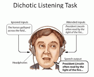
Many functions are usually more acute in one hemisphere, like hearing and repeating one speech while listening to two simultaneously. This is one of the dichotic listening tests and usually shows a right-eared advantage (the right ear is better at separating out the two voices), but the patients with AgCC have no ear advantage for this.

And it wasn’t just “earedness;” the AgCC patients were also much more like to be ambidextrous, showing no predilection for right or left hand in fine motor functions. Together, these findings suggest that one of the functions of the CC is to suppress some functions in each hemisphere to give lateralization. This brings up an important question. What’s the advantage to a functionally (and therefore structurally) asymmetric brain?


Small birds need to watch to find food to peck up and
eat, but also to pay attention for predators or angry
vegetarians. These things are done by different sides of
the brain and it is lateralization of function that lets us
do multiple things at the same time.
Evolution has resulted in many animals having specialization of functions in separate halves of the brain, so it must convey some advantage. It seems that multitasking is the answer. Lateralization of function allows for simultaneous brain function on different tasks, or at least makes it a lot easier. A 2004 study in baby chicks showed this.

Some chicks were lateralized in the shell (one hemisphere will do all the visual processing if you expose the shells to light 3 days before hatching). As compared to normal chicks, the lateralized chicks could find food just fine, but couldn’t pay attention to predatory birds while they pecked for corn. The two functions, which are normally controlled by different hemispheres could be carried out by the control chicks, but couldn't be done when both functions were experimentally forced into the same hemisphere.

Next week, we’ll see how sex hormones play a role in asymmetric development of the brain structure and function. And there are individual differences (fluctuating asymmetries) in our brains as well.


Ocklenburg S, Ball A, Wolf CC, Genç E, & Güntürkün O (2015). Functional Cerebral Lateralization and Interhemispheric Interaction in Patients With Callosal Agenesis. Neuropsychology PMID: 25798664

Rogers, L., Zucca, P., & Vallortigara, G. (2004). Advantages of having a lateralized brain Proceedings of the Royal Society B: Biological Sciences, 271 (Suppl_6) DOI: 10.1098/rsbl.2004.0200

Muckli, L., Naumer, M., & Singer, W. (2009). Bilateral visual field maps in a patient with only one hemisphere Proceedings of the National Academy of Sciences, 106 (31), 13034-13039 DOI: 10.1073/pnas.0809688106

Baizer, J. (2014). Unique Features of the Human Brainstem and Cerebellum Frontiers in Human Neuroscience, 8 DOI: 10.3389/fnhum.2014.00202


For more information or classroom activities, see:

Brain structures –

Lateralization of function –

Everybody Is Just A Little Twisted

$
0
0
Biology concepts – bilateral asymmetry, brain, ventriculomegaly, brain torque, fluctuating asymmetry, sex hormones


“Getting all your ducks in a row” actually comes
from bowling. The pins used to be known as
ducks….still are in duckpin bowling. Before pin
setting machines, people had to put the pins
down in even, straight lines so that the game was
predictable and fair. The “ducks” had to be
in “rows.”
When you are organized, you have “all your ducks in a row.” When you are calm and in control, you’re “thinking straight.” When you are doing the right things, you’re “on the straight and narrow.” When something is undiluted, like a story or whiskey, you get it ”straight.”

Basically, if something is straight, it is correct, unembellished, pure, or not perverted. So what would be the opposite of this? Twisted, of course.  Twisted tales are slightly weird or not true to the stories they are taken from. And when you're “a little twisted” you’re thinking is mentally unsound or a bit perverted. 

In modern internet slang, twisted means high and drunk, so again it means a perverted form of thinking. Not that it always has to be bad; Stephen King is truly a bit twisted, but he writes well and leads a relatively normal life. Some people bask in the sunlight of their twisted thinking.

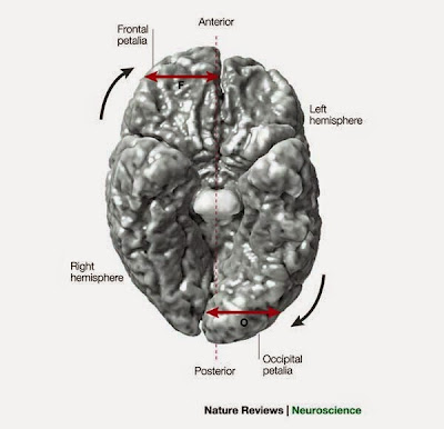
But what if I told you that everyone's brains, and therefore everyone's thinking, is just a bit twisted? In technical terms it is called a Yakovlevian torque, but let’s not kid ourselves, it means that our brain is twisted in our skull.

First recognized by the Russian born Harvard anatomist Paul Ivan Yakovlev, the midline fissure where our two cerebral hemispheres butt up against one another doesn’t exactly follow the midline of our skull. There is a slight twist to the left, so that the right cerebral hemisphere crosses the midline to the left behind your forehead and the left hemisphere crosses over to the right side of your skull in the posterior. Everybody’s brain is just a bit out of whack!


Here is a human brain cartoon as seen from
 below from a Nature Review paper. The right
hemisphere is actually on the left side. Notice how
the frontal lobe of the right hemisphere sticks out
further? It’s the same with the occipital lobe in
back on the left lobe. In each case, they twist so
they the midline of the brain crosses the midline
of the skull. The brain is torqued to the left. 
In other words, the right side of your brain is torqued slightly forward relative to the left and this twists it out of bilateral symmetry with your skull. At least part of this is due to the larger left parietal lobe areas for speech and language that most people have on their left side (see last week’s post), but that isn’t the only source.

Several sources of asymmetry go together to produce brain torque, and it can be greater or lesser in each individual – yes, some people are more twisted than others.

Yakovlev observed that different people had different size depressions on the interior surfaces of their skull in the right anterior and left posterior (called petalias). You can imagine that with a tofu-consistency brain sitting in an opened skull, it might be hard to quickly recognize a slight twist to the left, but Yakovlev wrote about it and hypothesized on its importance.

It wasn’t until 2009 that a couple of studies (here and here) confirmed the torque using very advanced imaging techniques and alot of math. The hard part is accounting for brain position internally and skull midline externally, each of which have fluctuating asymmetries within one individual and variances between individuals. But both studies concluded that Yakovlevian torque is real. Now we just need to figure out it’s significance.


This short clip shows the loss of grey matter
(unmyelinated neurons) through development in
a person with schizophrenia. As colors move to
yellow, that indicates more loss. The hemisphere
start out asymmetric and become more so. Maybe
cases can be diagnosed earlier by watching
for asymmetries.
Even if we don’t know why our brains need to torque, we can perhaps use the observation in medicine. Some torque is normal, but too much occipital bending may be bad. Two studies from the same group have correlated excessive torque with major depression and bipolar disorder. One even related atypical torque to developmental stuttering in boys.

On the other hand, a study in autism showed that there was no correlation between autism spectrum disorders and atypical Yakovlevian torque. Well, sort of. This single study only looked at high functioning, right-handed, boys in a narrow age range. So who knows if there might be some relationship between autism and torque in other affected groups. Always be sure to take into consideration the limits of any study you read.

So torque is the norm in human brains; guys and girls both have a leftward twisted to the brain within the skull. There are many more asymmetries in brain than just the ones we have discussed; there are many examples of left > right asymmetries and some of right > left. These differences are evolutionary and are apparent even during the second trimester. But the mechanism and reason for each asymmetry may be different; some are based on gender - boy brains and girl brains are different!

This exception in bilateral symmetry is two fold; there are gender-induced differences in the brain that are both sexual dimorphic – meaning that they depend on the sex hormones or sex chromosome genes of each individual, and they are asymmetric – meaning that the gender-induced structure or function changes affect one hemisphere more than the other.


Dr.s Ruigrok, Suckling and Baron-Cohen have
collated sexual dimorphisms in brain structures.
The red areas are larger in women; the blue areas
larger in men – the girls’ should have been pink.
These differences are due to genetic, hormonal, and
environmental factors all mixed up in a big soup.
Many studies have linked prenatal androgen (male sex hormones) levels to asymmetry, but some asymmetries appear before androgen even begins to be produced. These male asymmetries are probably due to the genes expressed on the sex chromosome. A 2014 study linked the two phenomena. Apparently regional asymmetries in the male cerebrum and cerebellum are exacerbated compared to female brains through the joint action of testosterone and X-linked genes.

Male brains are more lateralized than females. Functions are segregated more strictly in male hemispheres, so perhaps it’s true that only women use their entire brain.

A few weeks ago we talked about fluctuating asymmetries– those differences in structure size and position from individual to individual. We were talking then about external asymmetries, but peoples’ brains have fluctuating asymmetries as well. One in particular may help predict neurologic disease – and it isn’t even a brain structure.

Several studies go back and forth on whether the size or asymmetry of the lateral ventricles (CSF filled spaces in the brain, see this post) can predict schizophrenia or developmental delays. A 2010 case reportsuggested that alone they may have no particular effect, but if mild ventriculomegaly (bigger than normal ventricles) is accompanied by atypical lateral ventricular asymmetry (the left lateral ventricle is normally a bit bigger than the right), then these may be predictive of delay and/or schizophrenia later in life.

For a more historical example of fluctuating asymmetry in the brain let’s go straight to the top – Albert Einstein. While he was alive people wondered if his brain was different from all of ours. He just thought on a different plane; his concepts were Earth shattering, yet he used thought experiments with elevators in space and passing trains.


Einstein had a brain to be admired, and I’m sure
he wouldn’t use his powers for the dark side. But
thought experiments about elevators in space and
trains passing by aside, I’m wondering what he
may have gleaned about the universe from a duel
using the force with Darth Vader.
When he died in 1955, Einstein consented to have his brain studied. The pathologist, Thomas Stoltz Harvey, did no one any favors. He weighed it, took some pictures of it, made some measurements, and then cut it up into 240 pieces. He kept some and gave a few to other pathologists. This pretty much eliminated any decent study that could have been done at the time. Thanks a lot Tom.

In 1999, a qualitative and quantitative study of the data and measurements recovered from Einstein’s brain was carried out, comparing it to 5 male and 56 female brains. The researchers’ results were shocking for what they did and didn’t find.

First, Einstein didn’t have a huge brain, it was basically the same size as everyone else’s. There weren’t extra lobes or a million times more neurons – it looked like a regular brain on first glance. But that’s where science comes in; scientists don’t stop at a first glance.

For one thing, Albert’s brain was missing a certain landmark. Without getting technical, there are two fissures (sulci) that usually pass by one another and create a little island of tissue near the temporal/parietal lobe border. Well, Einstein’s didn’t pass by one another, they merged. This allowed more room for brain tissue since there was only one fissure instead of two.


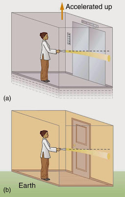
Long before he could measure the bending of light
by massive objects, Einstein thought about how
shining a light on Earth (force of gravity), and shining
a light in space elevator (force of acceleration) might
show how light illustrates general relativity. It took
long expeditions and many years, but his idea was
finally proved correct by measurement of
sunlight during an eclipse.
This is then related to the second difference they found in Einstein’s brain. His brain was about 15% wider than normal in the parietal lobes. He had more brain shoved into that area. As fluctuating asymmetries go, this is beyond huge, usually the difference might be 1-3%.

Why might this fluctuating asymmetry be important? Did it have a functional correlate? That would be hard to tell since the brain isn’t firing now. Wouldn’t it have been great if functional MRI had been around when Albert’s brain was still in Albert’s living head? I bet he could have lit up most of Princeton – but I digress.

The parietal lobe has many lateralized functions, but some of them are right in Einstein’s wheelhouse. This lobe is important mathematical reasoning, and for connecting visual, somesthetic and auditory stimuli together into a big picture.

Take all this together and what you get is that the parietal lobe is what creates mathematical relationships, conscious or unconscious, amongst the world and its moving parts. Professor Einstein was better at that than everyone else, so maybe his wider than normal parietal lobe was responsible.

Of course this doesn’t let the rest of us off the hook. Plenty of people do some awesome thinking and reasoning with very ordinary brains. As we have shown before in this blog – exercise your brain and it will become sharp. Use it or lose it.

Next week, more internal asymmetries in those bilaterally symmetric animal bodies. Your lungs are for breathing, but right and left don’t participate equally – and there’s some cool math involved, so warm up your parietal lobes.


Maller, J., Anderson, R., Thomson, R., Rosenfeld, J., Daskalakis, Z., & Fitzgerald, P. (2015). Occipital bending (Yakovlevian torque) in bipolar depression Psychiatry Research: Neuroimaging, 231 (1), 8-14 DOI: 10.1016/j.pscychresns.2014.11.008

Maller, J., Thomson, R., Rosenfeld, J., Anderson, R., Daskalakis, Z., & Fitzgerald, P. (2014). Occipital bending in depression Brain, 137 (6), 1830-1837 DOI: 10.1093/brain/awu072

Mock, J., Zadina, J., Corey, D., Cohen, J., Lemen, L., & Foundas, A. (2012). Atypical Brain Torque in Boys With Developmental Stuttering Developmental Neuropsychology, 37 (5), 434-452 DOI: 10.1080/87565641.2012.661816

Savic, I. (2014). Asymmetry of cerebral gray and white matter and structural volumes in relation to sex hormones and chromosomes Frontiers in Neuroscience, 8 DOI: 10.3389/fnins.2014.00329

Witelson, S., Kigar, D., & Harvey, T. (1999). The exceptional brain of Albert Einstein The Lancet, 353 (9170), 2149-2153 DOI: 10.1016/S0140-6736(98)10327-6


For more information or classroom activities, see:

Gender differences in brains –

Yakovlevian torque –

Albert Einstein’s brain –


Fibonacci Numbers And Odd Lungs

$
0
0
Biology concepts – respiratory system, bilateral asymmetry, evolution, estivation, Fibonacci scaling in nature


Worf, although raised by humans,was mostly Klingon.
He definitely adhered to their warlike nature. His
three lungs would make him a great fighter. More
oxygen exchange means more ATP production
and more energy for the muscles. 
Not that I’m a devout Star Trek fan, but did you know that Klingons are supposed to have three lungs? The third lung was supposed to give them extra endurance on the battlefield – makes sense.

Closer to home, there was a soccer player from South Korea, Park Ji-Sung, who played for Manchester United. His nickname was “Three Lungs Park” because he could run and run without tiring.  Maybe he’d still be playing if figured out where the ball was going to be and attacked there; a Klingon would have.

Continuing our stories of internal bilateral asymmetries, let’s look at the lungs. Most people have a general idea of what their lungs look like, they seem to be fairly symmetric – but they’re not, not by a long shot. Lungs are perhaps the most asymmetric of the paired organs. Knowing this, I started to wonder if there are any animals with three lungs, or maybe just one lung.

I wonder weird things, don’t I? As it turns out, we can find examples of both in the same animal group. Snakes come in several flavors with respect to lungs and breathing. Some snakes have just one lung. This is helpful for maintaining their svelte figures, losing a lung for them was strictly a space saving measure.


See in this picture how the trachea can stick out
and to the side while it swallows prey. As it
draws in the prey, the right and left sides of the
jaw will ”walk” over the body. As this happens,
the trachea will switch back and forth from side
to side to maintain an open airway.
Snakes have a trachea that is somewhat like ours. It has rings or partial rings of cartilage that carry air from the mouth to the lung(s). They mouth breathe, but occasionally their mouths are full, having unhinged to swallow something whole. So how do they breathe when they have their mouths, throats and bodies stuffed with a rat or a goat or overly curious next door neighbor?

I'd think they would have a hard time of it. Could you breathe through your mouth after stuffing it with the last four dinner rolls just so your brother wouldn't get them? I think not. But the trachea of snakes is better than ours. It isn’t anchored in the throat, so it can actually move forward like a snorkel (see the picture). The trachea will stick out the side of their mouth as they stuff it full of very surprised frog legs, allowing them to breathe and eat at the same time.

A snake’s trachea communicates with its lung or lungs, depending on how many they have. Many species have a long, skinny right lung and a very small, nonfunctional left lung. As far as I can tell, it’s always the right lung that functions. So for snakes, possibility number one is just a right lung; possibility two is having a right lung and a small, nonfunctional left lung.


Atretochoana eiselti is the only known species
of lungless caecilian, a legless type of amphibian
that looks much like a worm. You can see in the
lower image that it has sealed, nonfunctional
nostrils. It gets its oxygen through its skin in its
moist environment. A second lungless caecilian,
this one totally terrestrial, was supposedly found
 in 2009. But a later study showed that it does
have a well-developed single lung.
The exceptions are the boidesnakes. These are the boas and pythons. They have a left lung that exchanges gases just fine, it just happens to be a bit smaller than the right lung. That's possibility number three.

In the cases of snakes with nonfunctional or barely functional left lungs, some can have a tracheal lung. This is a sac that sits on the dorsal (back) side of the trachea, above the level of where the trachea enters the lung(s). Even though this sac may or may not have typical gas exchange epithelial structures, it can exchange oxygen for carbon dioxide because it is highly vascularized and has a very thin barrier. That's four - four different lung anatomies and numbers, and all within the snakes.  

If a snake swallows something wider than its body, it makes it very hard to expand the lung(s) to breathe. The tracheal lung comes directly off the trachea, so if the snake can get air into its trachea, it can breathe via the tracheal lung, even if it can’t expand its large right lung.

So there we have it, evolution resulted in some snakes with one lung, some with two, and others with three (big right lung, small left lung and a tracheal lung). This begs the questions – did snakes originally have two lungs and were reduced to a single lung and perhaps a tracheal lung? Or did primitive snakes have one lung and some have developed more over evolutionary time?

A 2015 paper has compared the anatomy and development of vasculature in three lineages of snakes lung numbers that represent the versions we have talked about here. This paper concludes that there has been a step-wise reduction in the functionality and size of the left lung through evolutionary time.

Most animals with lungs have two of them (snakes not withstanding). How about animals with no lungs? There are so many that you could hardly call them exceptions. Nematodes, flatworms, placazoans, sponges, cnidarians, some arthropods (insects) and some annelids (earthworms and such) – the animals of these phyla exchange oxygen for carbon dioxide via direct diffusion through skin and outer layers.


Mudskippers are not lungfish. They have functional
gills and can trap an air bubble in their gills to
breathe while they are out of water.
Echinoderms, some arthropods (crustaceans), some annelids (polychetes), most mollusks, and most fish have gills that extract oxygen from the water. So really, it’s just a few snails and most of the tetrapods (four limbed animals) that have lung(s). Maybe we’re the exceptions.

Amphibians present some exceptions. Most have lungs, but can also absorb oxygen through their skin. Frogs have gills as tadpoles, but adults can breathe air via lungs, diffuse oxygen through their skin, and even exchange gases through the lining of their mouth. Of course, there is an exception to the exception in frogs. In 2008, scientists discovered the first lungless frog, on Borneo. This species only exchanges gases through its skin and mouth; the rest of its organs have expanded to take up the space of the lost lungs.

Did you notice above that fish were on our list of lungless animals, since they use gills to exchange gases? Well, that’s not true for all fish. The lungfish are aptly named; they have lungs and can breathe out of water.


The lungfish can hibernate (estivate) for years
until the rain brings it out of stasis. It can only
do this because it is an obligate air breather.
Many species of lungfish (the Dipnoi subclass) are obligate air breathers even though they do have gills. Most lungfish have two lungs, but the Australian lungfish only has one (yet another animal with one lung). These lungs look more like ours than like a tracheal lung air sac of snakes. They aren’t just highly vascularized bags, they are divided and have terminal gas exchanging structures like our alveoli.

The lungs of lungfish are modified swim bladders. The swim bladder is a buoyancy device; by altering the amount of gas in the bladder, the density of the fish is altered and helps it to dive, surface or maintain a certain depth. Without a swim bladder, the fish would constantly be working harder to dive, surface, or maintain its place in the water column.

The lungs of all tetrapods evolved from the swim bladder of fish when their ancestors left the water for land. However, some just modified their swim bladder and stayed in the water. It was thought for a long time that the lungs of lungfish and birchi (another type of fish that have lungs) evolved to help them survive in water that had little dissolved oxygen.


The top image shows the three lobes of the right
lung and the two lobes of the left lung. Notice the
cardiac notch in the left lung. Each lung is divided
 into lobes, segments, and lobules. The bottom
image shows the bronchial tree. This tree goes
along with the caption in the next figure. Notice
how much longer the left bronchus is than the
right. Does it look like the tree branching in the
picture below?
This is possible, since many live in freshwater that can have very low oxygen levels (like in Amazon or some African rivers), but it may also be that the lungs developed to provide more oxygen to the heart. This in turn allowed them to have more energy, grow bigger and be more active – gars and tarponfall into this class. They have swim bladders that connect to the pharynx or esophagus, so they can gulp air for more energy.

Lungfish may have developed lungs for another reason. Many live in rivers that will dry up in the hot summer. If they relied only on gills, this would be the end of them. But by being air breathers, they can go into a state called estivation (aestas = summer, a dormancy in hot/dry period, like hibernation, see this video). They hole themselves up in the mud, excrete a layer of mucous to keep themselves moist, then breathe air in the dried up river hole until the rains come.

Regardless of their motivation, it was one of these bony fish that took to living on land and evolved some version of the various respiratory systems that today’s tetrapods can display. Most have developed the two-lunged version because symmetry of body plan makes things easier during development.

But that doesn’t mean that we two lunged animals have lungs that are perfectly symmetric – far from it. In the vast majority of cases, the right lung is bigger than the left lung, just like in the snakes. But I don’t think it’s for the same reason.

Most animals have a smaller left lung because the left lung has a cardiac notch, a space for the left ventricle of the heart. The size asymmetry is often reflected in the subdivisions of the lungs. Each lung can be divided into lobes by the fissures that are easily seen with the naked eye. The lobes are then divided into segments and lobules.


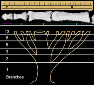
Fibonacci series and phi are found all over nature.
The bottom image shows how trees, and bronchial
trees branch in Fibonacci scaling. The top image
shows how phalanges are scaled in Fibonacci
numbers. If you divide the last Fibonacci by the
previous, the answer tends toward phi
(1:1.618033…). Divide 13/8 or 21/13 or 34/21;
see how it gets closer to phi? The distance
between branches of bronchial tree does
the same thing.
This branching system is asymmetric, complex, and varies from species to species. Because of this, many tetrapods have specific numbers of lobes for right and left lungs. The left lung almost always has fewer lobes. In humans, there are three lobes to the right lung and two in the left – some others (R/L): dog 4/2, cat 4/3, rat 4/1, cow 4/2, horse 3/2, gorilla 4/2, sheep 4-5/3, pig 4/3.

But some animals have an equal number of lobes in each lung – armadillos and harbor seals have three lobes to each lung, while wombats, whales, two-toed sloths, elephants and rhinos all have one lobe to each lung. I couldn’t find a single example of an animal that had more lobes in the left lung.

The number of lobes and lobules has to do with the branching of the bronchial tree. And this has to do with math. I'm sorry, but yes, there's math involved. The Fibonacci sequence (0,1,1,2,3,5,8,13…) is found often in nature. So is phi, a non-repeating decimal like pi. Phi is 1.618033…. and can be found as a ratio in many biologic measurements (1:1.618). The length of your forearm to arm will tend toward phi, as will the curve of a nautilus shell if you break down a phi rectangle into other phi rectangles.

The branching in a lung is also phi; the distance to one branch (1) will be shorter than the distance to the next (1.618). Also, the number of branches will be fractals (a scaling pattern, based on Fibonacci and phi). So, the lung is asymmetric but not random. There - that much math wasn’t too painful, was it?

Next week, the left/right asymmetry continues in your body. Why is the right version of so many paired organs bigger?


Wilkinson M, Kok PJ, Ahmed F, & Gower DJ (2014). Caecilita Wake & Donnelly, 2010 (Amphibia: Gymnophiona) is not lungless: implications for taxonomy and for understanding the evolution of lunglessness . Zootaxa, 3779, 383-8 PMID: 24871732

Bickford, D., Iskandar, D., & Barlian, A. (2008). A lungless frog discovered on Borneo Current Biology, 18 (9) DOI: 10.1016/j.cub.2008.03.010

van Soldt, B., Metscher, B., Poelmann, R., Vervust, B., Vonk, F., Müller, G., & Richardson, M. (2015). Heterochrony and Early Left-Right Asymmetry in the Development of the Cardiorespiratory System of Snakes PLoS ONE, 10 (1) DOI: 10.1371/journal.pone.0116416

Goldberger AL, West BJ, Dresselhaus T, & Bhargava V (1985). Bronchial asymmetry and Fibonacci scaling. Experientia, 41 (12), 1537-8 PMID: 4076397


For more information or classroom activities, see:

Lungs –

Lungfish –

Snake respiratory systems –

Fibonacci sequence and phi in nature -


The CPU In Your Head

$
0
0
Biology concepts – homeostasis, neuroendocrine system, hormone, pituitary, hypothalamus


Let’s face it, everything I know about computers I
learned from Tron and Tron Legacy. What I learned
most from the sequel is that we still can’t make a
decent avatar for Jeff Bridges. But I did learn about
the CPU and programs and users. I like to think there
are small motorcycle races going on in my laptop
while I write. It makes it more interactive.
A computer can be a wonderful tool. It can facilitate learning, entertain you, store information for future retrieval, and manage menial tasks to free your mind for higher thinking. But a computer can also create problems; it makes facts look like knowledge, it presents all content equally so it can be hard to discern validity, and it can distract you from true accomplishment or learning.

Basically, a computer is like any other tool, its worth depends on how it is used. But its a very complex tool with many parts that are easily broken or can wear out. A computer is like your brain in some ways; let’s look at one specific system in your body that can be likened to part of your computer.

The central processing unit (CPU) of your computer controls most of what your machine can do, but it’s functions fall into two broad categories – 1) calculations and information storage, and 2) monitoring the machine itself. The CPU has programs to manipulate information and present information in different forms, but it also has sensory and feedback systems to keep the machine working at optimum levels.

It is this second function that we’re interested in today. Let’s say you use your laptop for an extended time and it starts to heat up. Sooner or later you'll hear the fan turn on; this is your computer’s attempt to maintain the temperature within a small window of values so that function is maintained and there is no damage to the machine. Your body does the same thing.

Another example – you let your computer sit for a while without doing anything. Sooner or later the screen will go black and the machine will go into a sleep mode to save energy and not burn out the screen. Once you start to use it again, it will draw more power; if you stream a movie or 100 videos of cats playing the piano, it will draw a lot more energy and the battery will wear down faster.


Homeostasis is a little like riding a Segway. If you
stand perfectly still and upright, nothing moves; all
the sensors are working but they don’t have to make
any adjustments. If you lean forward, the gyroscopes
recognize it and move the Segway forward so that you
stay balanced; the same thing if you lean backwards.
Everything works to keep your body systems in
balance even when, especially when, something
changes in the system.
Your body also has a system for controlling how much energy is made and used by your body, depending on the demands you are placing on it. In your computer, a portion of the CPU is dedicated to these functions of maintenance and there are peripherals in the machine (the fan, the temperature sensors, the screen saver, the virus software) that carry out the maintenance functions called for by the CPU.

In your body, the brain is your CPU. You’re cerebral cortex controls your higher functions, but each hemisphere has older parts that help maintain the body systems the hypothalamus and hippocampus (so do you have dual processors?). The peripherals that help control your body and keep things on normal are the endocrine glands and the neuroendocrine cells. Together, they’re called the neuroendocrine system and they maintain homeostasis (homeo = like and stasis = standing still).

Homeostasis is one of the characteristics of life. To be alive, an organism needs a system to resistant changes in itself when there are short-term changes in its environment. Like a computer overheating and turning on the fan, your body is constantly sensing internal temperature and turning on and off discrete systems to maintain a constant 98.6˚F.

You have homeostatic systems that control reproduction, temperature, energy consumption and production, hunger, sleep/wake cycles, basal metabolic rate, osmolarity, drinking, and blood pressure. Some of the sensory systems run straight to the brain via hardwiring (another computer analogy). These are the neurons of the peripheral and central nervous systems.

The outputs might be neural, but many times they aren’t. Imagine trying to send a neuron to each cell that needs to get a chemical message (via neurotransmitters released at the ends of the neurons). That would be very cumbersome to maintain and would require trillions more neurons.


“To whom it may concern,” is the introduction to
every hormone message sent throughout your body.
Like bulk e-mails, the hormones contact every cell
via the blood system, but only those that can and
need to respond (have the right receptors) will read
the message and do something about it.
Instead, your brain controls a system where messages can be sent to all cells of the body. Those that can receive the messages by having the correct receptors or gene control elements can then respond to the messages sent out. Those chemical messages controlled by your brain (mostly but not always) are called hormones (from Greek horman = to set in motion).

Hormones are the hallmark of the endocrine (endo = within, and crine = sift) system. When released into the bloodstream, they travel to every part of the body so that all cells have the opportunity to respond. Not all will, and you wouldn’t want them to. Specific cells and organs have the ability to respond to specific signals because they have the right hormone receptors.

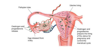
There are eight endocrine glands in a human body. The adrenal glands (2 of them), the parathyroid glands (4), the thyroid gland (1), the pineal gland, the pancreas, the ovaries (2) or testes (2), the hypothalamus, and the pituitary gland. They can be stimulated to release hormones by either neurons, other hormones, or other chemicals. Besides these well known glands, a few tissues will release hormones in specific situations. The placenta will release progesterone and estrogen, and the stomach can release gastrin to stimulate gastric juice (acid) and ghrelin to stimulate hunger.

In addition to endocrine glands, there are also some cells that can be directly stimulated by neurons to release hormones into the blood. These are the neuroendocrine cells. There’s a lot more of them you think, and they’re just about everywhere in your body.


The upper image shows the PNEC’s in your respiratory
tract (NE). They can be alone or be grouped into neuro-
endocrine bodies (NEBs). A 2008 study, and several
before it, has correlated a hyperplasia and hypertrophy
of PNECs with Sudden Infant Death Syndrome. Too
many and your respiratory control may get thrown out
of whack; it may just stop working. It might be good to monitor
the PNEC system in youngsters to try and predict
susceptibility to SIDS.
It’s hard to explain what neuroendocrine cells are and aren’t. They aren’t neurons, but they can look and act a lot like neurons. They can release hormones into the blood (endocrine) or to local cells (paracrine) but they aren't exactly endocrine cells. In other cases, they can release neurotransmitters that act on neurons – this often occurs in sensory situations, like with the Merkel cells in your skin that sense touch.

For one example, there are pulmonary neuroendocrine cells (PNEC) in every part of your respiratory tract, from your nose to your alveoli. While they seem to act like neurons in many respects, they are derived from epithelial tissue, not the neural crest tissue that all neurons come from.

Each PNEC spans the distance from basement membrane to the air conducting space. On the luminal side, they have microvilli that stick out into the lumen and sample the air as it passes. On the basal side (bottom) they communicate with neurons. Just what are they doing there?

They have several functions, including regulating the development of the respiratory system in the fetus. They also regulate the function of the respiratory cells by sensing oxygen levels, controlling the muscular tone of the bronchi, regulating pulmonary blood flow and modulating immune responses. The origin and function of PNEC’s is reviewed in a 2012 paper, which also highlights a problem with them – when PNEC’s go bad, they cause a deadly small cell cancer.

In response to what the cell senses, it will release a variety of chemicals, many of which can act as neurotransmitters – hence the reason they are considered neuroendocrine cells. But the PNEC’s may have another function, one hinted at in a 2014 study. The results of these experiments indicated that PNEC’s can detect chemicals in the air as it passes over their microvilli – they quite literally can smell the air, as their microvilli were found to have olfactory (smell) receptors, just like in your nose!


The hypothalamus is part of the diencephalon, a part
of the brain older than the cerebral cortex, but not as
old as the thalamus. The different nuclei each function
in different systems; the lateral nucleus senses
hormones and regulates thirst and hunger, the
dorsomedial nucleus regulates BP and HR. Several
nuclei are involved in making releasing hormones,
ADH and oxytocin.
The need to smell volatile chemicals in the lung may have something to do with responses to toxic chemicals; remember that the PNEC’s also regulate the pulmonary immune response. In chronic pulmonary diseases, like emphysema due to smoking, the PNECs are less responsive to volatile chemicals, maybe as a way to dampen the immune response.

So what controls much of this neuroendocrine system? Your brain of course. Well, a small part of your brain that's pretty old in terms of evolution (even primitive hagfish have a pituitary). The hypothalamus senses many of the inputs that tell your brain just how well your body is maintaining homeostasis. If something goes astray, or if there is a change in your environment that forces a change in your body chemistry, the hypothalamus will then spring into action (O.K., it’s always working, it will just work harder).

The hypothalamushas a neuroendocrine relationship with the pituitary gland (better named the hypophysis, where hypo = under and physis = growth – it is a growth under the brain). The hypophysis comes in two parts. Both parts release hormones, but where those hormones come from and the sources of the tissues of the two pituitary parts are very different.

The anterior pituitary isn’t part of the brain at all. The tissue for the adenohypophysis(anterior pituitary, adeno = secreting) comes from the roof of the embryonic mouth. It is epithelial in origin and has cells that produce hormones in response to signals from the hypothalamus (also hormones). Here’s how it happens.


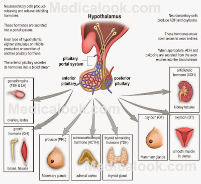
The pituitary gland, or hypophysis, has two small
portal systems that carry blood from one place to
another instead of to a particular tissue and then
back to the heart. The anterior system carries
releasing hormones from the hypothalamus to the
ant. pituitary and then carries stimulating hormones
from the ant. pituitary to the rest of the body. The
posterior system moves hormones from the
hypothalamus directly to the rest of the body.
Image credit to Medicalook.
Some portions of the hypothalamus sense changes from the body and produce hormones called releasing hormones. These stimulate the epithelial cells of the adenohypophysis to produce hormones that then act on the endocrine glands throughout the body. Confusing, yes?

Well, it gets worse. There are six releasing hormones from the hypothalamus that stimulate production and release of seven hormones from the adenohypophysis that then act on at least 20 endocrine glands and neuroendocrine cell types. Hypothalamus to adenohypohysis to endocrine gland – it’s called an axis, and there are several of them.

On the other hand, the posterior pituitary (neurohypohysis) is derived from brain tissue. Instead of the neurons of the hypothalamus producing releasing hormones that then act on the neurohypophysis, the hypothalamic neurons project right into the posterior pituitary where they deposit their hormones (oxytocin and antidiuretic hormone).  The neurohypophysis doesn’t make any hormones itself, it just stores what the hypothalamic neurons produce and then releases them to the circulatory system.

That’s a heck of an exception- part of your brain isn’t actually part of your brain. Cells from your mouth control most functions in your body! Some people I know have mouths that completely ignore their brains! Next week, let’s talk more about the neuroendocrine system. Your thyroid gland size correlates to which hand you use to write – say what?


Gu, X., Karp, P., Brody, S., Pierce, R., Welsh, M., Holtzman, M., & Ben-Shahar, Y. (2014). Chemosensory Functions for Pulmonary Neuroendocrine Cells American Journal of Respiratory Cell and Molecular Biology, 50 (3), 637-646 DOI: 10.1165/rcmb.2013-0199OC

Song, H., Yao, E., Lin, C., Gacayan, R., Chen, M., & Chuang, P. (2012). Functional characterization of pulmonary neuroendocrine cells in lung development, injury, and tumorigenesis Proceedings of the National Academy of Sciences, 109 (43), 17531-17536 DOI: 10.1073/pnas.1207238109

Porzionato A, Macchi V, Parenti A, Matturri L, & De Caro R (2008). Peripheral chemoreceptors: postnatal development and cytochemical findings in Sudden Infant Death Syndrome. Histology and histopathology, 23 (3), 351-65 PMID: 18072092


For more information or classroom activities, see:

Homeostasis –

Neuroendocrine system –

Hypothalamus –

Pituitary –

Thinking Asymmetrically About Hormones

$
0
0
Biology concepts – neuroendocrine system, bilateral asymmetry, internal asymmetry, hormones, endocrine glands


Jack Nicholson did some try asymmetric thinking
in The Shining. The fact that he was driven insane
by the ghost of a horrid past shouldn’t think less of
his accomplishments. Predicting that Shelly Duvall
would go for the radio – brilliant. Sneaking up on
Scatman Crothers – inspired. Following the boy
into the maze –oops.
Independent thinking; thinking outside the box; free thinking; lateral thinking; these are all terms for trying to come up with answers to problems through rejection of established logical methodologies. In today’s business, military and political worlds, they like to call it asymmetric thinking.

An asymmetric military engagement might be one where a traditional force is thwarted and confused by a nontraditional force of computer geeks hacking their communications and selling all their weapons on Etsy. Asymmetric thinking approaches what might be a traditional question at a creative angle, rejecting the tools and assumptions that are normally found “inside the box.”

Asymmetric thinking asks why outrageous solutions aren’t being considered. Many job interviews now include asymmetric thinking questions. A man goes into a restaurant and asks for a glass of water. The waiter points a gun at him and the man thanks the waiter and leaves. What’s your explanation for that? (see end of post)

Today we'll talk about asymmetric thinking in a different way – a lateral thinking approach to asymmetrical thinking, as it were. We have talked about the asymmetry of the brain hemispheres and how they sit asymmetrically in your skull. We have also talked about the neuroendocrine system and how it is controlled by a part of your brain that isn’t really part of your brain. Now let’s talk about the asymmetry of the neuroendocrine system as it begins in your brain and ends in the endocrine glands.


Most views show the hypothalamus from the side
(see last week’s post), so you don’t appreciate that
there are two of each nucleus. For those that dump
hormones into the pituitary gland, both sides
participate, but our story today shows that they don’t
necessarily participate equally.
The hypothalamus, as well as the paired endocrine and neuroendocrine glands (see last week's post), demonstrate significant asymmetry. I suppose that’s not so unusual - all the structures we’ve talked about in the past few weeks have structural and functional asymmetries. The weird part about it here is that we are talking about hormones being moved into the blood.

The whole purpose of the hormone system is that it can be used to bathe the entire body in functional hormones at the precise levels, so that all cells that can respond will respond. How does that jibe with an asymmetry where one gland of a pair does more than the other?

The hypothalamus is a good example. You have two halves of your hypothalamus, one in each hemisphere (sort of, see above), but they both deposit releasing hormones into the same pituitary vein complex so they can stimulate your single pituitary gland. Yet, studies show that the right hypothalamus makes more gonadotropin-releasing hormone than the left hypothalamus.

On the other hand, thyrotropin-releasing hormone (stimulates the release of thyroid stimulating hormone, TSH, from the pituitary) is higher in the left hypothalamus. Together, the results of several studies shows that the right hypothalamus plays a bigger role in controlling reproduction, while the left hypothalamus works more in metabolic rate. You have a lateralization of structure and function in your two hypothalami, just like in your two cerebral hemispheres, even though both halves work on a single pituitary gland.

How about some of the other endocrine and neuroendocrine glands?

Thyroid –  You only have one thyroid gland, which lies over the front of your windpipe in your neck. There are two lobes, one on the right side and one on the left, connected by the isthmus across the windpipe.


The thyroid receives stimulation from the anterior
pituitary release of TSH (thyroid stimulating
hormone). If for some reason you get to much
stimulation (maybe autoantibodies), or the thyroid
start making too much thyroxin on its own (tumor),
then you have Grave’s disease. In some cases, you also
get an autoantibody during Graves that attacks the
fibroblasts around the orbit of the eye. The
inflammation makes the lid retract and pushes on the
eyeball, making it bulge out of the socket. Which of these
two has thyroid eye disease?
The thyroid releases thyroxine hormones T3 and T4 into the blood that function to control your metabolic rate (see this post). Yet the right lobe of the thyroid is more vascularized and is almost always larger than the left lobe. The thyroid also has a sexual dimorphism, as it is usually bigger in women than in men, and the asymmetry of right > left is even larger in women.

The size difference may not be innocuous. Many studies have shown that thyroid diseases and cancer affect the right lobe more often than the left. And it gets weirder. A 2009 research paper from China showed that handedness may also play a role. They found that the right > left size difference was larger in right-handed people. However, the left lobe was about the same size no matter which hand the person preferred. So, does the hand you use influence the size of the thyroid, or does your thyroid predict which hand you will use? Or, is it a correlation without significance?

Parathyroid – You have four parathyroid glands – maybe. These are located on the backside of your thyroid gland (hence the name para = by). The parathyroids are important in regulating calcium levels in the body. This may seem weird, having four glands to control the levels of one element. But consider that calcium plays some major roles, from controlling muscular contraction, to neuron transmission, to at least a dozen different second messenger systems in every cell.

The parathyroids are small, only about 33 mg each, so they are easy to lose when people have surgery on their thyroid gland. A 2011 studysought to find out where they sit normally so surgeons would be able to find them and preserve them. Unfortunately, they found that position and number are quite variable. Forty-three percent of people have at least five glands instead of four. And the positions of the four common ones can be variable, they aren’t always in the same place. The extra ones can be just about anywhere! Good hunting Mr. surgeon.


If you soak bone in vinegar (acetic acid), it will remove
the calcium and leave the protein matrix. Notice that it
must be the calcium that gives bone rigidity. This isn’t
how PTH works. PTH stimulates osteoclast activity,
which removes the calcium AND the protein matrix. PTH
also decreases the amount of calcium lost in the urine,
but for some reason, we work just fine without PTH or
without its balancer hormone, calcitonin.
The thyroid and parathyroid seem to do different jobs, but they’re linked by more than just anatomy. The parathyroid hormone (PTH) made and released by the parathyroids works to increase calcium availability, by increasing bone break down (osteoclast activity, see this post) and increasing the amount of calcium recovered from the urine.

But wouldn’t you need a balancing hormone to decrease calcium levels if they get too high, so a balance could be established? This is how many hormones work; there are hormone pairs that have opposite stimulatory functions. The balancing hormone for PTH is calcitonin, made by the neuroendocrine parafollicular cells (C cells) in the thyroid gland.

But here’s the exception. Calcitonin is important in fish and birds, but it seems people and many other mammals can get along fine without it. Remove someone’s thyroid and they have to take thyroid hormone for the rest of their life. But they get along just fine without calcitonin. This is one instance where the balancing hormone isn’t necessary. It seems an asymmetry of function in calcium regulation is just fine in people.

Adrenal glands -  These glands sit on top of each kidney. You think of them as sources of epinephrine in the fight or flight syndrome, but they do much more. Adrenal (ad = of or near, renal = kidney) glands have a cortex, which is toward the outside (not the core, this always confused me), and a medulla, which is in the middle (makes more sense).


cortex of three layers and a medulla. The cells of the two
regions are of different origin, the medulla is nerve like,
while the cortex is epithelial in origin.
The medulla is neuroendocrine, the cells look like neurons and are stimulated directly by sympathetic (autonomic) nerves (see this post). The medullary chromaffin (they take up lot of stain) cells release epinephrine and norepinephrine in response to a fight or flight situation. Epinephrine and norepinephrine are neurotransmitters in many parts of the brain, and in this case they work on cells to increase heart rate, glucose availability to muscle and the like.

The adrenal cortex is made up of three layers, each produces hormones to be released into the blood. The outside most is the zona glomerulosa, which makes aldosterone to help control osmolarity and blood pressure. The zona fasiculata is the biggest, and makes cortisol that controls the metabolic rate. The zona reticularisis inner most and makes sex hormones, androgens specifically.

You have two adrenal glands – and they each do the same things in response to the same signals, either hormonal or neural. So why is the left adrenal gland almost always bigger than the right? Is it because the venous drainage of the right and left adrenals is different? In the right, the veins dump into the inferior vena cava, while the left drains into the left renal vein. I really don’t know if that would make a difference.


This is just weird. A paper from 2005 states that since
the autonomic (sympathetic and parasympathetic)
nervous system controls the neuroendocrine system,
asymmetries in the ANS can have ramifications on
endocrine function. Asymmetry in behavior can
affect ANS, which then can affect endocrines. This study
showed that feeding cows from the left side (affects right
side ANS) improved reproductive ability and lactation; so
the right ANS must have more influence on reproductive
endocrine function. Could I have some left-hand milk please?
But here’s the kicker, a group in 2002 studied the size of the adrenal glands and their functional abilities in wild animals and their domesticated counterparts; several species of foxes, minks, etc. They found that domesticated animals had a larger size difference in adrenals than did the wild versions.

What is more, when they compared aggressiveness, no matter whether the animal was wild or domesticated, the most aggressive animals had the largest size differential – always left bigger than right. This makes it sound like the medulla was involved – aggression being involved, but it wasn’t.

The increase in left adrenal size in domesticated animals was due to an oversized zona fasiculata (cortisol and other glucocorticoids), while the left adrenal asymmetry in the aggressive animals was due to a larger zona reticularis (sex hormones). Ah… now that makes some sense. Doesn’t it always come back to sex?

Next week, let’s look more into the neuroendocrine system and gender. The testes and ovaries have the most spectacular asymmetries.

(The man had the hiccups)


Ying M, & Yung DM (2009). Asymmetry of thyroid lobe volume in normal Chinese subjects: association with handedness and position of esophagus. Anatomical record (Hoboken, N.J. : 2007), 292 (2), 169-74 PMID: 19051270

Trut LN, Prasolova LA, Kharlamova AV, & Plyusnina IZ (2002). Directional left-sided asymmetry of adrenals in experimentally domesticated animals. Bulletin of experimental biology and medicine, 133 (5), 506-9 PMID: 12420075

Rizhova, L., & Kokorina, E. (2005). Behavioural asymmetry is involved in regulation of autonomic processes: Left side presentation of food improves reproduction and lactation in cows Behavioural Brain Research, 161 (1), 75-81 DOI: 10.1016/j.bbr.2005.01.007

Hojaij, F., Vanderlei, F., Plopper, C., Rodrigues, C., Jácomo, A., Cernea, C., Oliveira, L., Marchi, L., & Brandão, L. (2011). Parathyroid gland anatomical distribution and relation to anthropometric and demographic parameters: a cadaveric study Anatomical Science International, 86 (4), 204-212 DOI: 10.1007/s12565-011-0111-0


For more information or classroom activities, see:

Hypthalamus – see last week’s post

Thyroid gland–

Parathyroid glands–

Adrenal glands-

What the Heck Are Those Doing There?

$
0
0
Biology concepts – internal asymmetry, neuroendocrine, reproduction, hormone, absence symmetry


This is the Decatur County Courthouse in Greensburg
Indiana. A mulberry tree has been growing out of the
steeple for over a hundred years. Nature has things out
of place, sometimes for a good reason, sometimes
through sheer tenacity.
“That really shouldn’t be there… “ but it is. You can probably think of many examples of things that seem out of place, but with further investigation and reasoning, you figure out that there’s a plausible explanation for that particular thing being in that particular place.

A couple of examples - in December of 2013, a University of Arizona team announced that they had found a planet orbiting a star about 300 light years from Earth. No problem, we’ve found many exoplanets. But to our current understanding, it shouldn’t be there.

Planet HD 106906b is 11x the size of Jupiter and travels in a path 650x larger than Earth’s orbit around the sun.  This makes it the largest planet we have noted orbiting such a long distance from its star. Our current hypotheses of planet formation can’t account for this planet.    

Usually a planet will accrete from the left over material in the debris disk that forms a star. But a planet this large would take too long to form in that manner. And the outer edge of a debris disk, where this planet is located, doesn’t have enough material to build a large planet. So why is it there?

Perhaps it was a failed star, formed at the same time as its star. Many binary star systems form by quick accretion in two areas of the same disk. But binary star systems usually have a mass ratio of 10:1 or less. HD106906 (the star) and the presumed “failed star” (HD 106906b) have a ratio of >140:1. There’s no good reason for it – yet there it is.

Now for a less universe-shaking example. My family and I live in Indianapolis, the largest city in the United States without a navigable waterway. Yet, there's a US Naval Station located on the near north side of the city. Built in 1936 as a WPA project, the Heslar Naval Armory housed sailors that trained on Lake Michigan (200 miles away) each summer.


This is the Naval Armory in Indianapolis. There is a river
behind it, but the last time anyone tried to navigate the
White River from the Ohio to Indianapolis they ran
aground and the entire boat had to be disassembled.
It turned out to be a good idea. In WWII, a landlocked naval station was just the place to train naval communications officers without worrying about being bombed or spied on. The naval armory was also the single most important site for developing US naval war strategy, including the D-Day invasion. Sometimes out of place things are there for a good reason. And here we meet our topic for the day.

In terms of reproduction and neuroendocrine function, there’s a true disconnect with the testes and their function. Animal testes do a couple of important jobs. They produce the male gamete cells for reproduction, but they also produce sex hormones that control reproduction and secondary sex characteristics.

If the testes are so important, why are they housed in harm's way, outside the body cavities? You think you know the answer, but really you just know a portion of the story. The testes are housed in the scrotum instead of inside the abdominal cavity. Think how silly it would be to suggest that a woman’s ovaries should be housed in a sac outside her body – yet there are the male testes, in a sac outside his body.

One weird theory is that large testicles and scrotums are a sexual dimorphism ornament; it costs energy to make large testicles, so the larger they are, they better health the animal must have to be able to afford to invest so much in ornament size. To take advantage of this as a way to draw mates, they have to be seen. Therefore, they're outside the body. It's an example of the handicapping hypothesis– an organism handicaps itself by investing so much energy in one thing, but it pays off in a reproductive advantage.


Testicle size varies according to species and breeding
strategy. The right whale has the largest testicles,
about 500 kg (1100 lb) each, 10x larger than those of the
blue whale.  In general, monogamous males tend to
have smaller testicles. A paper from 2013 showed that
testicle size in humans is negatively correlated to
parenting ability. Those with smaller testicles were more
likely to be nurturing fathers. 
However, there isn’t any evidence to suggest that this is how it works in humans, or in most animals. If it were the case, then the genital size should get bigger and bigger, until it becomes a burden too great to overcome. And elephants and some other animals do have their testes packed into their abdominal cavity. In fact, the vervet monkey(Chlorocebus pygerythrus) is about the only example where the scrotum and testes are a sexual ornament, being neon blue (see picture).

This idea of scrotum as ornament is understandable, but if this isn’t the reason, then what is? Believe it or not, the answer explains several testicular asymmetries.

It’s all about temperature – male gamete cells survive only a short time at normal body temperature, so you save their active period for when they need to swim to the egg. By having them outside the abdominal cavity, they are cooler. The skin of the scrotum is thin, and the arteries of the scrotum and testicles lie right next to the veins; more opportunity to pass heat to blood that is moving away from the testicles. Everything is geared to lowering the temperature a few degrees.

Believe it or not, temperature is also the reason why they are positioned asymmetrically. In the majority of men, the right testicle doesn’t hang as low as the left. A 1997 paper actually studied this and showed that about 62% of right-handed men and 58% of left-handed men had a left testicle that hung lower. But the number of men with a lower right testicle was exactly the same in both right handers and left handers (around 21%). What changed was the percentage of men that had testicles that were positioned horizontally in the scrotum.


The vervet monkey (Cercopithecus aethiops) is an old
world monkey that lives Acacia woodlands of the
mountains of southern Africa. There are five subspecies,
all of which are threatened. They are used in research,
because they have hypertension, anxiety and alcohol
dependence as humans do. You can see why someone
might get the idea that testes and scrotum might be a
sexual ornament for mate selection.
An earlier paper had attributed this to better and larger muscular development on the dominant side. In abdominal muscles, this would mean more tension on the testicular cord from abdomen to testicle (a remnant of the descending testicle in fetal development). More tension means that it can’t hang as low. But this isn’t the reason.

Again we go back to temperature. There's a muscle called the cremaster. It responds to several signals, including temperature and adrenaline and testosterone. This muscle can raise or lower the testicles independently of one another. In a dangerous situation, the testicles are drawn up closer to the body to reduce chance of injury. In cold temperatures they are drawn up in an effort to keep them warm.

As we said above, the scrotum holds the testicles outside the body to minimize gamete activation until needed. But they don’t want to be too cold either. There is a small range of temperature in which function is optimal, and each testicle needs to be at that temperature. So, the body senses the temperature and the cremaster moves the testicle closer to and then farther away from the body to modulate temperature. Believe it or not, they both are constantly moving in independent orbits!


As with this statue of Hercules, the Greeks got it wrong.
Very accurate in their sculptures, they most always had
the left testicle hanging lower than the right in their
male nudes. But, they also had the left testicle as larger.
They fell prey to the idea that bigger would hang lower.
My thoughts on this – someone went around examining
and measuring the testicles on Greek sculptures?
According to a 2008 paper, if the testicles hung side by side, they would warm each other; the maximum surface area of the scrotum for temperature dissipation would not be utilized. Therefore, one hangs below the other for maximum independent temperature regulation. Apparently it isn’t a deal breaker, because 20% of men have testicles at equal heights; perhaps they have a slightly lower reproduction rate.

This leads to another asymmetry in testicles – one is usually bigger than the other. It varies amongst the species of the world, but in most mammals, the left is usually slightly larger than the right. But we humans are exceptions, in men, the right is usually larger than the left. It’s larger, but hangs higher – an example of antigravity?

The working hypothesis is that one testicle is dominant, usually the right in humans. This testicle contributes more to reproduction and plasma testosterone levels, and is affected more by changing levels of luteinizing hormone. But the other one is just as important. It is evolution’s back up plan. If the dominant testicle is injured, the other one takes over. By reducing it’s role in normal function, it reduces the risk of use problems developing; when it is needed, it is fresh and ready to go.


The leatherback turtle has a skylight in its skull (2014).
The bone is so thin that sunlight can reach the pineal of
the epithalamus. It has light sensing receptors, just like
the eye. In fact, most vertebrates have this ability. We
don’t since our cerebral expansion has buried the
epithalamus and hypothalamus deep on our brain.
In non-mammals, the testicles are usually similar in size, but there are exceptions in size asymmetry and even in number. Sharks usually have a much larger right testicle. In birds, one testicle may be larger, but it switches back and forth amongst the different families and species. The bufflehead duck has a left testicle that is four times the size of the right!

In animals that have a distinct breeding system, the sizes of the testicles can change. Just before the breeding season, the testicles may swell to double or triple their normal size. This occurs in the many bird species, and amazingly, their brain senses the day length directly. A 2000 paper looked at mallards, but other papers have shown in other species as well that the hypothalamus of many non-mammal vertebrates has light sensitive cells and can detect lengthening days right through the skull!

In some species, including a couple of birds, only one testis develops. The black coucal cuckoo has only a right testis. Jawless fish have a single testis; it forms in the midline and is probably a fusion of the two testes. The worm, C. elegans, has just a right testis. Its bowel is on the left side, so this is probably a space-saving mechanism.


This ground beetle is one of thousands of carabid
beetles. They are predators, fast and have nasty
secretions as a defense. And most them only have one
testicle. Interestingly, it’s always the right one.
Likewise, a 2005 paper showed that 174 species of carabid beetles are monorchid (one testis). This is also to save room in the exoskeleton. They hypothesize that the testes are the only of the paired organs not to be linked to the same mechanisms of development, so they are more likely to go off body plan. This is called absence asymmetry. The authors suggest that the trade off comes for maximizing some organs within the limited space.

So now you can ask not only, "why are they located there," but also, "does there really need to be two of them?" Next week, we shouldn’t leave out the girls. Ovaries have just as many exceptions as testes.


Davenport, J., Jones, T., Work, T., & Balazs, G. (2014). Pink spot, white spot: The pineal skylight of the leatherback turtle (Dermochelys coriacea Vandelli 1761) skull and its possible role in the phenology of feeding migrations Journal of Experimental Marine Biology and Ecology, 461, 1-6 DOI: 10.1016/j.jembe.2014.07.008

Kumar, A., & Kumar, C. (2008). Swinging high and low: Why do the testes hang at different levels? A theory on surface area and thermoregulation Medical Hypotheses, 70 (3) DOI: 10.1016/j.mehy.2007.06.023

Bogaert, A. (1997). Genital asymmetry in men Human Reproduction, 12 (1), 68-72 DOI: 10.1093/humrep/12.1.68

Will, K., Liebherr, J., Maddison, D., & Gali�n, J. (2005). Absence asymmetry: The evolution of monorchid beetles (Insecta: Coleoptera: Carabidae) Journal of Morphology, 264 (1), 75-93 DOI: 10.1002/jmor.10319

Mascaro, J., Hackett, P., & Rilling, J. (2013). Testicular volume is inversely correlated with nurturing-related brain activity in human fathers Proceedings of the National Academy of Sciences, 110 (39), 15746-15751 DOI: 10.1073/pnas.1305579110


For more information or classroom activities, see

Not many classroom activities involving testicles. Sorry.


Ovaries March To A Different Drummer

$
0
0
Biology concepts – neuroendocrine, ovary, bilateral asymmetry, internal asymmetry, absence asymmetry, hormones, ovulation


Many things we are taught in school just aren’t so.
The Salem witch trials, for example, did not result in
women being burned at the stake. Sure, some were
imprisoned and a couple dozen were hanged, but
none burned at the stake.
A lot of the things we think we know just aren’t so. I’ll give you a few examples. Vincent van Gogh cut off his ear in an insane rage, right? Nope, his “friend” Paul Gauguin cut it off as he drew his sword in a drunken fight with van Gogh. They made up the story so Gauguin could avoid jail. Not nearly so tragic, but it has the ring of truth if you knew Gauguin.

Engineering professors and even physicists at university will teach you that glass is an amorphous liquid. The reason that windows in extremely old buildings are thicker at the bottom is because the glass has had time to flow. Nope.

Glass doesn’t have a crystalline lattice when solid, but it doesn’t flow. The reason old windows are thicker at the bottom is because they used to make glass panes by pouring molten glass on a wheel and spinning it. The force would spread it out, but it would be thicker on the outside edges. When the panels were cut for panes, they installed the thick side at the bottom for stability. So there.

Finally, there were 13 original American colonies… or maybe not. Delaware was swapped back and forth between Maryland and Pennsylvania. Delaware didn’t come into existence as its own colony until the Revolutionary War. It was known as the ”Three Lower Counties” from 1664 until 1776 and shared a governor with Pennsylvania for the last 75 years of its existence.

Speaking of things that just aren't so, there are couple of things in the neuroendocrine system that most people think they know. There are asymmetries in every part of the endocrine system, ant the others are no exception to having exceptions.


The ovary produces hormones and releases ova. Notice
that the oviduct (fallopian tube) doesn’t connect directly
to the ovary. When the egg is ready to be released, the
estrogen and progesterone cause the fimbria at the near end
of the oviduct to swell and come closer to the ovary. When
the egg is released, the cilia on the fimbriae cells sweep
it into the oviduct.
The ovaries are the source of eggs to be fertilized; those eggs might become small people with wrinkly skin.  But they are so much more. As part of the neuroendocrine system, they are stimulated by hormones and neural impulses and respond by releasing hormones of their own. Depending on the time in a woman’s fertility cycle, they release various amount of estrogen, progesterone and even testosterone.

The ovaries are paired organs like the testes of males, but not all animals have two functioning ovaries. In the Greater Horseshoe Bat (Rhinolophus ferrumequinum), there is only one functioning ovary, the right one, and it's 3-4x the size of the non-functional left ovary. On the other hand, the Natal Clinging Bat bat (Miniopterus natalensis) has only a left functioning ovary and it is several times the size of the non-functional right ovary.

Single ovary examples also exist in the primitive fishes. Lampreys have only one ovary as result of fusion of the two gonadal primordial into a single functioning gland. Hagfishes have a single ovary simply because the other one doesn’t develop.


The hagfish is a primitive fish. The females only have one
ovary, but that isn’t the weirdest part. They produce
proteins and mucins that mix with water and form a slime
when they are disturbed. The filaments are 100x thinner
than hairs, but 10x stronger than nylon, so they are a
subject of much research.
Interestingly, sometimes it’s the right that doesn’t develop and sometimes it’s the left. On the other hand, sharks start out with two ovaries, but the left one atrophies over time, leaving one ovary but two oviducts.

Many birds have one ovary – almost always the left one. A study from 2013 made use of very rare early bird fossils that preserved the ovary tissue; preservation of soft tissue elements is indeed rare. They found that these early fliers had already donated one ovary to the cause of flying.  The hypothesis is that dinosaurs laid many eggs because they had two ovaries, but early birds sacrificed an ovary to reduce weight and make it easier to fly.

The survival advantage afforded by flight offset the disadvantage of fewer eggs, so it was basically a reproductive no harm, no foul. These basal birds had already moved away from the reproductive mechanisms of dinosaurs and present crocodilians toward more bird-like strategies. What this doesn’t explain is why many raptors – like hawks and eagles, have two functioning ovaries. A 2014 paper showed that the right ovaries were functional and capable of responding to, and producing, estrogen and progesterone.

Some birds of prey have two ovaries and some have one. In some, the two are both functional and in others the right is vestigial. This makes me wonder about the evolution of birds. Did the loss of an ovary occur independently several times in different lineages? Or did it occur once in the progenitor of all birds, but some of the descendants evolved the second on again?


The mountain viscacha is a rodent, but looks like a cross
between a rabbit and a chinchilla. They have short forelegs
and long fluffy tail to go along with the rabbit-like ears.
They live in dry places, so they almost never drink. They get
all their water from the plants they eat.
There are even a couple of mammals with a single functioning ovary. The waterbuck (Kobus ellipsiprymnus) lives in sub-Saharan Africa. It is related to antelopes, but differs in that the females of waterbucks have only a left functional ovary.

The mountain viscacha (Lagidium viscacia) is a rodent that lives in the rocky, high altitude, mountainous regions of South America. It starts out with two functional ovaries, but about the time of their first breeding season, the right ovary overgrows, the left shrinks a bit; only the right becomes functional. The exception in this exceptional animal is that if the right ovary is injured or diseased, the left will grow and take over its functions. This doesn’t occur in our other examples.

Similar to testes, there is also a functional asymmetry in ovaries, and this is where we get into the major fallacy that people are taught about the female reproductive system. We are taught that ovaries are good sharers, they take turns ovulating, right-left-right-left, one each month. No……it just ain’t so.

They can take turns, but they usually don’t. And women who want to have children should be glad that they don’t split the load equally. For humans (women mostly), there is slightly less than 50% chance that the opposite ovary will release an egg in the next cycle, according to a 2000 study. This means that side of ovulation is basically random for any given month, but this doesn’t mean that every ovulation has an equal chance of producing an embryo.


Notice that after ovulation, the fertilization of the egg takes
 place in the oviduct, not the uterus. The embryo already
has 32-128 cells by the time it hits the uterine wall. This is
why it is important for the ovary to be producing estrogen
and progesterone the whole time. The uterus must be made
ready for the incoming embryo.
Another 2000 study showed just how unequal ovulations can be. In thousands of ovulations tracked in fertile and infertile women, 64% of pregnancies occurred after right ovary ovulations. In infertile women treated with intrauterine insemination or in vitro fertilization, pregnancy rates were low, as they always are. But if coordinated with right-sided ovulation, they were twice as likely to produce pregnancies as when compared to left-sided ovulations.

However, it isn’t just a right-sided ovulation that produces the best odds of pregnancy; the series of previous ovulations matters as well. If you were to monitor which ovary ovulated over three cycles, there would be eight possible sequences: left-left-left, left-left-right, etc. all the way to right-right-right. This is exactly what a 2011 study tracked, along with pregnancy rates.

The researchers found that the pattern most likely to produce a pregnancy was left-left-right. These results would need to be repeated several times, especially since significant results are difficult to assess when there are eight variables, but their numbers were very convincing. So asymmetry in the function of the ovaries can have a very real affect on hormone levels and pregnancy rates.

Ovulation of a single follicle might be a 50/50 shot each month, but over time the right side of the reproductive system in women seems to be dominant. If the follicles were counted in each ovary (sort of a permanent record of ovulations) of a woman late in her reproductive years, about 62% of them will be on the right side.

Likewise, progesterone and estrogen blood levels are higher during a right-sided ovulation cycle. This data, along with the pregnancy data, indicate the female reproductive system is really right-side dominated. Why is the right side dominant?


The top image shows the corpus luteum that develops from
the follicle that released the egg. This is a huge source of
hormones. It also shows the follicle atresia, where primary
follicles degenerate before releasing eggs. The bottom image
is the timetable of the happenings in the ovary. Notice that the
degenerating corpus luteum is still bigger than the primary
follicle, so over time, the ovary does get bigger. Then as she
gets close to menopause, they get smaller.
A good explanation comes from the fact that the drainage of blood for each ovary is different. The left ovary is drained by the left renal vein, but the right drains in to the inferior vena cava (like the adrenals, see this post).  There tends to be higher venous pressure in the left renal vein and so this side drains slower.

If the blood moves out slower, then the corpus luteum (the leftover follicle of ovulated egg) stays around longer, and this makes it less likely that the left ovary will be ready to ovulate again the following month. Over time, the right will have more follicles from ovulations. The hormone levels would also be higher if carried out of the ovary faster, so this is probably why plasma hormone levels are higher after a right-sided ovulation.

The right side dominance is likely to switch to the left side later in a woman’s reproductive years because of the relatively lower numbers of ova left on the right side. This may be why it is less likely that a woman will become pregnant in her later years.

In most mammals, right and left ovaries are about the same size (given the exceptions of mountain viscacha and waterbuck we talked about above). But in humans, this is merely how they start out. Later in the reproductive years, there is often an acquired size asymmetry.


A dichotic listening test is for attention and picking out one
noise precisely. Most people have a right ear advantage (REA)
for speech because the speech centers are on the left side of the
brain. Research shows that women have a reduced REA compared
 to men and it is affected strongly by estrogen and progesterone
(2011). This may be so that they will focus in less on one sound
and might then be able to pick up on distress from their baby.
After ovulation, the follicle expands and becomes the corpus luteum. This structure produces hormones that would stabilize the system to prolong the pregnancy. If the egg is not fertilized or the pregnancy is not carried for a long time, the corpus luteum reduces in size and hormone production falls. However, the leftover follicle is larger than the pre-ovulatory situation. 

Over time, the number of follicles increases and the size of the ovary increases. More right-sided ovulations (since the right side drains faster and therefore means it can be ready to ovulate again the next month) means that it enlarges more than the left, and a size asymmetry develops.

Hormones, number of ovulations, pregnancy rate – there isn’t anything about the ovaries that isn’t asymmetric – and yet people are taught that they have a symmetric size and function. Right-left-right-left.... yeah sure.

Next week, asymmetries abound in the human body, but they're usually a little here a little there. But what if every organ in you body turned out to be on the wrong side?


Cowell, P., Ledger, W., Wadnerkar, M., Skilling, F., & Whiteside, S. (2011). Hormones and dichotic listening: Evidence from the study of menstrual cycle effects Brain and Cognition, 76 (2), 256-262 DOI: 10.1016/j.bandc.2011.03.010

Rodler D, Stein K, & Korbel R (2015). Observations on the right ovary of birds of prey: a histological and immunohistochemical study. Anatomia, histologia, embryologia, 44 (3), 168-77 PMID: 24895012

Zheng, X., O’Connor, J., Huchzermeyer, F., Wang, X., Wang, Y., Wang, M., & Zhou, Z. (2013). Preservation of ovarian follicles reveals early evolution of avian reproductive behaviour Nature, 495 (7442), 507-511 DOI: 10.1038/nature11985

Fukuda, M. (2000). Right-sided ovulation favours pregnancy more than left-sided ovulation Human Reproduction, 15 (9), 1921-1926 DOI: 10.1093/humrep/15.9.1921

Ecochard, R. (2000). Side of ovulation and cycle characteristics in normally fertile women Human Reproduction, 15 (4), 752-755 DOI: 10.1093/humrep/15.4.752

Fukuda, M., Fukuda, K., Tatsumi, K., Shimizu, T., Nobunaga, M., Byskov, A., & Yding Andersen, C. (2011). The ovulation pattern during three consecutive menstrual cycles has a significant impact on pregnancy rate and sex of the offspring Fertility and Sterility, 95 (8), 2545-2547 DOI: 10.1016/j.fertnstert.2011.02.010


For more information or classroom activities, see

Not too many classroom activities for ovaries and hormones, but here's a link to a series suggested by Matthew Knoepke:

http://nubio.northwestern.edu/labs/28-days-later


 I am also impressed by the idea of using a pomegranate as a model for the human ovary.


Organs Don’t Always Follow The Plan

$
0
0
Biology concepts – internal anatomy, asymmetry, symmetry breaking, primary ciliary dyskinesia, situs solitus, situs inversus


You hold your right hand over your heart for the national
anthem, correct? So what happened here?  Photoshop - they
even reversed the buttons on his jacket and the lapel pin to
sell it.  But notice the medals on the marine behind him; the
photoshopper didn’t switch them back to the left side.
Michelle came much closer to covering her heart. He missed
by a mile. He must be honoring the pack of cigarettes in his
breast pocket.
The national anthem starts. You rise from your seat and place your hand over your heart. But where’s your heart? Most people place their right hand over their left pectoralis major, halfway between the sternum and the armpit. Don’t put your hand in your armpit; nobody wants that.

But the heart is actually located in the middle of your chest, protected by the bony sternum. True, it’s tilted so some of it sticks out to the left side. This is why your left lung is smaller and has the cardiac notch (see this post). The reality is that some of the heart is located right along your midline and some of it is just barely in the left half of your thoracic cavity.

So if you want to cover your heart when the national anthem is played, put your hand over the center of your chest – but your going to look silly next to all those people who don’t really know their own anatomy. Or maybe they do; the heart isn’t always where the anatomy textbooks say it is supposed to be. And neither are the rest of the internal organs.

We’ve talked a lot in the previous weeks about internal and external asymmetries. Despite the fact that most animals are mostly bilaterally symmetric, there are parts of animals that just can’t be symmetric; we only have one of them. Unless they position themselves right along our midline and each half looks exactly like the other, internal asymmetry just isn’t possible.

Even for single organs that are on the midline, they're often not completely symmetric – see our posts on the brain (here and here). The heart itself isn’t symmetric, with the left heart ventricle being much thicker and a stronger pump than the right ventricle. We talked about the thyroid being larger on the right side; the pancreas crosses the midline but the head of the pancreas is much larger than the tail.


The abdomens of most mammals look pretty similar. The rat
on the left has a slightly more centrally located liver, and the
uterus is very different, but you can’t see it here. The spleen
(#6 in rat) is larger and wraps around to the front in the rat
as compared to the human.
The organs of the thoracic (chest) and abdominal (GI) cavities are examples of the asymmetry inherent in most animals. In your abdomen you have one stomach, liver, pancreas, gall bladder, spleen, and one set of small and large bowels. Each is located in a very particular place; like packing for vacation, you have to make the best use of the room you have.

Your thoracic organs are indispensible – you might be able to live without one lung, but every try getting along without both of them? And forget about going without a heart; although the Grinch did fine for a while with one that was three sizes too small.

Paired organs aren’t necessarily symmetric either, but they are most often located symmetrically within their cavity. Each kidney is located pretty much the same distance from the midline. And even though your lungs are asymmetric, their location on each side of the thoracic cavity is fairly symmetric. Part of the advantage of paired organs (kidneys, lungs, adrenal glands, testes, ovaries) is that you have a back up if something goes wrong with one of them.

For single organs, there is no back up. If they go bad, you’re in trouble. You don’t have another source of insulin outside your pancreas, no other organ makes bile besides your gall bladder, and your spleen is crucial for removal of old blood cells and immune complexes. Yet, you can get along without some of your abdominal organs.


If Hannibal Lecter had eaten just part of the census man’s liver and
left him living, it is very possible that his liver would have grown
regeneration. The hepatocytes start to split and regrow the
mass. But they don’t have the signals to produce the original
shape; they just form a functional mass of tissue.
A bunch of people are walking around without a spleen. Some lose it surgically after an injury. The spleen is located to the left of and behind your stomach.  It isn’t protected by bones as your ribs protect your lungs, and it has a thin, brittle covering that is vulnerable to damage. An accident that involves sudden sideways forces can tear the spleen and lead to substantial bleeding.

The injury often involves an automobile, but football players (American football) and rugby players have also lost spleens after tackles. One clinical case detailed a splenic rupture that didn’t occur until 70 days after an abdominal injury. You can get along without your gall bladder too, but you’ll have to read about that yourself - and don't eat potato chips while you read about it.

So how are our pieces fitted together – your heart points to the left of center, the liver and gallbladder are on the right. The stomach is on the left, and so is the spleen. The pancreas is about in the middle. For the digestive tract, the situation makes sense and is driven by the position of the stomach. Food exits the stomach to the right side, so all the things that help digest the food should be on that side – liver for glucose storage from small bowel, gall bladder for fats, head of pancreas for proteins and other things.

The normal arrangement of internal thoracic and abdominal organs (stomach, spleen and heart on left; liver pancreas, gallbladder on right) is called situs solitus. This is the picture you see in your anatomy textbooks and in bodies ripped open by movie werewolves and zombies. But sometimes the pictures aren’t right…. or left.

Some people have mirror image internal organs; what is usually on the right is found on the left, and what usually points to the left now points to the right. This situation is called situs inversus totalis. Luckily, the inversion of organs doesn’t really present a medical problem on its own. Since everything is reversed, the connections from organ to organ and from blood supply to organ are maintained.  Sometimes there is a reason for the inversion, but other times it’s random.

A study from Norway indicated that the chance of situs inversus increases with maternal age, and it is also more common in cultures with small gene pools and higher incidence of inbreeding. However, it is not more common in twins than in multiple single births of a family.

In the cases of situs inversus that are not sporadic (random), what controls the differentiation of right and left and how does it go wrong?


The nodal cilia rotate rather than beat. The third image shows
how they tilt backwards while positioned in the posterior of
each cell.
The identification of how left and right are determined in the embryo has been a long and arduous road, starting in the 1700’s. Different pieces of evidence, usually from mutant animals or from people with diseases, have been added together to form a picture of left-right determination.

The system goes back to the cilia that we talked about last Fall and Spring. The node cells that run along the midline of the embryo have monocilia(one cilium/cell). These are specialized cilia that don’t have the central pair of microtubules, instead they have a 9 + 0 configuration.  They gyrate instead of beating.


Follow the debris to see the leftward flow 
of the fluid.
Nodal cilia gyrate with a long, curved stroke to the left and then a slightly bent return stroke. This movement generates a strong leftward flow of the thick liquid that surrounds the embryo. Normally, gyrating cilia would just create a circular vortex, but these cilia are positioned posteriorly on the nodal cells and tilted posteriorly. Flow studies show that the position and tip are responsible for the leftward flow instead of a whirlpool.

The leftward flow is somehow responsible for turning on different genes in the embryonic cells that will be on the right and left sides, but people and other animals with a defect in nodal cilia are just as likely to exhibit situs inversus as situs solitus. It isn’t that non-functioning nodal cilia cause situs inversus, they just don’t promote situs solitus as they normally would.


Nodal is important for the growth of some cancer cells. If you
harvest lefty from embryonic stem (ES) cells, you can use it to
inhibit nodal and stop the cancers cells from proliferating. This
could be one of the new therapies for cancer in the next few years.
The initial thought was that the leftward flow generated by nodal cilia created a concentration of some morphogen (a shape [morpho] generating [gen] molecule). The morphogen would have a high concentration on the left side and lower on the right, and this would signal for left side genes to be expressed on the left side.

It’s true that there is strong asymmetric gene expression on the left and right sides. The controllers are a couple of proteins related to an immune system protein called TGF-beta (reviewed here and here). The nodal protein is expressed only on the left side, while lefty protein is expressed on the right side. Nodal drives development of a left side, while lefty is a feedback inhibitor of nodal and therefore prevents a left side development on the right side. The asymmetry of nodal and lefty expression was supposedly driven by the concentration gradient of some morphogen generated by the cilia.

However, no one could find the morphogen. Meanwhile, it was discovered that mutations in another type of cilium also resulted in situs inversus. These cilia are immotile; they’re the sensory cilia we also talked about last fall. This led to the two cilia model to compete with the morphogen concentration hypothesis.


In some people with primary ciliary dyskinesia, or immotile
cilia syndrome, the mucociliary elevator isn’t operable, so they
are subject to recurrent respiratory infections.
The two cilia model supposes that the leftward flow generated by the nodal cilia bends the immotile sensory cilia and this sets off a signaling cascade in the cell. The signaling is different on the left and right sides because the left side receives a much stronger current that bends the immotile cilia more, while the right side cilia don’t bend much less or not all.

All this was discovered by accident; different researchers were looking several different diseases. Primary ciliary dyskinesia (PCD) is a rare genetic disease where the motile cilia don’t move. About half the people with PCD have situs inversus, if they do then this PCD is called Kartagener’s Syndrome. Other researchers were looking at polycystic kidney disease (PKD). These people had a defect in immotile cilia in the kidneys, but also had a high incidence of situs inversus.


In polycystic kidney disease (PKD), the defective cilia don’t 
sense flow and they trigger overgrowth of epithelium to 
form cysts. A normal kidney is on the right.
While situs inversus totalis isn’t a problem on its own, patients with PCD and PKD often have other problems. These problems are related to the cilia; it isn’t just the nodal cilia or sensory cilia that are defective, it will be other cilia as well, as well as the organs in which they work. Next week we’ll look the other side of the coin; sometimes organs in the wrong place cancause very big problems.


Babu, D., & Roy, S. (2013). Left-right asymmetry: cilia stir up new surprises in the node Open Biology, 3 (5), 130052-130052 DOI: 10.1098/rsob.130052

Nonaka S, Yoshiba S, Watanabe D, Ikeuchi S, Goto T, Marshall WF, & Hamada H (2005). De novo formation of left-right asymmetry by posterior tilt of nodal cilia. PLoS biology, 3 (8) PMID: 16035921

Shiratori H, & Hamada H (2014). TGFβ signaling in establishing left-right asymmetry. Seminars in cell & developmental biology, 32, 80-4 PMID: 24704359

Resteghini, N., Nielsen, J., Hoimes, M., & Karam, A. (2014). Delayed splenic rupture presenting 70 days following blunt abdominal trauma Clinical Imaging, 38 (1), 73-74 DOI: 10.1016/j.clinimag.2013.09.003


For more information or classroom activities, see:

Situs inversus –

Liver regeneration –

Primary ciliary dyskinesia –

Polycystic kidney disease –


It’s 11 PM, Do You Know Where Your Organs Are?

$
0
0
Biology concepts – situs inversus, situs ambiguous, dextrocardia, dextroposition, isomerism, canalization


Catherine O’Hara has situs inversus, but she doesn’t
have any strange stories to tell about it as do Dr. No
and Donny Osmond. On the other hand, a weird
movie like Beetlejuice deserves a star with a
strange anatomical quirk.
Being an arch criminal has its good points and its bad points. You have a lot of disposable income, but then again a lot of people are trying to kill you. Criminality is sort of a mirror image of polite society, so perhaps it fits into our discussion of situs inversus (mirror image organs) last week.

Dr. No of the James Bond stories was the quintessential bad guy, but he still had a congenial relationship with 007. During a discussion over tea, or perhaps as Bond hung over a pool of sharks, Dr. No explained to James that he once survived an assassination attempt when a his would be murderer tried to stab him in the heart.

Little did the assassin realize that Dr. No had situs inversus. Even though he was stabbed on the left side, right where his heart should have been, Dr. No laughed it off and then cut up the poor guy with a laser. It helps to have an edge that people don’t know about.

Then again, sometimes situs inversus can be a pain. Donny Osmond, of the Osmond Brothers and then later of Donny and Marie, had situs inversus. As a child he had a bout of lower left abdominal pain. Nothing special is located in the lower left abdomen, so his family ignored it - so did the doctors. When they arrived in England, he had to have emergency surgery because of appendicitis. His situs inversus meant that his appendix was on the opposite side.

In the cases mentioned above, the condition was situs inversus totalis, but it isn’t always totalis. This is rare, but it happens, and most often it involves the heart. Even when it comes to location, the heart wants what the heart wants.


We’re talking about looking for horses instead of zebras
and we’re talking about hearts, so why not zebrafish
hearts? It turns out that zebra fish can regenerate their
heart after injury, as can many animals. A new paper is
showing how the cells can change into the cell type they
need and then proliferate.
One of the tenets of medical training is that when you hear hoof beats behind you, think horses not zebras. Another way of saying the same thing – common things occur commonly. So if a patient presents in your clinic, you should expect that their organs are where they most commonly are. However, they could be situs inversus totalis; this would be a Shetland pony instead of a horse.

But zebras do exist, and you have to know that they exist and what they look like. One zebra is called isolated levocardia (levo= left, and cardio = heart). If situs inversus is rare (1in about 10,000 live births), then isolated levocardia is very rare (in about 25,000 live births). Levocardia means that the apex (the bottom of the heart comes to a blunt point) is directed to the left – this is normal. But in isolated levocardia, the rest of the organs are situs inversus, only the heart is in the normal orientation.

Remember that the lungs are designed to fit around the heart. The left lung has a cardiac notch for the apex of the organ. If the lungs are switched, then the cardiac notch is on the right lung; this is fine if the heart is mirrored as well. But in isolated levocardia, the right lung has room for the heart apex, but the heart is point to the left.


The top left image is the normal arrangement. B shows
something called dextroposition. This happens with
congenital malformations of the chest or sometimes in
injury. The heart points left but is moved to the right side
of the chest. C is dextrocardia without situs inversus, there
are often congenital heart defects in this condition. D is situs
inversus totalis. The heart works fine because
everything is switched.
This in itself is a problem, but not a huge one. The problems really come because there are often congenital heart defects that accompany the change of orientation. The valves of the heart may be misshapen so they don’t close completely (called a heart murmur), the muscle fibers may not align for efficient pumping. None of these problems allow for maximal blood flow through the body.

The prognosis of patients with isolated levocardia is not good, only 5-13% live beyond the age of five years. This is usually due to severe cardiac defects. The problems with the vessels may be a part of it, but these patients often have defects of the heart organ itself. The chambers may be transposed or the septa between the chambers could have holes or other defects.

Situs inversus with a normal heart position is one exception, but how about the opposite exception? Can all the organs be in the normal orientation except the heart? Yes – it’s called dextrocardia. If you want to be picky, dextrocardia refers to any situation where the apex of the heart points to the right instead of to the left, but the vast majority of these cases occur in situs inversus. What we are talking about is an isolated dextrocardia, although the terms in dextrocardia get somewhat muddled. I prefer dextrocardia without situs inversus or dextrocardia with situs solitus (situs solitus is the normal organ arrangement).

Dextrocardia without situs inversus does occur on its own (sporadic), but it seems that it occurs more rarely in mammals as compared to lower forms (fish, amphibians). This may be an incidence of canalization, the tendency for a conserved trait to become more resistant to mutation or environmentally induced perturbation over evolutionary time.


The normal heart s on the left. The aorta is connected to the
left ventricle (the connection is hidden behind the
pulmonary artery. The right image transposition of the
arteries. The aorta empties the right ventricle and the
pulmonary artery empties the left ventricle. This sets up two
different circulatory systems: heart-body-heart,
and heart-lungs-heart.
It’s no wonder heart position is canalized in humans; individuals with dextrocardia without situs inversus also have a very high incidence of severe cardiac malformations and defects. Most patients with isolated levocardia or dextrocardia without situs inversus die before they can reproduce, so any mutations that lead to it are not passed on; therefore most cases are in fact sporadic.

One of the most common congenital heart defects seen in dextrocardia with situs solitus is called transposition of the great arteries. There are five great vessels that carry blood to and from the heart. The superior (1) and inferior (2) vena cava bring blood from the body back to the heart. The pulmonary artery (3) is an exception, being one of the few arteries that carries deoxygenated blood, takes the blood from the right ventricle to the lungs to pick up oxygen. Soon after the pulmonary artery (PA) leaves the heart, it splits in two, one for each lung.

The pulmonary veins (4) bring blood back to the heart from the lungs. Yet another exception – it’s a vein but it carries oxygenated blood. There are actually four pulmonary veins, two from each lung, but they all join together as the get to the left atrium of the heart.

Finally there is the aorta (5). This vessel carries oxygenated blood from the left ventricle to the entire body. This is why the left ventricle has to be so thick and strong. It has a long way to pump the blood. Of course, our descriptions above are of the way the vessels are supposed to fit together.

In transposition of the great arteries (TGA), the aorta and the pulmonary artery have switched connections. The aorta is attached to the right ventricle instead of the left, and the reverse is true for the pulmonary artery. Do you see the problem? In TGA, the aorta receives deoxygenated blood from the SVC and IVC via the right atrium and pumps it right back out to the body. The pulmonary artery receives oxygenated blood from the PVs via the left atrium and pumps it right back to the lungs!


These are the holes in the heart. The left image is the patent
ductus arteriosus from the aorta to the pulmonary artery.
The middle image is the atrial septal defect with a hole
between the two atria, while the right image shows the hole
between the two ventricles in the ventricular septal defect.
There are in fact two small circulatory systems in TGA: 1) heart – lungs – heart, and 2) body – heart – body. This isn’t a recipe for long-term survival. The only blessing is that we can often see another heart defect in these babies, a patent ductus arteriosus. In utero, a fetus doesn’t need to breathe air, the oxygen for the blood is supplied by mom via the umbilical artery. There is a connection between the pulmonary artery and the aorta called the ductus arteriosus. The blood can travel from the aorta to the pulmonary artery or vice versa through the ductus depending on which side of the heart just contracted.

Usually the ductus arteriosus closes on its own a couple days after birth. In the rare case, the ductus will remain open (patent). These are some of the babies you hear about with a “hole in their heart” (the hole could also be from a patent septal defect). The PDA is the only thing that keeps TGA babies alive – some oxygenated blood from the heart – lung – heart system can mix with the deoxygenated blood of the body – heart – body system.

Patent ductus arteriosus is more common in babies with other congenital heart defects, but doctors often give the infants prostaglandins to ensure that the ductus will stay open until they can get in there and switch the vessel connections surgically. The key is to find out there is a problem before the baby is born.


The two middle images show situs ambiguous. The heart is
in the center and has defects, the liver is all across the
abdomen, and the spleen is absent in one and too
numerous in the other. 
One last exception for the day, something even more rare than isolated levocardia or dextrocardia. In situs ambiguus (sometimes called heterotaxy, where hetero= other, and taxis = arrangement), there is a bad arrangement of the organs. The condition comes in two forms, right and left isomerism.

As with all sciences, terminology is important, and there is a discussion now as to whether isomerism or even heterotaxy are appropriate terms to uses for situs ambiguous (reviewed here and here).  Isomerism (iso = same and mer = part) means that parts are duplicated, like two right halves of the heart, but it isn’t all the organs or even the whole heart, just the atria. So is this an appropriate term?

Right isomerism usually comes with more heart defects than left isomerism, and there is no spleen in right isomerism as opposed to several spleens in left isomerism. So if I had to have one or the other, I would pick left isomerism for my heterotaxy. There are also malrotation problems in the intestines and the stomach is sometimes on the wrong side. If you’re ever given the choice, just take situs solitus; it’s boring, but it’s safe.

Next week - how similar are twins? Sometimes they are so similar because they didn't completely separate.


Kikuchi, K. (2015). Dedifferentiation, Transdifferentiation, and Proliferation: Mechanisms Underlying Cardiac Muscle Regeneration in Zebrafish Current Pathobiology Reports, 3 (1), 81-88 DOI: 10.1007/s40139-015-0063-5

Yousif, M., Elhassan, N., Ali, S., & Ahmed, Y. (2013). Isolated subpulmonic fibrous ring, mirror-image dextrocardia and situs solitus in a young lady unreported and a near miss Interactive CardioVascular and Thoracic Surgery, 17 (6), 1043-1044 DOI: 10.1093/icvts/ivt278

Anderson, R., Brown, N., Meno, C., & Spicer, D. (2015). The importance of being isomeric Clinical Anatomy, 28 (4), 477-486 DOI: 10.1002/ca.22517

Loomba RS, Hlavacek AM, Spicer DE, & Anderson RH (2015). Isomerism or heterotaxy: which term leads to better understanding? Cardiology in the young, 1-7 PMID: 26088959


For more information or classroom activities, see:

Situs inversus –

situs ambiguous –

transposition of the great vessel

One Egg, Two People, A Bunch of Reasons

$
0
0
Biology concepts – twinning, placenta, amnion, monozygotic, conjoined twins, in vitrofertilization


Twins are always a subject of interest. Quicksilver and
Scarlet Witch are twins in the Avenger and X-Men series.
Would they be fraternal (dizygotic) twins or identical
(monozygotic) twins? The answer – either. Stick around
for a few posts and find out why.
You may not know it, but mankind has achieved human cloning. We can produce two or more individuals from a single organism, each with exactly the same genetic material. What's more amazing, the people who first achieved this feat had no scientific training what so ever.

How can this be? Cloning humans is a highly technical affair, with all kinds of molecular biology and developmental biology knowledge required. It would require a team of scientists with a handful of PhD degrees ….. or …. a woman could just had monozygotic twins.

Twins come in two flavors – monozygotic and dizygotic. You may know them by other names. A zygote is a fertilized egg, so monozygotic twins come from one (mono) egg and dizygotic twins come from two separate fertilized eggs.

Monozygotic twins (MZ) are commonly called identical - they aren’t - but more on that later. Dizygotic twins are known as fraternal, but fraternal means brother, so this name is bad too.

You see a set of twins on the street. Do you think you would know if they are monozygotic or dizygotic? I bet you can’t.

For many years scientists were sure they could tell the difference by just looking; sure enough that they used the sight test to enter twins into studies on genetic traits. When DNA testing became more common, they found that many of the twins they studied were actually dizygotic. Science had to throw out forty years of twin research.

We’re going to talk about many exceptions amongst twins, but perhaps we should start with just what MZ twins are and aren’t, and how they might come about. First, there is an egg that is fertilized by a male gamete cell – a zygote is formed. The zygote splits two to three times over the first couple of days.


The twinning process isn’t as simple as monozygotic or
dizygotic. There is also how many placenta (chorions)
and amniotic sacs to deal with. If they have one placenta,
they mix blood, so genetic testing is going to be harder.
Which type they are depends on when the zygote split
or if there were two fertilized eggs.
The egg eventually implants into the wall of the uterus and develops an amniotic sac and a placenta. The amniotic sac contains fluid that makes the developing baby buoyant. This cushions the baby and allows it to move freely in the uterus. The placenta (chorion) connects the developing individual to the uterine wall and supplies the baby with oxygen and nutrients via the umbilicus (see this post). Inside the amnion inside the placenta is where the embryo develops into a fetus.

This how things normally happen, but about 3.5 times in 1000 live births, the zygote or embryo splits and becomes two embryos. This is possible because the cells of the early embryo are totipotent stem cells; they have the potential to become any type of human cell. Later on, the cells at different parts of the embryo are merely pluripotent– they can become several different types of cells. Even later, the cells begin to differentiate into the tissues that the fetus will need.

Therefore, those 3.5 embryos that split and become MZ twins have to split early in development. The placenta (part of the chorion) develops around day three, and most zygotes don’t split that early; therefore, 75% monozygotic twins are monochorionic(they share one placenta and chorion).

To become a dichorionicset of twins, the split must occur within those first two days, and this only occurs about 25% of the time. On the other hand, the amniotic sac develops around day nine. If the split occurs later than day nine, then the MZ twins will be monochorionic and monoamniotic(MoMo).

This is also rare because most splits occur before day nine. The result? – most MZ twins are monochorionic, diamniotic (MoDi). Can you explain why there can’t be a dichorionic monamniotic set of MZ twins? Of course you can.


The top image shows a time lapse of the blastocyst
emerging from the zona pellucida – hatching. The bottom
left image shows the components of the blastocyst before
it hatches. The right bottom image shows how the zona
can be nicked with a laser to assist in hatching.
Did you know that human eggs hatch? There is a zone of extracellular matrix proteins and “stuff” that surrounds the egg as it matures in the ovary. This zona pellucida continues to surround the egg even after fertilization. About eight days after fertilization, the egg “hatches” out of the zona pellucida so that it can implant into the wall of the uterus.

By this time (8-14 days), the embryo is more than 15-30 cells total, called a blastocyst. It is hollow and the cells are just starting to assume roles, meaning they are no longer totipotent. So the split needs to come before the end of that second week. One theory is that the blastocyst collapses and the cells on each side of the pinch becomes an individual.

This is the traditional view of when MZ twins form and how to account for the placenta and anmniotic sac number. But there are other views. A newer theory contends that the split occurs much closer to fertilization, maybe in the first couple of divisions after fertilization. Under this hypothesis, the chorionicity and amnionicity have more to do with random fusion of separate structures rather than their development based on time of split. By the way – there’s no evidence for this yet, just some twins that may not fit the traditional pattern - if, in fact, the recorded timing was correct.

The question you shouldbe asking about MZ twins is just why the egg splits into two (and very rarely, three) in the first place. It’s rare, but it has happened throughout history, so there is a cause. Or causes.

The short answer is science doesn’t know yet, but it has many hypotheses. The rate of MZ twinning has been stable over the years and doesn’t show a predilection for a race or area of the world. These all point to MZ twinning having no genetic basis. So what might be causing it?


Which twins are monozygotic and which are dizygotic?
Sometimes it isn’t easy to tell. This is why 40 years of
twins research before genetic testing had to be thrown
away. By the way, Mary Kate and Ashley Olsen on the top
are fraternal twins. The two women below are identical.
One smoked most of her adult life, the other didn’t.
An egg might be more inclined to split if it is older, meaning more time before it is fertilized. There might also be hormonal imbalances. Hormones play a big role in fertilization and continuation of an embryo’s development. Older women and younger women have more hormonal imbalances, and these are the women who are more likely to have MZ twins. A clue may come from reproductive medicine.

The increase in in vitro fertilization (IVF) therapies in the last decades has paralleled a slight increase in MZ twins. This isn’t because of more than one embryo implanted in IVF - those would be dizygotic twins, we’re talking an increase in MZ twins only here.

IVF therapies involve more than just implanting embryos. The reproductive system has to be made ready for the zygotes, and this means giving the lady lots of hormones. Might this throw off the balance and promote zygote splitting? Could be, but there are other possibilities.

The age of the embryo when implanted makes a difference. Five-six day embryos have a higher splitting rate than those that are implanted when they are only 2-3 days old. However, both age group embryos had a higher rate of MZ twinning than did control embryos from natural fertilizations. So perhaps we need to look further.


For in vitro fertilization. The eggs are removed from the
ovary, and each in turn is held in place by a slight suction
on a pipet (bottom right). The egg is pierced and one male
gamete cell is injected through the zona pellucida and into
the cytoplasm. Then, several are allowed to divide a few
times and then they are deposited into the uterus.
Maybe the tendency to split comes from manipulating the zona pellucida. Sometimes the clinicians will weaken the zona by laser (called laser assisted thinning) to promote hatching and implantation, and this may promote splitting, while those that don’t have the zona manipulated may have a delayed implantation. Each situation is accompanied by increased embryo splitting (weakened zona and delayed implantation in uterine wall) so perhaps its all zona regulated.

Then again, the idea that MZ aren’t linked to genetics may be wrong. There have been observations of MZ twins running through the males in some families, perhaps through a y-linked gene. Another study has documented seven families with higher than normal rates of MZ twinning.

It is through these mechanisms, or others we have no idea about yet, that an embryo might split to become MZ twins. But we have been assuming that the split embryo becomes two individuals. What if that doesn’t happen? What if the split embryos remain attached to one another? You get conjoined twins.


Chang and Eng were the most famous of the Siamese twins.
In fact, they are why we call conjoined twins Siamese. They
became rich plantation owners in the American South, but
lost all their money when the North won the Civil War. They
were each married and had 21 children total. There are
nor Eng had twin children themselves.
The incidence of conjoined twins is very low, only 1/45,000 to 1/200,000 births, and 40-60% of them are still born. It is a hard thing to carry or deliver conjoined twins.

There have been famous examples of conjoined twins, sometimes called Siamese twins because of the most famous pair – Chang and Eng of Siam in the 1800’s.

Conjoined twins can also be called imperfect twins, because the embryo split imperfectly. If the split occurs very late, perhaps after day 12 post-fertilization, the rate of conjoined twins might be increased. At this point the cells are already past the point of starting to differentiate, so they are becoming tissues and organs. The ones that split become organs on their own, but the cells that are shared will become organs and structures that the twins will share. In all, we don’t know the risk factors for creating conjoined twins, but it is interesting to note that Latin American women have a much higher rate of conjoined twins than anywhere else in the world. I can't imagine why.

Conjoined twins are described based on where they are joined. The majority are joined at the chest (called thoracopagus, pagos is from Greek = something joined) or the buttocks (pyopagus). The number and extent of organ and structure sharing can vary greatly, but thoracopagus twins often share a heart, while pyopagus twins might share a lower GI tract.


Dr. Ben Carson was the lead surgeon in the successful 1987
operation to separate occipital craniopagus conjoined twins.
He has also been lauded for his hemispherectomy method to
stop seizures and craniofacial reconstruction in
achondroplastic dwarfs. Now he is running for president.
What one has to do with the other, I have no idea.
Omphalopagustwins are joined near the belly button, rachipagustwins share a majority of their spine and ischiopagustwins are connected at the pelvis. In the later case, there may be two, three or four legs between the two individuals. Lastly, there are craniopagus twins. These twins are joined at the skull, and some brain tissue connection may also occur. Quite literally, these twins might know what each other is thinking.

Needless to say, there are many more papers describing them and their problems than there are solutions and treatments. Surgery is getting better, but it depends on what they share. What we have described are the equally conjoined twins. But sometimes, one twin dominates over the other. One will be smaller, weaker, and less able to survive. This is called a parasitic twin. Taken to the extreme ……. Well, that will have to wait for next week.


Herranz, G. (2013). The timing of monozygotic twinning: a criticism of the common model Zygote, 23 (01), 27-40 DOI: 10.1017/S0967199413000257

Kanter JR, Boulet SL, Kawwass JF, Jamieson DJ, & Kissin DM (2015). Trends and correlates of monozygotic twinning after single embryo transfer. Obstetrics and gynecology, 125 (1), 111-7 PMID: 25560112

Knopman JM, Krey LC, Oh C, Lee J, McCaffrey C, & Noyes N (2014). What makes them split? Identifying risk factors that lead to monozygotic twins after in vitro fertilization. Fertility and sterility, 102 (1), 82-9 PMID: 24794318

Machin G (2009). Familial monozygotic twinning: a report of seven pedigrees. American journal of medical genetics. Part C, Seminars in medical genetics, 151C (2), 152-4 PMID: 19363801


For more information or classroom activities, see:

Monozygotic twins –

Zona pellucida –

Chorion and placenta –

Amnion –

Conjoined twins -


Twins That Share More Than Clothes

$
0
0
Biology concepts – mosaicism, mosaic twins, chimera, anaphase lag, non-disjunction, polar body twins, oogenesis, preimplantation genetic screening


Card stunts are a modern form of mosaic art. North Korea
is the master of the card stunt, followed by the University
of Iowa in a distant second place. Each individual block is
a child with a book of colored pages. They changes pages
in eerie synchrony, but it’s not like they have
a lot else to do.
Mosaics were a popular art form in ancient Rome and Greece. Individual tiles might be one color, but put them next to one another in the right pattern and a picture would emerge. Nowadays people can do the same thing with photographs. Manipulate the photos or use photographs of the right overall darkness and you can create a large picture from hundreds or thousands of smaller ones.

In organisms with chromosomes, this isn’t the normal case. Each individual is made up of many cells, but each cell has exactly the same complement of chromosomes. But not always. Mosaicismin genetics refers to an individual where not every cell has the same chromosomal makeup. Some cells have genetic makeup "A" and some have genetic makeup "B." But it doesn’t have to be limited to two genetically different cell populations, there could be more.

We have talked about mosaics before (this post and this post). They can come from a chromosome loss in an early cell that then divides and becomes a whole population of cells with that different chromosome number. It can also come from an endoreplication event (see this post) where a cell ends up with too many of a certain chromosome. There could be a non-disjunction event as well; this is how Down syndrome or Klinefelter’s syndrome come about.

Anaphase lag is yet another mechanism to create a cell with different chromosome number. In this case, the spindle apparatus (see this post) in a mitotic or meiotic cell division doesn’t connect completely with a chromosome or doesn’t pull it efficiently. As the spindle pulls the chromatid pairs apart and the two daughter cells start to form nuclei, the tardy chromatid is left out.


Here is an example of anaphase log in meiosis I. The
chromosome pairs line up and are pulled by spindles to
two new nuclei. If on pair lags, it doesn’t make it into the
new nuclear envelope and is lost. Now the two daughters
have different chromosome profiles. The phenomenon
could occur in meiosis II or mitosis as well.
Now you have a nucleus with one too few chromosomes, and one with the original number (or perhaps one too many depending on if the nucleus forms around the lagger). Either way, you now have daughter cells that are not genetically equal to each other. In an embryo, time and cell divisions will give altered cells in greater number and type, but the normal cells expand as well, so you end up with tissues or organs systems that have two different chromosomal profiles.

Now consider what would occur if the early embryo undergoes a mosaic producing event, and then splits into two embryos – mosaic monozygotic (MZ) twins. A mosaic embryo, no matter what mechanism leads to the mosaic could become mosaic twins, as long as the chromosome change in some cell occurs before the split of the embryo into two embryos. Each twin will end up with some cells of one chromosomal profile, and some of the other. Given all that must happen to create them, is it any wonder that mosaic MZ twins are rare?

But there is another way for a mosaic individual, or MZ twins to form as well. Consider the case of two male gamete cells fertilizing the same egg (called polyspermy). When the zygote cell divides, one cell may get some of male gamete #1 chromosomes and some of male gamete #2 chromosomes. Male gametes carry either an X or a Y, so it is conceivable that some cells will be XX and some will be XY. If the embryo split, could you get MZ twins that are one boy and one girl?


Invertebrate animals have a fast and slow block to prevent
more than one male gamete from reaching the egg. The
fast one occurs within 10 seconds. But mammals have a
couple of slow methods only, including a growth in the
thickness of the zona pellucida by the degranulation of
cortical granules. In mammals, all the mechanisms are
slow (about 1.5 hr), so it is possible for more two male
gametes to get in. This is especially true in IVF where
one in ten embryos undergo polyspermy.
Yes!!! A case in 2007 is purported to have occurred in just this way, except that the split didn’t leave all the cells of one embryo XX and all the cells of the embryo XY, they were a mix. In this case, one embryo’s cells that formed sexual organs that were male, while the other embryo had a mixture of XX and XY cells in the places where reproductive organs would develop – this embryo formed a true hermaphrodite.

Some people might want to call these twins chimera twins, but chimeras come from the fusion of two embryos into one (two complements of mom’s DNA). Mosaics form from one zygote only. For instance, another mosaic twin case formed after an XXY Klinefelter syndrome embryo lost the Y chromosome in some cell(s) (maybe from an anaphase lag). Then the embryo split and you had an XXY male and an XX female.

Can you think of another way to end up with mosaic twins? What would the result be if a mosaic zygote split so that all the cells of one type ended up in one embryo, while the other embryo ended up with all the cells of the second genotype? Would those be mosaic twins? Would they be MZ twins; would they be genetically identical? How could you tell them apart from dizygotic twins? And yes, there would be a way to tell if they were DZ or started out MZ, can you figure it out?

Imagine the case where a zygote splits into MZ twins and then a cell of one embryo (or both embryos) undergoes an endoreplication or anaphase lag. Now one (or both) embryo is/are mosaic, but they’re different from one another. Would they still be considered mosaic twins?

As was the case with MZ twins in general, the incidence of mosaic twins may be increasing because of assisted reproductive technologies (ART, see this post). There are several different kinds of ART methodologies, but the one we have talked about most is in vitro fertilization (IVF). Where IVF and mosaic may twins cross paths is in something called preimplantation genetic screening (PGS).


In one type of preimplantation genetic screening, a single
cell of an embryo is harvested and tested for it’s chromosomal
profile. Individual genes can be probed too as a way of
identifying recessive or dominant mutations that might lead
to disease in the baby. In an oocyte, a polar body would be
assessed instead of the oocyte itself.
It is the hope of every potential parent that their child/children will be born healthy. And more and more couples are using ART to try and become parents. In cases where there is a family history of disease or of the mother is older and therefore more prone to non-disjunction events, it may be suggested that the oocytes or embryos undergo PGS before they are used for IVF or delivered to the uterus.

To look at the genetic make up of an oocyte, you can’t analyze the nucleus, it’s just one cell and you destroy the nucleus as you do PGS. You have to use something called a polar body. During oogenesis (formation of the egg by mitosis and meiosis) there is the production of a smaller cell that usually isn’t capable of being fertilized; this is the polar body.

The first round of meiosis of the primary oocyte results in the polar body #1 and the secondary oocyte. In meiosis II, the secondary oocyte divides unequally and produces the ovum and the polar body #2. Each division is unequal in terms of cytoplasm, so the polar bodies are smaller than the oocyte.

Sometimes the first polar body will divide again in meiosis II; therefore, you can get three polar bodies for every egg produced. The first polar body (and any daughters of it) will have a different genetic makeup from the egg, but the second polar body will have the same chromosomes as the oocyte.

In PGS of an oocyte, the first or second polar body is harvested for genetic testing before the embryo is implanted in the uterus (not every follicle will retain each polar body). The goal is to identify genetic diseases that the individual willhave, or might be predisposed to. If they find a problem, they don’t use that egg for IVF.

In the case of embryos that have undergone IVF already, they will harvest a single cell of the growing embryo to do PGS analysis on that. If that embryo shows chromosomal anomalies, they won’t deliver it to the uterus.


You can also check for trisomy 13 (Down syndrome) in a
fetus by amniocentesis (upper cartoon) or by a new maternal
blood test (bottom image) that assesses child DNA fragments
in the mother’s blood. But of course, this is after the pregnancy
is well along. PGS, on the other hand, assesses the possibility
before the embryo is implanted and the pregnancy proceeds.
PGS in oocytes might help for non-disjunction syndromes like Down Syndrome or Klinefelter’s. In embryos PGS can also be used to identify individuals that have acquired two copies of a recessive gene that will guarantee a genetic disease. This is especially sad in cases where the disease doesn’t manifest until the victim has already had children and passed on a possible death sentence – something like Huntington’s disease (see this post) or fatal familial insomnia (see this post).

Unfortunately, most studies indicate that PGS of the oocyte isn’t very helpful in predicting that the embryo will be OK; too many things can go wrong after PGS is done. In one case, two PGS-approved embryos were implanted, but they ended up with triplets. Two babies were mosaic MZ twins and one of them had Down Syndrome.

There is also the suggestion that some oocytes harvested from ovaries for ART are conjoined, made within one follicle. This may increase the chances of mosaicism from fusion. And several instances suggest that PGS can induce twinning, since pulling the cell from the embryo is a lot like induced hatching – and we saw that messing with the zona pellucida is associated with increased MZ twinning.


Here is how the polar bodies are formed. After the haploid egg
is formed, the first polar body may divide and leave three polar
bodies that might be fertilized. It would be rare, but it might
happen. The second polar body is genetically identical to the
ovum. The two daughters of the first polar body
are identical to each other.
Our discussion of PGS highlights one last type of MZ twinning – potentially. It is theoretically possible that a daughter of the first polar body (if it undergoes meiosis II) could be fertilized. The polar body and the oocyte have different chromosomal makeup because of random assortment in meiosis I, so these might be considered dizygotic twins if the polar body and the oocyte were both fertilized.

But if the secondpolar body and the oocyte were both fertilized, each embryo would have the same maternal DNA but different paternal chromosomes.  Would they be MZ twins? Dizygotic twins? The theoretical names are polar body twins or ½ twins, but science hasn’t proven it can happen yet.

One case of half or semi-identical twins is known, the 2007 case of sex discordant MZ twins with hermaphroditism we talked about above. But since they are mosaics, we know they didn’t come from two separate fertilizations of a polar body and an oocyte. The search continues.

Next week – given the nine types of MZ twins we have talked about, wanna bet that there’s more than one type of dizygotic twins?


Wei, Y., Zhang, T., Wang, Y., Schatten, H., & Sun, Q. (2014). Polar Bodies in Assisted Reproductive Technology: Current Progress and Future Perspectives Biology of Reproduction, 92 (1), 19-19 DOI: 10.1095/biolreprod.114.125575

Gardner AJ, & Evans JP (2006). Mammalian membrane block to polyspermy: new insights into how mammalian eggs prevent fertilisation by multiple sperm. Reproduction, fertility, and development, 18 (1-2), 53-61 PMID: 16478602

Souter, V., Parisi, M., Nyholt, D., Kapur, R., Henders, A., Opheim, K., Gunther, D., Mitchell, M., Glass, I., & Montgomery, G. (2006). A case of true hermaphroditism reveals an unusual mechanism of twinning Human Genetics, 121 (2), 179-185 DOI: 10.1007/s00439-006-0279-x

Taylor, D., Thum, M., & Abdalla, H. (2008). Dichorionic triamniotic triplet pregnancy with monozygotic twins discordant for trisomy 13 after preimplantation genetic screening: case report Fertility and Sterility, 90 (5), 201700000-2147483647 DOI: 10.1016/j.fertnstert.2008.01.095

Rosenbusch B, & Hancke K (2012). Conjoined human oocytes observed during assisted reproduction: description of three cases and review of the literature. Romanian journal of morphology and embryology = Revue roumaine de morphologie et embryologie, 53 (1), 189-92 PMID: 22395521

Tachon, G., Lefort, G., Puechberty, J., Schneider, A., Jeandel, C., Boulot, P., Prodhomme, O., Meyer, P., Taviaux, S., Touitou, I., Pellestor, F., Genevieve, D., & Gatinois, V. (2014). Discordant sex in monozygotic XXY/XX twins: a case report Human Reproduction, 29 (12), 2814-2820 DOI: 10.1093/humrep/deu275


For more information or classroom activities, see:

Mosaic twins –


Oogenesis/polar bodies –


PGS/PGD -
Be careful, much of the information on PGS/PGD is from companies that do it for money.

Don’t Disrespect The Dizygotic

$
0
0
Biology concepts – dizygotic twins, assisted reproductive therapies, in vitro fertilization, oogenesis, follicles stimulation, hormones, body mass index


In the 2015 movie Spy, Melissa McCarthy looks like a
housewife, but gets to prove herself as an international
agent. Sometimes the things that look boring really
aren’t. Fraternal twins (dizygotic) are really much more
interesting than identical twins (monozygotic).
Sometimes the thing that seems least interesting has some hidden facets that make it the most riveting thing around. Superman was a geek as Clark Kent. Sure, he was still tall and muscular, but put on the glasses and everyone just saw something boring. As Superman, he turned out to be a rather interesting character. The lesson, don't dismiss what seems uninteresting.

If you consider monozygotic and dizygotic twins, the identical version appears to be more interesting on the face. Two people that look exactly alike? That’s so cool. But on the other hand, their just a mistake of embryologic division, and that happens to lesser or greater degrees all the time.

It’s amazing that that something as difficult as making another person can ever go right, but what if it happened twice – at the same time in the same uterus?! Dizygotic (fraternal) twins are the truly amazing feat, and I’m going to prove it to you.

The rates of monozygotic (MZ) twinning are the same all over the world, but dizygotic (di = two, and zygote = fertilized eggs, DZ) twinning rates vary from place to place. What’s more, DZ twin rates are exploding worldwide. In the US, they are up from 1 in 58 births in 1980 to 1 in 35 births in 2008. And no, it’s not just because of assisted reproductive therapies (in vitro fertilization and things like that).

African women have more DZ twins, so it probably has a genetic component, but tall women have more DZ twins too - so do older women, overweight women, and women who smoked before pregnancy. Just what the heck is going on here?

Let’s take a look and see what a female would have to do to give her the best chance of having DZ twins. First things first - to have DZ twins, a women must release more than one egg in one fertility cycle - duh. But what controls how many eggs are released? One factor is race and the genetics that go often travel along with race.


DZ twinning is so common in west Africa that is has
been put into their mythology. The first twins supposedly
came about because a farmer was killing monkeys, so the
monkey deities put two babies in his wife’s belly. To keep
them safe and healthy, he had to stop killing monkeys.
Twins are reported to carry the fate of the family, good or
bad, so they are treated especially well.
Worldwide, the rate of DZ twinning is about 13/1000 live births. But Nigerian women have twins in 60 of 1000 live births, because they have the highest basal levels of follicle stimulating hormone (FSH). The ovaries are under some intense hormonal controls, including the FSH that stimulates a follicle to start developing and to release an egg. With more FSH, it is more likely that more than one egg will be released.

Interestingly, Nigerians are different in another subtle way. European and American women have about the same FSH levels with women who twinned having just a bit more than women that don’t have twins. In contrast, while all Nigerian women have much higher FSH levels, the ones that were more likely to have twins had much higher levels of FSH than those that were not as likely to have twins. It seems that something more might be involved, though I don't know what it is.

On the other hand, DZ twins in East Asia are exceedingly rare. Japanese women are the least likely to have DZ twins, just under 2 births of every 1000 in Japan are twins. And low and behold, Japanese women have the lowest basal serum FSH levels.

But the genetic link to DZ twins doesn’t stop at race and locality. DZ twins can run in families, while MZ twins are pretty random. Even more perplexing, if any woman has a set of DZ twins, she becomes 4x more likely than the control population to have anotherset of DZ twins. It turns out that having DZ twins creates a permanent bump in her FSH levels, so the release of multiple eggs is more likely after having had a set of twins.


A disputed idea about twinning in different parts of the
world has be to relate it to ecology. R type species have
more offspring but invest less in them because the harsh
environment means more will not survive. K type species
have fewer offspring but invest more in their nurturing.
Under Philipe Rushton’s theory, Africans have more twins
because they act more like an R type species. I reject this;
humans re a single species.
FSH is certainly a big factor in DZ twinning, and we will see more of it below, but there are other genetic factors that can play a role in creating DZ twins. One protein, growth differentiation factor-9 (GDF9) seems to have a dual role in fertility. A primary function for this protein is to make sure that follicle and egg development proceed normally. GDF9 prepares and maintains the ovary cells and their functions so that an egg has the best chance of developing and being released.

But on the other hand, it seems that reducing GDF9 function can promote DZ twinning. A 2006 study showed that there can be several mutations in the GF9 gene, some in the upstream region that controls the expression level and some in the coding region that mcould create a shorter protein. These mutations are found more often in women that have had DZ twins.

Just having a mutation doesn’t mean that there will be less of the protein – sometimes a mutation can make the protein more efficient at its job, or a mutation in the upstream region can lead to a higher expression level. But in the case of GDF9 mutations, they seem to reduce expression.

A 2014 study showed that women (and sheep) that were heterozygotic for a mutation that eliminated GDF9 expression had both, 1) lower serum GDF9 levels, and 2) more DZ twins. Being heterozygotic means that had one copy of the regular gene and one of the mutant, nonfunctioning gene, so they had 50% of the normal blood levels of GDF9. So, a protein crucial for making sure good eggs are developed and released can also regulate how many eggs will be released.


Cow’s milk has high IGF-1 levels, so drinking milk may help
to promote DZ twinning. I don’t think cat really cares about
that; it just wants a drink without being kicked out
of it’s nine lives.
Genetics is fine, but let's talk about the weird reasons for having DZ twins. Women over 35 are 4x more likely to have DZ twins than a 20 year old mom. Yet, as women age, their fecundity(fertility) is reduced. It’s back to FSH we go; older women have higher levels of FSH. They are less likely overall to get pregnant but more likely to have twins. What gives?

It turns out that FSH levels are generally stable over the reproductive years of a woman (monthly pulses excluded), but after she hits about 35 or so, the levels start to rise. After menopause, the levels of FSH are very high in all women, so it seems that an small increase in basal FSH promotes twinning and fecundity, but a larger rise leads to infertility and is a sign of menopause. It’s when FSH levels have started to rise, but before they go too high that older women are more likely to have DZ twins.

See, I told you that DZ twins were more interesting than MZ twins – and we’re just getting started. It has been proven that tall women are more likely to have DZ twins as well. It just so happens that the countries that have the tallest women, inside and outside Africa, also have the highest DZ twinning rates.


No one should smoke, period. But most certainly don’t smoke
when pregnant. It leads to increased fetal heart rate,
miscarriage, and reduce birth weight. However, a study in 2010
indicated that smoking before pregnancy can
increase the chance of having DZ twins. I have no idea why.
Do taller women have more FSH? No. But the answer is related to FSH. There is a protein called insulin-like growth factor-1 (IGF-1). This protein is important for long bone growth, both in length and width. Tall women have longer bones, so they do have higher levels of IGF-1.

A secondary function of IGF-1 is to make the ovary more sensitive to FSH. If two women have the same levels of FSH, but one has a higher level of IGF-1, then she is more likely to have multiple ovulations in a single cycle. Hence, tall women are more likely to have natural DZ twins – so are cows. Cows that have more twins have 2x the level of IGF-1.

But there is another part, one that makes less sense as of now. In women that undergo IVF and have two embryos transferred to the uterus, the ones that are more than 1. 74 m (5 ft. 8 in.) tall are much more likely to have both embryos implant, develop and to deliver DZ twins. This isn’t associated with multiple ovulation, so it isn’t related to ovary sensitivity to FSH through increased IGF-1. Do taller women have uteruses of greater volume so that there is more room for two developing fetuses? I don’t know, ask a tall woman's twins if they felt less cramped.


Body Mass Index is supposedly a measure of healthy weight
(mass (kg)/ height (m)2). A BMI of over 30 or so means you’re
obese. BUT – it doesn’t take body type or muscle mass into
account. At his body building best Arnold Schwarzenegger
would have been considered morbidly obese.
The IGF-1 explanation might work for parts of another phenomenon as well. Women with a BMI (body mass index) of over 30 tend to have natural DZ twins at a higher rate. Other studies have shown that overweight women do have higher levels of IGF-1. But on the flip side of this, overweight women also have lower levels of FSH. So there is evidence to explain why high BMI women have more twins and evidence to say it shouldn’t be happening.

And the same study that showed that double embryo transfer in tall women resulted in more twins didn’t show the same thing for high BMI women.  If one wanted to make an evolutionary argument, you could say that being overweight is a signal that the environment is good enough to support more babies, so more DZ twins might result – but that’s just speculation.


The sextuplets from John and Kate + Eight are now 11 years
old. Their show is off the air, which is great for the kids, but
there are more just like it. So, we have learned today that if you
want DZ twins or multiples - be a tall, overweight, more than
35 year old African woman who used to smoke – or have
multiple embryos implanted like Kate Gosselin.
Finally, assisted reproductive therapies (ART) like IVF have increased the rate of DZ twinning. If you implant 6-8 embryos, some women are going to have twins or higher multiples. The TV is chock full of reality shows about quads and quints and sextuplets. BUT – ART can’t explain the size of the increase in DZ twinning; there just aren’t that many women having IVF. Can you think of another reason?

It’s not smoking (see image above); smoking rates, even among teenage girls are going down. Any other reason come to mind? Yep, obesity rates are skyrocketing. This may be the biggest single reason for the great leap in DZ twinning (along with increased access to prenatal healthcare that may preserve more DZ twin pregnancies).

Next week – let’s dive into DZ twinning. We saw for the past few weeks that there are many kinds of MZ twins (mosaic, parasitic, conjoined, vanished, polar bodysex discordant) – is the same true for DZ twins? You bet.


Simpson, C., Robertson, D., Al-Musawi, S., Heath, D., McNatty, K., Ritter, L., Mottershead, D., Gilchrist, R., Harrison, C., & Stanton, P. (2014). Aberrant GDF9 Expression and Activation Are Associated With Common Human Ovarian Disorders The Journal of Clinical Endocrinology & Metabolism, 99 (4) DOI: 10.1210/jc.2013-3949

Palmer, J., Zhao, Z., Hoekstra, C., Hayward, N., Webb, P., Whiteman, D., Martin, N., Boomsma, D., Duffy, D., & Montgomery, G. (2006). Novel Variants in Growth Differentiation Factor 9 in Mothers of Dizygotic Twins The Journal of Clinical Endocrinology & Metabolism, 91 (11), 4713-4716 DOI: 10.1210/jc.2006-0970

Steinman G (2006). Mechanisms of twinning: VIII. Maternal height, insulinlike growth factor and twinning rate. The Journal of reproductive medicine, 51 (9), 694-8 PMID: 17039697

Aladashvili-Chikvaidze N, Kristesashvili J, & Gegechkori M (2015). Types of reproductive disorders in underweight and overweight young females and correlations of respective hormonal changes with BMI. Iranian journal of reproductive medicine, 13 (3), 135-40 PMID: 26000003

Hoekstra, C., Willemsen, G., van Beijsterveldt, C., Lambalk, C., Montgomery, G., & Boomsma, D. (2010). Body composition, smoking, and spontaneous dizygotic twinning Fertility and Sterility, 93 (3), 885-893 DOI: 10.1016/j.fertnstert.2008.10.012

Groeneveld, E., Lambers, M., Stakelbeek, M., Mooij, T., van den Belt-Dusebout, A., Heymans, M., Schats, R., Hompes, P., Hoek, A., Burger, C., van Leeuwen, F., Lambalk, C., & , . (2012). Factors associated with dizygotic twinning after IVF treatment with double embryo transfer Human Reproduction, 27 (10), 2966-2970 DOI: 10.1093/humrep/des258


For more information or classroom act ivities, see:

Dizygotic twins –

Body Mass Index –


When You’re Not Just Yourself

$
0
0
Biology concepts – chimeras, twins, immune tolerance, ABO blood groups, random assortment


The latest Wrath of the Titans movie had a chimeraas one of
the monsters that had to be defeated. It had two lion heads,
breathed fire, wings, and a tail that was the head of a snake.
Those aren’t the chimeras we’re talking about today.
A strange tale of child support played out in Washington state in 2002. While she was pregnant with her third child, Lydia Fairchild and her long time boyfriend split up. She sued for child support. As a part of the proceedings, Lydia had to take a blood test to show that she was in fact the mother of the two children she already had. The test showed she wasn’t!

The partner was indeed the father, but something about Lydia was funky. Was she trying to game the system? Had she secretly been a surrogate mother? At the birth of the third child, the judge ordered an immediate genetic test for the newborn and the mother, just as she delivered. Once again, the test determined that she wasn’t the geneticmother. Certainly she was the gestational mother, the baby had just come out of her!

On a tip from another lawyer, her attorney asked that other cells be tested. They looked at several cell types, and those from Lydia’s thyroid and cervical smear demonstrated maternity for all three children. She wasn’t a surrogate, she hadn’t stolen embryos; she was their mother.

Lydia was a chimera; she had two populations of cells in her body, each with a different set of chromosomes. Most likely she had started out as a dizygotic (DZ) twin and there was a fusion or exchange event. In fact, Lydia recalled that she had a twin brother who died in infancy. We’ll talk about that possibility, but we’ll also discuss how it could have come from something much weirder.

There are a couple of ways to end up a chimeric twin; blood group chimerism and tetragametic chimerism. Blood chimerism is slightly simpler so let’s talk about it first.


People who get organ transplants are iatrogenic (medicine-
induced) chimeras because they now have two populations
of cells with different genetic profiles. If you get a blood
transfusion, you will transiently be a chimeric, but you’ll
soon be your old self. What if a chimeric
donated blood for you?
You have antigens on your red blood cells; type A cells have antigen A and the individual makes an anti-B antibody. Type B cells have B antigen on them and the individual makes antibody to the A antigen. If you are type AB, you have both antigens on your RBCs and you make no anti-blood group antibody. Finally, people with type O blood have neither antibody on their cells, but make both anti-A and anti-B.

When you receive blood in a transfusion, it is important that you receive blood that doesn't contain antibodies to the antigens on your RBCs. For instance, a type A person can’t use type B blood because it has anti-A immunoglobulin in the serum. Type O is the universal donor, while type AB is the universal recipient – can you explain why?

Apparently, it’s much more common than previously thought that DZ twins will mix blood systems, including hematopoietic(blood making) stem cells. This can occur in monochorionic twins (one placenta, see this post) where the placental vessels of each twin have a common portion where the blood can mix. Each twin has their own tissue cells and bone marrow cells, but they also have some bone marrow cells from their twin.  The weird part? A case from 2013 and one from 2014 show that one placenta is possible and not uncommon for dizygotic twins.

It’s estimated that 8% of DZ twins are blood chimeras, and the actual number may be much higher. With the incidence of DZ twins increasing (see last week’s post) so will the number of blood group chimeras.


Science depends on accurate definitions, so we all know
exactly what we’re discussing. This makes it hard because
so many people use chimera and mosaic incorrectly. A
chimera comes from two zygotes (even if from different times)
while a mosaic results from genetic changes in a single zygote.
Sharing part of a common placental artery isn’t the only way a twin might end up with blood group chimerism. If two twins are in the same amniotic sac (monoamniotic, see this post), they can exchange cells, including some stem cells, through the amniotic fluid. If the stem cells end up in the bone marrow, then a twin would produce some blood cells of the other twin’s blood group type. However, the 2013 case above was in a pair of monochorionic, diamniotic twins, so the chimerism must have come from the mixed blood in the placental system.

This is exactly what is believed to have happened in the first example of blood group chimerism investigated, in 1953. A woman tested for both blood type O and blood type A. Sixty percent of her cells were O, and her cheek cells didn’t have A antigen, so O was her blood type and A was the type of her twin, who had died in infancy – same as Lydia Fairchild's twin brother.

This chimerism is usually of little importance, but in the 1953 case, a benefit was possible. While her own immune system didn’t attack O cells because they were self, it also didn’t attack A type cells because it had become tolerized to them (saw them as self). So the adult female could use type A blood and even receive transplants from a type A donor. The tolerance of one set of cells for another of different genetic profile is the subject of much study now (we’ll see why next week). Can you think of a case where it would be harmful to be a blood group chimera?


We don’t know how embryos fuse to become a chimera. It
must happen early, but wouldn’t they still be surrounded 
by the zona pellucida before they hatch (see this 
post)? You figure that one out for a Nobel Prize.
Another mechanism for twins which would include chimerism is called tetragametic chimerism. The name kind of gives away the mechanism, and also hints at how much more rare this type of chimerism is. Tetragametic chimerism isn’t so much twinning as it vanished twin, likewe saw in our monozygotic (MZ) twin posts.

Tetra- means four and -gametic means from gamete cells. To get four of them, imagine two oocytes fertilized by two male gamete cells – DZ twins. But in this case, the two embryos fuse; just the opposite of MZ twins where one embryo splits. The result is one developing fetus made of two genetically different populations of cells.

Why do they have two different chromosomal profile cells if come from just one mom and one dad? It goes back to meiosis and random assortment. When replicated chromosomes (two chromatids each) line up in meiosis I, they arrange themselves as pairs – but mom’s can be on the right orthe left.


Random assortment ensures that all eggs (and all male gametes)
will have different genetic profiles – or will they? There is a 1 in
4 million chance that two eggs will have the same random
assortment patter, same for male gametes. That means a 1in 16
million chance that two identical eggs could be fertilized by
identical male gametes. But they would have to occur in the
same cycle – now we’re getting into really large numbers.
They get pulled apart to make the primary oocyte and first polar body (see this post) or primary male gamete cell, so the version (Ma's or Pa's) for each chromosome in each cell is random – ie, random assortment. The second split (meiosis II) then splits the chromatid pairs, so each daughter will be the same, genetically.

The picture (right) shows six possibilities if we only had two pairs of chromosomes, but we have 23! What are the chances of two eggs with exactly the same random assortment being fertilized by two male gametes that also just happened to have the exact same random assortment? Pretty low, so any fusion event is going to produce a chimeric individual.

It’s possible to get a blood group-confined chimera via embryonic fusion; in fact there was a case of it described in China in 2011. The baby was type AB, but formation of antigen-antibody complexes (agglutination) was mixed. Looked at parents blood types – AB and O. They assumed there was a parentage issue – why? Why can’t an AB and an O have an AB baby? Molecular cloning showed that the baby's hematopoietic cells were AO and BO, and other sites showed the same allele patterning, so the baby was a definite tetragametic chimera, but the mixed cells were confined to the blood compartment.


A chimera might show subtle signs, depending on where the two
populations of cells land and starting propagating. A shaft of
curly hair on a straight haired girl – maybe a chimera. Max
Scherzer of the Detroit Tigers might be a chimeric or a mosaic –
you can see it in his eyes - but there many other ways to end up
with two different colored irises.
Tetragametic chimerics usually show more diverse populations of chimeric tissues – like Lydia. Depending on where the cells of each twin take up residence in the fused embryo, they will become different kinds of tissues in different parts of the fetus. Think about the right and left orientation we discussed a month ago and how it determines what embryonic cells become what tissues (situs solitus versus situs inversus).

It wouldn’t have to be just right versus left; consider the Fairchild case. Blood cells and at least some of her cheek cells were from here twin, while her thyroid cells and her ovarian and cervical cells were her own. You can’t predict where the chimeric cells will end up because you don’t know how many cells fused and what positions they were originally in.

You usually don’t see the effects of tetragametic chimerism phenotypically, the two versions of mom’s chromosomes and the two versions of dad’s are usually close enough that the fused embryo fetus looks like a normal kid. But there could be indications – one ear lobe that hangs down and one that doesn’t - one big thumb (it’s genetic, ask my wife) and one thin thumb - patchy color of skin, eyes, or hair (heterochromia). If the two twins were of different sexes, then where the XX cells and the XY cells end up in the reproductive tissue will determine the sex of the baby - the offspring could be male, female, or intersex (true hermaphrodite).


Lines of Blaschko follow the same pattern in everyone. The bottom
right image of dermatitis is following the lines because the cells of
each patch are slightly different and may react to an allergen or
infection differently. In a chimeric, you might be able to see a
difference in the skin cells under UV light, or if the pigmentation
expression is very different, with the naked eye (top right).
One last topic for today, people with stripes! Chimeric humans can be sometimes be identified by UV light. Skin cells grow in a very specific pattern. A few cells propagate and spread in a common pattern for everyone. Named for dermatologist Alfred Blaschko, most people have genetically identical skin cells so you can’t see the line pattern. But there are subtle differences between the cells of each area, so an infection will often move along the Blaschko lines.

In tetragametic chimeras, there may be small differences in the pigmentation of the cells of the different areas, so a UV light can illuminate the patterns. If the genetic pigmentation expression is very different, you can see them easily.

Next week – it’s highly possible that we’re all chimeras. Maybe not twins, but it is likely that we all carry cells from our mom and she has some cells from us. You might even have cells from your older sister living inside you – oh yuck!


Yu, N., Kruskall, M., Yunis, J., Knoll, J., Uhl, L., Alosco, S., Ohashi, M., Clavijo, O., Husain, Z., Yunis, E., Yunis, J., & Yunis, E. (2002). Disputed Maternity Leading to Identification of Tetragametic Chimerism New England Journal of Medicine, 346 (20), 1545-1552 DOI: 10.1056/NEJMoa013452

Yu, Q., Li, Q., Gao, S., Su, Y., & Deng, Z. (2011). Congenital Tetragametic Blood Chimerism Explains a Case of Questionable Paternity* Journal of Forensic Sciences, 56 (5), 1346-1348 DOI: 10.1111/j.1556-4029.2011.01794.x

Kanda, T., Ogawa, M., & Sato, K. (2013). Confined Blood Chimerism in Monochorionic Dizygotic Twins Conceived Spontaneously American Journal of Perinatology Reports, 03 (01), 33-36 DOI: 10.1055/s-0032-1331377

Lee, H., Yoon, S., Ko, J., Seong, M., Park, S., Choi, J., & Oh, S. (2014). Monochorionic dizygotic twins with discordant sex and confined blood chimerism European Journal of Pediatrics, 173 (9), 1249-1252 DOI: 10.1007/s00431-014-2312-8


For more information or classroom activities, see:

Chimeras –

Blood groups –


You And Mom Are Never Apart

$
0
0
Biology concepts – chimeras, microchimerism, autoimmunity, tolerance, self, rejection, graft vs. host disease, HLA, Rh factor


Medicine can now accomplish many types of transplants – 
face, hand, multiple organs. What we can’t do yet is a head
transplant, although they speculated on it in this 1962 movie,
The Brain That Wouldn’t Die. A scientist goes looking for an
appropriate body for his girlfriend’s head.
Modern medicine and science have developed treatments that we would have thought impossible 40-50 years ago. Gene therapies, gamma knife radiation, and organ transplants are just a few amazing advances. Need a new kidney? That’s routine nowadays. How about a liver and a set of lungs – we can do that too. We can grow you a new ear on your back!

We all know about the dangers of organ transplant; the replacement organ isn’t yours, so your body might try to destroy it (immune rejection). Your cells have human leukocyte antigen (HLA) proteins in a pattern that identifies you; cells with a different pattern of HLAs is non-self and will be attacked by your immune system (see this post).

In order to reduce the chance of organ rejection, doctors look for a donor that has similar HLAs to the recipient. You have six different HLA proteins on each cell (A, B, C, DP, DQ, DR). For just A, B, and C, you have over 25 billion possible combinations, although some are rare and some are much more common.

The take home message is that the closer the match between donor and recipient, the less chance there will be of rejection. Over time, science has found out that A, B and DR are the most important for organ rejection – of course you have two alleles of each, one from mom and one from dad, so it can still be tough to find a six antigen match.

For two siblings, there is a 50% chance that they will have three alleles (antigens) match, and a 25% chance that all or none will match. For a non-related donor, a six-antigen match is about 1 in 100,000. Of course, nothing is guaranteed; six antigen matches have been rejected, while some zero antigen matches have worked out perfectly.


Graft versus host disease is a bad way to go. There are cramps,
vomiting diarrhea, liver problems, rashes, itching, breathing
problems, chronic pain. It is basically rejection that just keeps
going. The chances go way up if the donor and recipient are not
related. Here we see the tissue injury and inflammation
in the skin.
Let’s consider the other direction – what if the transplant rejects the recipient? This can be a deadly problem call graft-versus host disease (GVHD). The immune cells remaining in the donated organ attack the recipient. This is more probable in bone marrow transplants, because you are delivering a new immune system to a recipient. GVHD is a bad way to go.

Now let’s try to mesh our discussion of rejection and GVHD with what we talked about last week – some dizygotic twins carry cells from each other; they are chimerics. Dizygotic (DZ) twins are no more related to each other than any two siblings, and they often can’t donate organs for one another. So, why doesn't a chimeric person reject some of his/her own cells, just like in rejection or GVHD?

We talked about many cases of people with different genetic profile cells in their body – this would mean they had different HLA profiles as well, yet they're not rejecting each other. There must be more to it.

Just when does a body decides what is self and what is non-self is important in why chimerics don’t attack, or are attacked by, their twin’s cells. The fetus starts to develop T lymphocytes around 14 weeks of gestation and this is much after the formation of chimeras.

The immune system develops tolerance to self over time, and a chimera has different cells before tolerance is determined. The developing fetus sees the chimeric cells as self. But can you think of a situation where the organism already has decided what is self and then cells with a different profile show up? It’s a lot like a transplant, but it’s naturally occurring. The answer - pregnancy.


Gestational immune tolerance is where the mother’s body
leaves her slightly immunosuppressed in order to protect the
baby. That is completely different from my intolerance for bad
portraiture. Does he really need to be shirtless? Did this
picture need to be taken at all?
Why doesn’t a mother see her fetus, who has a different genetic profile, as non-self and attack the baby? Well, in some cases she does – it is most often when she is Rh- and the baby is Rh+. Rh is a blood cell antigen like A, B, and O. If you don’t have the Rh antigen, you likely make anti-Rh antibody. If the blood of an Rh+ baby mixes with mom’s (birth canal during delivery, miscarriage, injury), then mom’s anti-Rh could attack the baby’s blood.

This is especially dangerous in the next pregnancy, if that next baby is also Rh+. The mom has been sensitized and antibodies will cross the placenta and attack the baby’s RBCs. Mom is given RhIg (anti-anti-Rh antibodies; think about it) to bind up her anti-Rh antibodies and keep them from attacking the baby. Yes, antibodies cross the placenta; that’s how babies have a bit of immunity immediately after they are born. They start to make their own antibodies about 3-6 months after delivery (except IgM, they make a little of this in the womb).

Most people believe that the placenta is a barrier that keeps all the mom’s immune cells (not just the Rh recognizing ones) from attacking the genetically different baby. And to a certain extent this is correct. The placenta is an immune privilegedsite. Most things don’t get through and this protects the baby from the mom’s immune system. It works well enough that some women can choose to be a gestational surrogate– an egg from a different mom, fertilized with a male gamete, is transferred to her and she carries the baby to delivery. The baby is nothing like her genetically, but the pregnancy most often goes off without a hitch.


The legal issues in surrogacy are many, mostly because money
 and kids are involved. If the surrogate uses her own eggs it is
called natural surrogacy, but if a donor egg or the prospective
mother’s egg is used, it’s called gestational surrogacy,
But if we say most cells don’t traffic through the placenta, then some do, right? And this is our “exception that isn’t an exception” for today – microchimerism.

In just about every pregnancy (maybe every one) some of mom’s cells end up n the fetus and some of the fetus’ cells end up in mom. The number is low, less than 1% of the baby’s cells will be genetically mom’s, so it is called microchimerism.

Some of the cells that get through are likely to be stem cells, and since we can find them in people many years later, they must take up residence and live there – this isn’t like getting a blood transfusion and having a few cells that are different genetically for just a short time. The cells can live there at least 40 years (probably longer). There are different types of microchimerism, depending on where the cells come from and where they end up, and they might have a big impact on health. Let’s look at the types –

Fetal Cell Microchimerism (FCM) – It is a well known fact that women who give birth are less likely to have breast cancer. The reasons for this are a bit up in the air, but one hypothesis is that reproductive hormones increase your chance of breast cancer, and women who have had a baby had an interruption of those hormone cycles while they carried the baby. This reduces their overall chance (breast feeding prolongs the disruption, so it might reduce chances even more).


The easiest way to discover microchimerism? Look for a Y
chromosome in women who have given birth to boys (X on the
mom’s brain! And microchimerism may mean something.
A 2014 study found higher FCM in mom leads to longer survival
– less cancer and less heart disease.
But there is another hypothesis; one that concerns fetal cells in the mother. One report showed that mother’s that did get breast cancer after a pregnancy had significantly lower FCM – mom’s with more cells from their baby in their own system were less likely to get breast cancer. Is it a correlation or a cause? Too early to tell. But the thought is that the fetal cells put the mom’s immune system on higher alert and they are then more likely to recognize the breast cancer cells and destroy them.

There may be other effects as well. The first studies on FCM and mom’s health were done while investigating scleroderma, an autoimmune disease. Scleroderma hits more post-menopausal women, after they have had kids. Early studies found that women with scleroderma were more likely to have higher levels of FCM, and they found that the fetal cells were often in the skin, where scleroderma strikes.

So, is FCM helping or hurting mom? A later study stated that FCM might actually protect mom from scleroderma, but that if the women had cells from their own mothersthey were more likely to contract scleroderma. And a newer study of maternal thyroid autoimmune disease found that the healthy controls had more FCM than in women with Grave’s disease or Hashimoto’s thyroiditis. The fetal cells were also more likely to be in the vessels and the thyroid follicle cells. Are they there to repair damage from the immune system? Or to induce more tolerance?


Microchimerism could be a great thing for people who have
lost a mother or lost a child. Their cells are alive in you, so
when we say that they will always be with you – it’s true. I find
that very comforting.
So the answer is, we’re not sure if FCM helps or hurts. It probably depends on more than just presence/absence of cells; it might be environment, immune state of individual, source and type of cells that persist.

Maternal microchimerism(MMc) - Yes, you read that correctly above, babies (even when grown up and are moms themselves) can harbor cells from their mothers. In some babies, this may not work out so well. It may be why they end up with juvenile (type I) diabetes, since some studies show kids with diabetes have more MMc.  

Mom’s stem cells might infiltrate the pancreas and differentiate to become the islet cells that make insulin. This may induce an immune reaction to mom’s cells which then, through molecular mimicry(one looks enough like the other), switches to an attack on the baby's own islet cells.  It can’t just be from attacking the mom’s cells as islets because not every islet cell is from mom – there must be a switch in the attack to cells that look similar.


A 2014 study says that while FCM should promote fitness in
the baby and MMc should promote mom’s fitness, but there
can also be issues when it comes to limited resources and
sibling rivalry. More FCM could put off another pregnancy
and keep more food for the first baby. There be more as well
in tying the mother more to one offspring than the other.
Twin microchimerism– we talked last week about how some twins can exchange hematopoietic (blood) stem cells or other cells and become isolated chimeras – this is a type of microchimerism too. But the effects are limited because this is when they are developing tolerance.

Maternal Transfer Microchimerism– This is the weird one. Imagine cells transferred from baby to mom. Then later mom gets pregnant again, and some of the cells of the first baby end up in the second baby. Now the siblings are microchimeras to each other. One study showed that DZ twins with two placentas and two amniotic sacs still had cells from one another – they must have been passed through the mother.

Another study showed a woman who had not given birth had cells of different profile, but not her mom’s cells; they were from an older sibling. The cells must have moved from sibling to mom to her. And yet another paper found male (XY) cells in umbilical blood of female child – they could only have gotten there by transmaternal passage. Are we all carrying cells from somebody else??

Next week – the weirdness of DZ twins continues. Just what determines if two babies are twins? Over the next three posts we'll see that no decent definition exists.


Cirello V, Rizzo R, Crippa M, Campi I, Bortolotti D, Bolzani S, Colombo C, Vannucchi G, Maffini MA, de Liso F, Ferrero S, Finelli P, & Fugazzola L (2015). Fetal cell microchimerism: a protective role in autoimmune thyroid diseases. European journal of endocrinology / European Federation of Endocrine Societies, 173 (1), 111-8 PMID: 25916393

Ye J, Vives-Pi M, & Gillespie KM (2014). Maternal microchimerism: friend or foe in type 1 diabetes? Chimerism, 5 (2), 21-3 PMID: 25093746

Kamper-Jorgensen, M., Hjalgrim, H., Andersen, A., Gadi, V., & Tjonneland, A. (2013). Male microchimerism and survival among women International Journal of Epidemiology, 43 (1), 168-173 DOI: 10.1093/ije/dyt230

Haig, D. (2014). Does microchimerism mediate kin conflicts? Chimerism, 5 (2), 53-55 DOI: 10.4161/chim.29122

Eun, J., Guthrie, K., Zirpoli, G., & Gadi, V. (2013). In Situ Breast Cancer and Microchimerism Scientific Reports, 3 DOI: 10.1038/srep02192


For more information or classroom activities, see:


HLA system –

Rh factor –

Microchimerism -

Twins Versus Siblings, Where’s The Line?

$
0
0
Biology concepts – twins, genetic linkage, racial phenotypes, skin pigmentation, single nucleotide polymorphism,  random assortment, discordant race twins


A great story came out this past July about a twin Earth. Well, almost. In looking for exoplanets, the holy grail is a planet that looks a lot like Earth. It should have the possibility of liquid water, it should be about the same size so that the gravity would be similar, and it should be rocky, so people would have a place to stand.


The movie Prometheus had a very twin-like planet that apparently
had life on it that seeded and reseeded Earth with life – then was
going to destroy it. This was the prequel to the Alien movies. We
just found a close twin to Earth, but there’s no indication of disco
balls there. And traveling our fastest right now, it would take 25
million years to get there. Maybe disco will be dead by then.
The Kepler Space Telescope team found our closest twin yet, and she’s only 1400 light years away (8.23 x 10¹⁵ miles). Kepler-452b is 1.6 times as large as Earth when most of the exoplanets are 6-20 times the size of Earth. It has about twice the gravity - still in a decent range, but will drive every teenage girl nuts. She’s going to weigh twice as much!

The planet is right on the edge of the habitable zone where liquid water would be possible, and she is about 6 billion years old (Earth is 4.55 billion years old). Kepler-452b has an orbit time of 384 days. Everybody will be just a bit younger, so now we have the women back on board. The star it orbits is of the same type as our Sun (G2) and gives off about 10% more energy – tanning will be easy. That’s about the best you’re going to do when looking for a twin planet. Astronomers aren’t sure, but they suspect that it has a rocky core and surface, so building shopping malls will be easy – thank goodness.

This is the best possibility science has found yet for a planet that might harbor life that looks like us. It’s age means that it's had time to evolve life to a significant stage. And it could be a second home to us if we keep destroying our world and we learn how to travel that far. On the other hand, it could be a gas planet and have a toxic atmosphere so that nothing like us could live there. Could there be twin species out there staring at us through a big telescope?

This got me to thinking – how do you define twins? Monozygotic is easy, they’re conceived at one time from one egg and one male gamete. But what about dizygotic twins? They’re no more closely related as any two siblings, so what makes them twins? Is it because they were conceived in one attempt? Is it because they have the same mother and father? Is it because they are born at the same time?


There are 600 proteins used to make and distribute melanin
in skin, hair and eyes. There are at least 50 genes that control
skin pigmentation to some degree. Each has many different
versions (alleles). A new forensics test can predict accurately
how dark a person’s skin is just by testing any DNA
from the individual.
Well today we’re going to begin to redefine your idea of dizygotic twins. Almost everything you think makes a person a twin has an exception. Let’s start with an easy one. Dizygotic twins are of the same race, or at least they look like it ----maybe, no.

If one child is fair skinned, blue eyed, and straight haired and another is brown skinned, brown eyed and woolly haired, can they possibly be twins? The answer may surprise you.

Some of the heritable systems that control obvious “racial” phenotypes like skin color or hair texture can be quite complex. Many genes that contribute to the variability in skin pigmentation – there are over 600 proteins that work together in melanosome (see this post) production of the eumelanin that colors the skin brown.

One gene, solute carrier family protein member 5(SLC24A5) has a significant effect on whether a person will be light or dark skinned. Its gene has only two known alleles, one confers dark skin and the other light. Dang near 100% of northern European descent have the light skinned allele of SLC24A5.

But despite the strong contribution of this gene and a close family member (SLC45A2), SLC24A5 accounts for only 30% of the pigmentation difference between Western Africans and Northern Europeans, so many other genes are also involved.  One such gene product is the melanocortin 1 receptor (MC1R). There are several variant alleles, some of which have no function and result in much lighter skin color.


This chart is simplified greatly, because we know now that
there are many genes that control eye color. OCA2 controls
brown to some degree, the B= brown, dominant allele. The
gey gene works in blue and green coloring. If you have just
one B, your eyes will still be brown.
It is believed that when humans left Africa for more northerly latitudes, there was less genetic pressure to keep a functional MC1R for protection from the solar radiation. The non-functional alleles came from genetic drift (lack of pressure) and became predominant in the northern populations – light skin.

The key is that different alleles for all the skin pigmentation genes get passed on and it is the combination of alleles in each male and female gamete that join which determine the skin color of that individual.

Because so many genes with so many alleles all work together to control skin coloration, most biracial kids turn out to be a shade somewhere between mom and dad; the odds of getting a certain set of alleles to look more like one than the other are low. But it does happen. Now add in the chances of having twins (about 1 in 50 or so), and then multiply all this together with the odds that both twins would receive a set of alleles to code for a very dark skin pigmentation and that the other twin would get a complete set for very light skin.


Here are Lucy and Maria Alymer (left) and Lauren and
Hayleigh Spooner-Durant. They are sets of dizygotic twins.
Makes you ponder the genetic linkage of hair texture and hair,
skin and eye color. It also makes you wonder if it might be X
linked, all the sets I have seen are girl/girl.
Like skin pigmentation, eye color is controlled by several genes, some of which overlap with skin color regulation. Single nucleotide polymorphisms (SNPs, single base changes in gene sequences that might lead to small or large changes in function) are known in the OCA2 and HERC2 genes are known to control blue eye coloration, it being the recessive trait where loss of function leads to the lighter color.

Northern Europeans have several possible eye colors – blue, brown, green, hazel, while people of African descent almost always show dark brown irises. The alleles for variant eye color (like skin) have evolved only recently and are recessive. The dominant alleles are those for dark skin and dark eyes.

Then there are the genes that control hair texture. By in large, the shape of the hair follicle regulates the coil of the hair. The flatter the follicle, the tighter the coil of the hair. On the other hand, the diameter of the follicle determines how thick the hair is. Africans universally have tight coil and thick hairs. They make enough oils but it's hard to coat all the way down the hair shaft due to the coils.

With random assortment in the production of male and female gamete cells (see this post), the chances are high that two eggs or two male gametes will have different alleles for skin color, hair texture, and eye color. In biracial couples, the chances are high that since these phenotypes are controlled by several genes each, the combination of alleles will leave a child somewhere in the middle for each trait. But something called genetic linkage can skew the chances of some combinations of traits showing up together.


This is Kian and Remee Hogdson. They are from the UK, just
like the two sets above. Will more interacial couples and
babies strengthen the species by increasing diversity, or would
reproductive isolation like most often happens (you marry
someone who looks like yourself) allow for divergence and the
ability of one group to have the best chance of surviving some
evolutionary catastrophe? I don’t know.
The relationship between skin, eye, and hair color makes sense. Some of the same genes that contribute to skin pigmentation are also involved in iris pigmentation. Alleles that would make skin dark go along with alleles that promote dark eye color, so it is not surprising to me that people of Africa descent have a combination of dark skin, dark irises, and dark hair.

What I wonder about is the linkage between skin pigmentation and hair texture. People of African descent almost universally have tightly curled, ie. woolly, hair. Why are the dark skin and the woolly haired so strongly associated with one another? If the genes are located close to on another on the same chromosome or if one gene affects the other, then they might be inherited as a package – this is the essence of genetic linkage. The closer two genes are on a chromosome, the more likely that those alleles will always show up together.

There’s no evidence (yet) that hair texture and skin color genes are linked to one another; in fact, the many genes involved in these traits makes linkage less likely. But don’t tell that to Kian and Remee Hodgson. Despite the long odds (maybe one in 2 million), there have been several cases of discordant race twins recently, mostly in the UK (see pictures above). Lucy and Maria Alymer, as well as Lauren and Hayleigh Spooner-Durant. Looking at their pictures, it sure seems that these traits are inherited as a package.


There is no absolute when it comes to phenotypes and race.
There are blue-eyed Africans, and Europeans with kinky hair
(ask your parents who Bob Ross was, with his happy little trees).
It goes the other way as well, some Africans can have
straight or wavy hair. 
On the other hand, a set of black and white twins born in 2010 were ascribed to one twin having inherited a form of oculocutaneous albinism. One twin had white skin with red hair and red irises, while the other twin had dark skin, dark hair and looked like the parents. So, different looking twins doesn’t always mean a strange allele package.

If these exceptions weren’t weird enough, how about some exceptions to the sets of traits? Woolly hair syndrome is what it's called when fair skinned people have tightly coiled hair like people of African descent. There are blue-eyed Africans – usually from a spontaneous mutation in the OCA2 gene, or from a disease like Waardenburg syndrome, or perhaps they have Caucasian ancestors on both sides of the family (because blue eyes are recessive). There are also African tribes with high percentages of people with straight or wavy hair.

Just remember, we’re all in this together and we’re all playing with the same genes. It’s just the combination of alleles you get that will tell the tale. With the advent of more interracial couples, we are moving toward a time when all alleles will be equally possible in all people. This won’t mean everyone will be the same color, just that more shades of pigmentation will be possible in each baby.

Next week we take on another possible definition of twinning – are all twin babies born at the same time?


Sturm, R., Duffy, D., Zhao, Z., Leite, F., Stark, M., Hayward, N., Martin, N., & Montgomery, G. (2008). A Single SNP in an Evolutionary Conserved Region within Intron 86 of the HERC2 Gene Determines Human Blue-Brown Eye Color The American Journal of Human Genetics, 82 (2), 424-431 DOI: 10.1016/j.ajhg.2007.11.005

Duffy DL, Montgomery GW, Chen W, Zhao ZZ, Le L, James MR, Hayward NK, Martin NG, & Sturm RA (2007). A three-single-nucleotide polymorphism haplotype in intron 1 of OCA2 explains most human eye-color variation. American journal of human genetics, 80 (2), 241-52 PMID: 17236130

Maroñas, O., Phillips, C., Söchtig, J., Gomez-Tato, A., Cruz, R., Alvarez-Dios, J., de Cal, M., Ruiz, Y., Fondevila, M., Carracedo, �., & Lareu, M. (2014). Development of a forensic skin colour predictive test Forensic Science International: Genetics, 13, 34-44 DOI: 10.1016/j.fsigen.2014.06.017

Gerstenblith MR, Goldstein AM, Fargnoli MC, Peris K, & Landi MT (2007). Comprehensive evaluation of allele frequency differences of MC1R variants across populations. Human mutation, 28 (5), 495-505 PMID: 17279550


Twins of Different Seasons

$
0
0
Biology concepts – twins, dizygotic, delayed interval delivery, steroid hormones, lung development, time of birth


Leonardo DiCaprio starred as the twins Louis and Philipe in
the latest iteration of The Man In The Iron Mask. I’m partial
to Gabriel Byrne, Gerard Depardieu and Jeremy Irons as the
three musketeers. Louis and Philipe were monozygotic
twins, and they are more often born in close proximity,
but not always.
Dateline Monaco, December, 2014 - Princess Charlene and Prince Albert announce the birth of royal twins. The girl, Gabriella, arrived two minutes before the boy, crown prince Jacques Honore. Gabriella is the older, but Jacques is the heir and will inherit about 1 billion dollars. This is according to a state constitution that dates to the 1600’s.

This isn’t the first time twins have thrown a cog in the works of succession. Consider the case of The Man In The Iron Mask. Alexandre Dumas gives us a story of intrigue, murder, revenge and affairs of state in 17th century France. King Louis XIV isn’t the ruler that France deserves, but he’s the one in power and he is cruel.

The musketeers obey him, but begrudgingly. When it comes to be known in the halls of the palace that a young man in the country looks just like the king, we learn that Louis XIV is a twin. The prior king had sent the younger twin (by minutes) to the country to avoid the problems of succession. Louis XIV finds him, imprisons him and forces him to wear the iron mask.

Needless to say, everything turns out just fine. Philipe, the younger twin replaces Louis on the throne with the help of the musketeers and saves the populace from starvation. So, yes, in the end it seems that twins can be a bother on succession.

The real story of the iron mask most likely includes an older brother, not a twin. No one really knows who he was, but he was a prisoner of Louis XIV and was hidden behind a mask. He died in 1703, and his treatment while in prison suggests that he was a royal. Hence, the idea that he may have been a brother of the king.


This is the birth of a baby orca at Sea World in San Diego
last year. When is the baby born? Some people say it’s when
the baby takes its first breath. This is tough to judge for a
whale; it is born underwater, but it has to breathe air. The
first thing mom does is lead baby to the surface. So, the
most common determination is when the whole body
is outside mama. 
Such is the mess that a few minutes can make. If the other baby had pushed his way in front, the king would have been different all along. A few ticks of the clock make all the difference in twins. But how long can the gap be? Let’s continue our discussion of what makes twins more than just siblings. Last week we discovered that "race" isn’t a barrier to twindom – but how long apart can two babies be born and still be considered twins?

The first thing to consider is what defines time of birth. Because certain actions, like delivering the body after the head is out, or cutting the umbilicus after delivery can be delayed by complications, time of birth is usually defined as when the complete body hits the air. For a natural birth, this means a decent time gap between twins. They’re coming down a one lane road and there’s no room to pass or drive side by side. In C-sections, it’s almost always just a minute or two difference, but with natural delivery the average is 17 minutes.

If that time gap comes at the right moment, twins can be born with different birthdates. How many twins this affects in the US is low – and here’s why. About 98% of US births take place in the hospital, and this is probably higher for twins because the risks during delivery are higher.

Hospitals like to deliver babies when it is more convenient for doctors and staff, so they induce labor plan caesarian section births for day time. Therefore, the hours of the day when most babies are born in the United States are between 8:00 am and 9:00 am and between noon and 1:00 pm.


Most mammals are born during the inactive period for
their species – diurnal animas are born at night and
nocturnal animals are most often born in the day. Pigs, for
example are born in the nighttime, so are horses – just
ask any veterinarian.
But, if you have your baby at home, or somewhere other than the hospital, the most likely time is in the wee hours of the night - between 12:00 am and 4:00 am. Evolution may play a role here, a history of nomadic movement meant that mom and baby might be left behind during a daytime delivery, but nights were a time to hide and protect each other – a better time to deliver a baby.

The time of midnight is an artificial construct, made by man to order his day, so having one twin born on the left of midnight and the other born on the right side doesn’t really make a difference. Their birthdays differ by one digit and mom and dad might feel compelled to plan two different parties, but that’s about it.

Likewise, if the twins are born across a midnight that occurs on the last day of a month, then twins could be born in different months – astrologists might consider this important, but I don’t know who else would. Your accountant might point out that if the two children were born across the midnight hour on December 31st, then you’ll get the tax exemption on just one of your two kids for that first year. OK, now I’m listening.

Aaron and Luke Hegenberger were born on the night of December 31st, 1999. Aaron was born at 11:53 pm, while Luke was born just a few seconds after midnight. So, even though they entered the world separated by only eight minutes, the boys were born on different days of different months of different years of two different millennia!


Twins born by C-section have shorter gaps between births
for obvious reason. The short time to second birth is
important, as second twins do better with shorter gaps.  One
study showed how much more stressed the second
twins are while another discussed the idea that
C-section is suggested if the time between births is getting
low, even though C-section has its own risks.
In general, if the babies are near full term, a short birth interval is better, but there can be times when leaving one bun in the oven is advisable. With the use of more assisted reproductive therapies like in vitro fertilization, there are more twins being born, but also more problems – twin births always have more chance for problems.

There can be growth discordance, where one fetus just matures faster than the other. Delivering twin A might be warranted, but twin B could use another few days of maturation. This brings us to the idea of delayed interval delivery (DID) of multiples. Medical professionals now have ways to deliver one baby and delay the birth of the second.

It is a sad thing to contemplate, but twin pregnancies are associated with higher rates of miscarriage. When one twin dies in utero and must be delivered, all efforts are made to save the second twin. This would include delaying its delivery until it is past the 25th week. Just a few days can make all the difference.

The lungs are the primary concern when delivering preterm infants. At about 25 weeks of gestation, the lungs begin to change morphology. They go from the canalicular stage (tubal) to the saccular stage, meaning they start to develop alveoli, the air exchanging sacs of the lung.


Retinopathy of prematurity is caused by using high tension
oxygen with preemies. The high pressure oxygen helps with
immature lungs, but it causes overgrowth of blood vessels
in the retina and can lead to complete blindness. This
occurred a lot in the 1960’s before they realized the risks.
At this same time, the lungs start to produce surfactant, a phospholipid and protein concoction that allows better gas exchange across the fluid/air interface by reducing the surface tension of the lung tissue. Surfactant also keeps the lung surfaces from sticking to one another during exhalation so that inhalation is easier.

Many studies have shown that if one twin is delivered before 25 weeks, there is a lower chance of survival. Delaying the second twin’s delivery is met with a much improved prognosis. One study indicated that in one health center, 25% of first twins born between 15-25 weeks of gestation survived, but delaying the second delivery to 25-31 weeks improved the survival of the second twin to 75% percent.

A six year study in a different birthing center showed a 14.3% survival for 22-25 week first twins, but delaying the birth of the second twin by just nine days on average improved their survival to 57.1%. This is astonishing.

I can think of two main issues when delaying birth of the second twin; 1) how to stop the second twin from coming out, and 2) how to make the most of the delay time. Both are critical and both can be managed in some cases.


Oxytocin is the hormone that induces labor contractions. They
are described as wave-like. They start at the top and contract
down the sides of the uterus. This forces the baby’s head
toward the birth canal.
To delay the second delivery, doctors can give tocolytic (toco from Greek = childbirth and lysis = release or dissolve) drugs. These compounds, like Nifedipine and Terbutaline, work to suppress labor. That means that they relax the uterine walls and counteract the body’s hormone (oxytocin) induced reaction to create contractions and push the baby out. But there are other ways as well.

Doctors can close the birth canal with sutures (called cervical cerclage); this often has the same effect on contraction production, or they can use an endometrial loop.

But the main issue is to get the second baby’s lungs to mature so that they can survive on the outside. You could just wait as long a possible to deliver the second baby, but giving them a push is better. Antenatal corticosteroids (ante = before and natal = birth) corticosteroids are a way to help out.


Baby Stewie from Family Guy got into some steroids and bulked
up in one episode. These steroids are different from the
glucocorticoids used to induce lung maturation. Stewie took
anabolic steroids, but both are lipid and have receptors inside
the cells, not on the surface. Warning, steroids DO NOT give you
big muscles, they just let you recover faster, so you can work
harder to get big muscles – and they cause major health problems.
Glucocorticoids like cortisone induce a quicker maturation of the lungs. Steroids are often associated with differentiation of cells, and here it proves mighty helpful. The steroids are lipids, so they travel right through the cell membrane. Their receptors are located in the nucleus and control the expression of genes – steroids change mRNA production levels for certain genes. In the lungs, it is the genes that help the cell differentiate into mature lung cell types and to make surfactant.

Just a few days of steroids will significantly improve the outcomes of premature births, and all of these techniques together allow for some amazing delays in twin delivery. The record? Well, the longest ones I could find in the literature were 104 days in one caseand 101 days in another (that's 3.5 months!) – these were with cervical cerclage and tocolytic treatment. But it also occurs in natural deliveries. A 2012 case in Ireland occurred where the contractions just stopped after the delivery of a first premature twin. Eighty seven days later her sister was born.  So – do we still define twins as babies born about the same time?

Next week – let’s shatter the last of the twins definitions. Do they have to come from the same father? Do they have to be conceived at the same time? No and no.


Reinhard, J., Reichenbach, L., Ernst, T., Reitter, A., Antwerpen, I., Herrmann, E., Schlösser, R., & Louwen, F. (2012). Delayed interval delivery in twin and triplet pregnancies: 6 years of experience in one perinatal center Journal of Perinatal Medicine, 40 (5) DOI: 10.1515/jpm-2011-0267

Padilla-Iserte, P., Vila-Vives, J., Ferri, B., Gómez-Portero, R., Diago, V., & Perales-Marín, A. (2014). Delayed Interval Delivery of the Second Twin: Obstetric Management, Neonatal Outcomes, and 2-Year Follow-Up The Journal of Obstetrics and Gynecology of India, 64 (5), 344-348 DOI: 10.1007/s13224-014-0544-1

Lewi, L., Devlieger, R., De Catte, L., & Deprest, J. (2014). Growth discordance Best Practice & Research Clinical Obstetrics & Gynaecology, 28 (2), 295-303 DOI: 10.1016/j.bpobgyn.2013.12.003

Hjortø, S., Nickelsen, C., Petersen, J., & Secher, N. (2014). The effect of chorionicity and twin-to-twin delivery time interval on short-term outcome of the second twin Journal of Maternal-Fetal and Neonatal Medicine, 27 (1), 42-47 DOI: 10.3109/14767058.2013.799657


For more information or classroom activities, see:

Delayed interval delivery -

Twin Sons Of Different Mothers…… Or Fathers

$
0
0
Biology concepts – twins, superfecundity, superfetation, hormones, reproduction


Best In Showis one of the great movies; it also gives you
a good idea how crazy some people get about their dogs.
I applaud that they love them that much, but they expect
you to love them that much as well. I like old movies too,
but I don’t need to see dogs recreating the iconic scenes.
When it comes to their pets, some people can really come unglued. People have had them stuffed after death so they can pet them forever. There has even been paternity suits concerning the offspring of said pets.

True, in some cases a lot of money might be involved in stud fees and in selling purebred pups, but that just goes to show how crazy things can get when pets are involved. One case concerned a female Shih Tzu was bred to two different males in the same estrus cycle – why they did that I have no idea. What was dumber, the two male dogs, a Shih Tzu and a Coton de Tulear, look very similar.

When the pups were born, each owner claimed that they were the pups of his male dog. A DNA fingerprinting method called barcoding was new at the time, and was used to determine that one pup came from each male.

Giving birth at one time to offspring fathered by more than one male is called superfecundation(super = beyond, and fecund= fruitful). Dogs, cats, and many other mammals that have litters are capable of superfecundity; although it is usually seen in stray animals that may mate several time in a single day. Raccoons have had superfecundation litters, and I’m sure it has happened with other animals as well, but whose watching.

With superfecundity in dogs as an example, let's ask our last two questions concerning the definition of twins. We saw in the last two posts that twins can be born months apart and don’t even need to be of the same "race." Now let’s ask – do twins have to be conceived at the same time, or even by the same father?


A litter of puppies where one looks a lot different than the
others. Different breeds mating will give something in the
middle, and even pure breds can have puppies of different
colors, but I don’t think a mom and dad hound will ever
produce a huskie (right) on their own. This is definitely
a superfecundation litter.
The definition of superfecundity is two or more eggs released in the same estrus cycle fertilized by one or more males and implanted and developed in the uterus. Superfecundity can be seen in two different situations. The first is homopaternal superfecundation, when the eggs are fertilized by the same male, but at different times of the same cycle.

The question that immediately popped into my mind when read about homopaternal superfecundation was – how would you know? Run of the mill dizygotic twins would be from the same cycle, the same father, delivered at the same time (usually). How would homopaternal superfecundation twins look any different? How could you tell the two situations apart? And if you can’t tell them apart, how do you know if they can happen in humans?

Consider the following possibility: a husband and wife undergo in vitro fertilization because they have not been able to have a baby in several attempts. Her eggs and his male gametes are used. Two embryos are transferred and a several weeks later they do ultrasound to see if they implanted and are developing. Low and behold – there are five fetuses in there!

Is this proof of superfecundation? No, but the couple did attempt to make a baby the natural way again after the embryos were transplanted – no, that’s not proof. Maybe the two transferred embryos split into a set of twins and a set of triplets. We know that it is possible from our discussions of monozygotic twinning rates during assisted reproductive therapies (see this post).


If you isolate and amplify DNA from several different areas
of the genome (loci, each is a locus), then mom will have
two alleles and dad will have two alleles. For the child, every
allele should match one from mom and/or dad. If not, you
may have the wrong guy.
The only way to tell would be to test the DNA of the parents and all five kids. If the results say that the husband is the father of all of them – is that evidence of superfecundity? No – they could still be monozygotic twins and triplets. But what if the genetic profiles of all the kids are different; what would this mean?

Dizygotic twins and trizygotic triplets don’t have the same chromosome profiles, just like regular siblings do not. Random assortment in the production of gametes means that the odds of two fetuses having exactly the chromosomes is millions to one (see this post). Monozygotic twins or triplets will have the same genetic profile – that’s sort of the definition of monozygotic multiples (unless they are chimerics, see this post and this post).

So, if all the kids have the same mother and father and none are monozygotic multiples, then where did the other three come from? Yep, a fertilization by the father separate fromthe IVF procedure. This is proof of homopaternal superfecundity. Do you think this is just a hypothetical case and the odds are too long to ever have it ever happen? Well it did, not once, but twice in the literature of the last 15 years, once in 2001 and once in 2011.

Remember that IVF isn’t just an egg harvest and return. Lots of hormones have to be given to the woman to make her ovulate several eggs and to prepare the uterus for implantation after the embryo transfer. This makes it possible for her to ovulate again, and makes it more probable that any fertilized eggs released later might implant and develop as well. And there you have it.


FSH rises just before ovulation and is the reason women
may ovulate more than one egg. Progesterone is produced by
the follicle left from the egg (corpus luteum) and keeps FSH,
LH and estrogen lower, so no other eggs will be released. If the
embryo implants (right side of chart) then progesterone would
be produced by the placenta and stay high – this would prevent
cycling until the pregnancy is over.
A study in 1993 suggested that superfecundity might be responsible for up to 0.5% of dizygotic twins, but that would be a hard thing to prove. The cases that come to light are usually when a question of paternity arises, and that arises much too often.

The second possibility is that eggs can be fertilized by gametes of two different males; the term is heteropaternal superfecundation. There are several cases where this has been proven by genetic testing (1997 and 2000), and in ancient Greek accounts, it has been cited as the reason for any set of twins – they were an untrusting, and apparently, philandering lot.

But you can see how one might assume that there are two dads – remember our discussion of different "race" twins a few weeks back – one paper states that in the case of black and white twins, heteropaternal superfecundity has to be ruled out before a case of different race twins can be proposed. And why does it matter – paternity suits show that heteropaternal superfecundation is present in 2.4 % of the cases that come to court concerning multiples.


Romulus and Remus were raised by wolves – their dad
(Zeus) had sent someone to kill them, but they banished
them to the wild instead. They were twins, and twins in
Rome and Greece were admired and feared. They might be
offspring of the gods, and they might be a sign of infidelity.
If your definition of twins includes a condition that they have to be conceived at one time or by one father – then your definition just got shot down. But don’t feel bad, I’m getting the idea that it’s impossible to pin down just what the true definition of twinning might be. Case in point – we haven’t even gotten to the weird exception yet.

Have you ever heard of superfetation? It is like superfecundity, but stretched out through time. To define it sounds like a riddle – can a pregnant woman get pregnant? Believe it or not, the answer is yes.

After an egg is released from the ovary, the follicle becomes the corpus luteum (see this post). This structure releases progesterone hormone which acts on the hypothalamus, so does the progesterone released from the placenta. In both cases, the progesterone down-regulates the action of the hypothalamaus on the pituitary, so the pituitary releases less hormone that stimulates ovulation in the ovary.

Basically, the reproductive system is telling the brain, "Wait a minute, we may have an implanted embryo, don’t release any eggs in the next estrus cycle." This is why women who are pregnant more often have eggs and will ovulate longer in their life; they go nine months without releasing any eggs every time they are pregnant.

But if there is an over abundance of a hormone called human chorionic gonadotropin (hCG), the signal from the brain may get overridden. HCG is the only hormone known to stimulate ovulation in a woman who might be is releasing progesterone from the placenta and follicle.


Several types of fish, like this black molly, are the first
animals to produce a placenta and give birth to live offspring.
They lost and regained the placenta many times, so some
fish have it and some don’t. Even in the same family, so
species will give birth to live young (viviparity), and some
will scatter eggs to be fertilized outside.
If the first embryo isn’t taking up too much room, the egg released the next month is capable of being fertilized and implanting in the wall of the uterus. Now you have two developing fetuses that may differ in gestational age by as much as five weeks! You can imagine that this might be mistaken for growth discordance (see last week’s post) or growth discordance might be mistaken for superfetation.

Superfetation occurs in other mammals. Placental fish– yes, some fish have placentas and give birth to live young instead of releasing eggs into an underwater nest – are notorious for having immature eggs fertilized while they are carrying a brood.

In brown hares (Lepus europaeus), the same thing occurs; the hypothesis is that it is a way to increase reproductive success in one breeding season for animals that don’t know how long they have before something big might eat them. Still, it has been hard to document superfetation in humans.

In 2007 there was a case of growth discordant twins, but it was the smaller one that was the right size for the gestational age – sounds promising. And in 1999 there was a case where the ultrasound early in the first trimester shows very different sized embryos. Usually growth discordance wouldn’t be seen until much later. The predicted gestational ages for the two embryos differed by four weeks. hmmmm


One big sign that our species may survive – reality shows that
announced paternity testing results used to be a big deal, but
we moved past them. Finally, humans show some good taste.
I wonder if any ever tackled the problem of superfecundity or
superfetation? I think the Jerry Springer’s head would
explode when he contemplated the ratings.
I ask you one last time – how do you define dizygotic twins? Same father, same conception time, same delivery, same gestational age, same size, same "race"? It seems none of them apply concretely. The next time someone tells you that they are a twin, you’ll have a lot to talk about.

And just to blow your mind a bit more – consider a case of heteropaternal superfecundation where the two embryos merge either totally or partially to create chimeric(s). Could you actually end up with a person who has two biological fathers?

In the next weeks, we go way back into the vault to look at a couple of posts about Halloween. It turns out that nearly everyone is a vampire - and then we'll see all the different ways one might end up being buried alive. 


Pollux, B., Meredith, R., Springer, M., Garland, T., & Reznick, D. (2014). The evolution of the placenta drives a shift in sexual selection in livebearing fish Nature, 513 (7517), 233-236 DOI: 10.1038/nature13451

Peigné, M., Andrieux, J., Deruelle, P., Vuillaume, I., & Leroy, M. (2011). Quintuplets after a transfer of two embryos following in vitro fertilization: a proved superfecundation Fertility and Sterility, 95 (6), 2147483647-2147483647 DOI: 10.1016/j.fertnstert.2011.01.029

James WH (1993). The incidence of superfecundation and of double paternity in the general population. Acta geneticae medicae et gemellologiae, 42 (3-4), 257-62 PMID: 7871943

Baijal N, Sahni M, Verma N, Kumar A, Parkhe N, & Puliyel JM (2007). Discordant twins with the smaller baby appropriate for gestational age--unusual manifestation of superfoetation: a case report. BMC pediatrics, 7 PMID: 17239246

Claas, M., Timmermans, A., & Bruinse, H. (2010). Case report: a black and white twin Journal of Perinatology, 30 (6), 434-436 DOI: 10.1038/jp.2009.156


For more information or classroom activities, see:

Superfecundity –

Superfetation –


Blood --- Not Just For Vampires Anymore

$
0
0

“Nosferatu” was the first film (1922, directed by F.W. Murnau)
made about the blood sucking undead. It followed the Stoker
novel so closely that his estate sued and a court ordered all the
copies destroyed. Only five survived, and were used to restore
the film in 1994. One area where did deviate from the novel
was in the way the vampire dies. Murnau introduced the idea
of sun sensitivity, which caught on and was accepted as part
of the myth.
Itmay not be surprising, but there’s a lot of pathology in Halloween. Since the holiday is coming up soon, let's take a look at some of the gory details.

Pathology (pathos = disease, and ology = study of) is the study of disease, and being dead is the worst disease - O.K., maybe being undead is worse. Between life and death is where the vampires live, so maybe this is a good place to start.

One prerequisite for being a vampire is that you have a taste for blood, but if that was the only rule, then almost everyone would be a vampire. Hematophagy (hemo = blood, and phagy = eat) is as common as bad Dracula impressions. Almost every culture consumes blood.

Many people eat cooked blood. The Poles eat blood soup (czernina), and the Brits love their blood pudding as much as the Chinese gobble their fried blood tofu. The next time you go to a French restaurant for the coq au vin, remember that the sauce is made with rooster blood!

There are also those cultures that drink blood. The inuit peoples drink fresh seal blood, and the Maasi in Africa rely on a mixture of cow’s milk and cow’s blood as a staple of their diet. And why not, blood is a decent source of nutrition.

Blood has a lot of protein and is a good source of lipids. Of course it is iron rich, and is a source of fluid and salt if you happen to be caught in the desert. If a vampire happens to pick out an uncontrolled diabetic, a drink of blood could also be a good source of carbohydrates.

These are Finnish blood pancakes. You have to wonder about 
a recipe whose first ingredient is 40 ml of blood. But 
the lingonberry jam on top is a nice touch; you would 
hardly remember that you are eating blood.
Manyanimals practice hematophagy. Female mosquitoes consume blood; both sexes of the Cimicidae family (bed bugs) survive solely on blood, as do arachnids of the Ixodida order (ticks). Some of the 700 species of leeches feed on blood only, but most eat small invertebrates as well. There is even a vampire finch on the Galapagos Islands that bites the rumps of other birds and licks off the blood. And then there are the vampire bats.

As members of the Chiroptera order (chira = hand, and ptera = wing), vampire bats are members of a grand biologic exception. Bats are the only mammals that truly fly. True flying requires lift, being able to sustain a rise in altitude by mechanical means. Closest to this is soaring, which is the use of upwelling air currents to gain altitude. But gliding is the most common type of aerial motion in reptiles, amphibians, mammals. Gliding is really controlled falling; it means moving at less than a 45˚ angle to the ground.

Bats are so finely evolved for flying that they have lost most of their ability to walk, but vampire bats are an exception even in the world of bats. They often approach their victims by walking or running up to them from behind. Vampire bats were quite the biologic discovery.

The vampire bat wasn’t named as such until 1774, but vampire legends (4000 BCE) and the word vampire (circa 1734) had been around much longer. Therefore, the bat was named after the undead, blood-drinking person, not the other way around.

Three species of bat, ranging from Mexico to Chile, subsist exclusively on blood. Each has evolved tricks to help them secure the blood they need. Their noses house special thermoreceptors to help them find areas of flesh where blood vessels lay close to the surface. The way their brain perceives and interprets this information (see this post) is very similar to the way pit viper snakes sense live prey (see this post).

Common vampire bats like to bite and lick blood from around 
the hooves of cattle and such. They are so sneaky, they run 
up to the animals from behind instead of flying. Their wings 
are stronger than most bats, so they can help support their 
body weight when they run or hop.
Two species (Diphylla ecaudata, Diaemus youngi) feed on the blood of birds, while the other (Desmodus rotundus, a.k.a. common vampire bat) feeds on mammals, including humans, but they all feed exclusively at night. This may have helped to link the bats to the monsters, as vampires are supposedly harmed by sunlight.

The common vampire bat will shave away the hair away with its teeth and then plunges its incisors in about 7-8 mm to bring blood, as its incisors are conical and are designed for cutting. Vampire bats are an exception in that they are the only bat species that do not have enamel on their incisors.

Enamel is very strong in compression and wear, but is brittle and rounds off the points of the teeth. Vampire bats need very sharp incisors, so they have forgone the enamel. Broken enamel would blunt their teeth, a lethal problem for a bloodsucker (although they don't suck).

The Swiss Federal Institute for Technology at Lausanne has
developed a drone that can walk and fly, based on the movement
of the vampire bat. When it goes terrestrial, it pulls in the the
middle section of wing and the rotates the wing tips
to propel itself (2015).
Importantly, vampire bat salvia contains anticoagulants to keep the blood flowing and vessel relaxants to keep the local blood vessels from constricting.  A new study has shown that bat saliva may have potential in human medicine. The common vampire bat is the source of a new clot-dissolving compound called desmoteplase; it activates an enzyme called plasminogen, which breaks down early clot formation.

Desmoteplase is structurally similar to a currently used clot buster called tPA (tissue plasminogen activator), but has some differences that make it more selective for fibrin. Importantly, it doesn’t cause nearly as much neuronal apoptosis or breakdown of the blood-brain barrier as does tPA. Desmoteplase is in phase III clinical trials for use in ischemic stroke patients (a brain blood vessel is blocked by clot). I wonder if human vampires have such useful saliva.

Ischemic stroke occurs when a blood vessel in the brain
is occluded so oxygen rich blood can’t reach the brain
tissue beyond the occlusion. The middle cerebral artery is
a common site for these cerebrovascular accidents. 
Desmoteplase appears to be effective against occlusions
caused by blood clots, but there can be other occlusions,
name scar tissue from infection or atherosclerotic plaques.
Vampire bats usually slice open a small vessel with their incisors, and then lick the 20-25 ml of blood that flows out. This is very different from the idea of vampires sucking out all the blood from a human; something not consistent with long life. But could losing blood ever be considered a good thing? You know there has to be an exception.

In certain diseases, removing excess blood is beneficial. We talked earlier about excess iron in hereditaryhemochromatosis, for which bloodletting is an appropriate treatment, but there are others.  Polycythemia vera is a genetic disease in which too many red blood cells are produced, leading to high blood volume and pressure, excess bleeding and clotting. To bring the volume closer to normal, a pint of blood may be removed once a week.

Finally, in chronic hepatitis C infection there is damage to the liver, a major storehouse of iron. This releases iron into the blood, and causes a secondary hemochromatosis. Small amounts of blood can be removed to help lessen the iron overload. Maybe old-timey medicine didn’t have everything wrong.

These same old cultures had myths about the undead that would feed on human flesh, but our current vampire myths date from early 1700’s Southern Europe. There are diseases that could be mistaken for some or all of the aspects of vampirism, but are they the chicken or the egg? In many cases, myths and folklore have some basis in fact, but in these cases hindsight is hardly ever 20/20.

Tuberculosis and rabies have a few aspects that are similar to the common tales of vampires. TB leaves its victims emaciated; they end up pale with swollen eyes that make them sensitive to light. They might cough up blood, and the first victim often gave the disease to other members of the house, so it have might appeared that the first was draining the others.

Similarly, people with rabies may exhibit a bloody froth from the mouth because lesions on the throat make it very painful to swallow. They may also be driven to bite people due to the encephalitis (encephalo = brain, and itis = inflammation) that the rabies virus causes. Other behaviors associated with rabies are sleeplessness (night time activity) and fear of looking at one’s own reflection.

Rabies spreads through the nerves, and the brain is the main
organ affected by the infection. Without vaccination or
treatment rabies is 100% fatal. Animals with the infection lose
fear of man, and become very aggressive, and then so do people
who contract the virus. Two cases of human bit rabies have been
confirmed (both in Ethiopia in the 1990’s).
Vampirebats are carriers of rabies, and this may contribute to their use in vampire lore, but recent evidence says bat rabies may not be such a bad thing. A 2012 CDC study shows that many Peruvian natives have a natural immunity to rabies, a disease that kills 55,000 people each year. The vampire bat maybe helping drive this immunity. It’s bite can deliver a sub-pathogenic dose of virus, enough to convey immunity, but not enough to cause disease. A case of vaccination by bite!

Another disease that mimics some vampire characteristics is xeroderma pigmentosum (XP). XP leads to an extreme sensitivity of the skin to the radiation of the sun. XP was first described in the scientific literature in 1874, just a couple of years before the first tales of sun sensitivity in vampires. There are several different types of XP, but all are autosomal recessive genetic diseases. Most involve mutation and inactivation of nuclear excision repair enzymes.

Sunlight contains UV radiation that causes DNA mutation. Excision repair enzymes usually fix the DNA damage. Without them, afflicted individuals manifest hundreds of skin cancers, and acquire others that are lethal (malignant melanoma). The patients’ eyes are very sensitive to light; they sunburn almost instantly, and must be kept out of sunlight. The children from the 2001 film, “The Others” had XP (while they were alive).

Congenital Erythropoietic Porphyria (CEP) is by far the disease most often associated with vampirism. Exceedingly rare, this autosomal recessive genetic disease has only been diagnosed in about 200 people, but there are many variants of porphyria that carry some or most of the same symptomology as CEP.

Porphyria can lead to deposits of porhyrins in the enamel
of developing teeth. The word porphyrin comes from the
Greek word for purple, so the discoloration is often darker
than what is shown here. Interestingly, tetracycline use in
pregnant women and children can lead to a similar
deposition, but for very different reasons.
The mutation common to the porphyrias is in the gene for an enzyme called uroporphyrinogen cosynthetase. Involved in heme synthesis, the loss of this enzyme leads to the buildup of heme intermediates called porphyrins. The porphyrins accumulate in the skin and organs and act as a sun-activated toxin.

The symptoms of the porphyrias do make you think of vampires: sun sensitivity with extreme burning, white skin, bloodshot eyes, sensitive eyes, anemia (low number and therefore a need for red blood cells), reddish tears, reddish urine, red pigment in the enamel of the teeth (erythrodontia).

The redteeth really bring to mind feeding on flesh or blood, and porphyrias also bring increased body and facial hair (hirsutism), so they may contribute to the werewolf legend as well. This is interesting because Medieval Europeans would burn the corpses of people who were thought to be werewolves, so as to prevent them from returning as vampires - better safe than sorry! 
Next week we will continue our look at Halloween by investigating death – how likely is that you might be buried alive?

For more information or classroom activities, see:

Hematophagy –

Vampire bats –

Xeroderma pigmentosum –

Congenital Erythropoietic Porphyria –


Medcalf RL (2012). Desmoteplase: discovery, insights and opportunities for ischaemic stroke. Br J Pharmacol. DOI: 10.1111/j.1476-5381.2011.01514.x

Amy T. Gilbert, Brett W. Petersen, Sergio Recuenco, Michael Niezgoda, Jorge Gómez, V. Alberto Laguna-Torres and Charles Rupprecht (2012). Evidence of Rabies Virus Exposure among Humans in the Peruvian Amazon Am J Trop Med Hyg DOI: 10.4269/ajtmh.2012.11-0689

 
Viewing all 288 articles
Browse latest View live