Quantcast
Channel: As Many Exceptions As Rules
Viewing all 288 articles
Browse latest View live

Look Who’s Talking

$
0
0
Humans have the ability to make language, although too 
many choose not to use it well. Our speech involves 
anatomy, genes, and our higher brain functions that 
allow us to attach meaning to words and abstract 
concepts. Donkeys only speak in ogre movies.
Question of the day – Why do humans speak and use a vocal language when other animals don’t?

To begin to answer this question, you first must decide what a language is. Linguists have four criteria for sounds to be a language. One, each vocalization has a certain order – the short "i" sound always precedes the "en" sound in the word “in.” Second, the must be order between vocalizations – this is syntax. Three, the vocalizations can not be tied to or defined by a specific emotional state – you can yell the word “Hey,” either to let someone know to stop doing something, or to call out to a friend you haven’t seen. And four, novel localizations are understood – you can say something that has never been said before, but those people listening to you will understand its meaning.

If sounds follow those four rules, then they are an oral language. So humans have spoken language and other animals don't - although the majority of people don’t use it very well. A discussion could be had as to whether whale song is language, whether American Sign Language is true language, and whether parrots can really talk.

But the question remains, why are humans so much better at making sounds and language. We share 98% of our genes with chimpanzees, but they can make only three dozen or so vocalizations. Humans can make hundreds of different sounds – every noise required for every language on Earth. Where did we separate from apes in terms of speaking?

Current hypotheses focus on two areas; brain molecular biology and body anatomy.  First the anatomy – we can make more vocalizations because of how our throats and chests have evolved.

The hyoid is the only human bone that is attached
to only muscles, not to another bone. Our hyoid is attached to 
tongue muscles, throat muscles, and jaw muscles. They  all
work together to help us produce thousands of vocalizations.
Next week we will look at a bone in cheetahs that attaches to
only muscles.

To make sounds, you must be able to expel air in a controlled manner, this requires rib muscles and innervation to allow controlled exhalation – we got it, apes don’t. The air that is expelled passes over the vocal folds and vibrates them – this produces sound waves. The wave that is produced is based on the way your muscles change the shape of the vocal fold cartilage, and one way to alter the laryngeal muscle tone and shape is by moving your tongue.

The tongue is a muscle, and ours goes further back in our throat as compared to that of apes. Theirs is housed completely within their mouth, but ours is attached much deeper, and we can change the shape of our voice box by using our tongue. You can stick out your tongue and move it side to side and feel your Adam’s apple move.Your adam's apple is NOT the same thing as your hyoid bone; the adam's apple is the laryngeal prominence associated with your voice box, but you can see that moving your tongue can modulate the vocal folds.

The other characteristic of the tongue that makes a difference is that it is our most sensitive touch appendage. We can make small and discrete moves with the tongue, and sense where it is in relation to our teeth and cheeks. This is another reason we can make so many different sounds, and is also why babies put everything in their mouths.

The intercostal muscles between the ribs are arranged
in several diagonal layers. The external muscles help with
inspiration, while the internal intercostals help with forced
exhalation. Humans have much more innervation of these
muscles (see the nerve traveling with the artery and vein in
Fig. B), so we can control exhalation for vocalization. It is
said that the human thoracic nerves allow for as much
control as the innervation of the hand and fingers.
Anotheranatomical difference is that humans have a free-floating hyoid bone; it is the only bone in the human body that is not anchored to another bone. By attaching to the pharyngeal and tongue muscles, our hyoid helps us to make more sounds than just hoots and grunts. While apes do have a hypoid bone, it is not located as deep in their throat as is ours. In fact, humans infant larynx and hyoid bone anatomy looks a lot like ape anatomy, but as we grow, our voice box and hyoid bone descend in our throat, while those of the apes do not. This is one reason it takes babies a while to learn to speak, muscle tone being another.

Your ribs muscles, your tongue attachment and your hyoid bone are all good reasons why humans can make more vocalizations as compared to no human animals, but our brains matter too. The shear size of our brain means that we can devote more neurons to abstract thought, assigning meanings to vocalizations – this is the basis of a large dynamic language. But there is a molecular issue as well.

The brain has several areas that work in language.
Broca’s area is involved in making sounds, while
Wernicke’s area is important for understanding
speech. The understanding comes from integrating
the sounds with memories and feelings in other parts
of the brain.

The fox2p protein is involved in vocalization and in understanding language. In songbirds with a mutated fox2p, their song is incomplete and inaccurate. In humans, defects in fox2p activity lead to severe language impairments in both speaking and in understanding. Two small mutations in the human fox2p as compared to the animal version is much of separates our language ability from theirs.

The fox2p protein acts in just about every cell, so it does have other functions. A very recent study has looked at the role of foxp2 function in auditory learning. Proper speech is closely related to auditory cues, these cues educate motor processes that are used to form sounds. In the 2012 study, mice heterozygous for either of two fox2p mutations showed significant defects in learning the motor processes to mimic auditory cues.

In similar fashion, language disorders are a hallmark of some mental diseases. Another 2012 study sought to determine if fox2p changes were associated with schizophrenia. In a population of Chinese Han, a one individual change in fox2p (of 12 studied) was significantly associated with schizophrenic patients, but was not found in normal individuals. This single nucleotide polymorphism was rare, but was associated with both depression and schizophrenia. It is evident that normal fox2p function is necessary for speech as well as other cognitive functions. And having a normal fox2p means we are able to talk about fox2p amongst ourselves.

On the down side, our ability to speak and make language also makes us vulnerable to choking to death. The lowering of the voice box in humans puts the vocal folds and larynx very close to the esophagus. This means that your hotdog is much more likely to get lodged somewhere that will block your air flow and suffocate you. Doubly bad, the act of having a hotdog stuck in your throat prevents you from using your spoken language abilities to tell your tablemates that you are choking - one of nature's cruel jokes.

Next week we will take a quick look at the speeds at which organisms can move - is distance per unit time the best way to measure this?


Kurt, S., Fisher, S., & Ehret, G. (2012). Foxp2 Mutations Impair Auditory-Motor Association Learning PLoS ONE, 7 (3) DOI: 10.1371/journal.pone.0033130

Li, T., Zeng, Z., Zhao, Q., Wang, T., Huang, K., Li, J., Li, Y., Liu, J., Wei, Z., Wang, Y., Feng, G., He, L., & Shi, Y. (2012). FoxP2 is significantly associated associated with schizophrenia and major depression in the Chinese Han Population World Journal of Biological Psychiatry, 1-5 DOI: 10.3109/15622975.2011.615860


How Fast Is Fast

$
0
0
The cheetah is one quick cat, but it often confused
with the leopard. Here is a guide. The cheetah has
single black spots, while the leopard has rosettes -
dark spots or circles with light fur in the middle. The 
cheetah is taller and skinnier, with a barrel chest.
Finally, the cheetah has “tear stains” dark lines that
run from its eyes to its mouth.

Take a lookat these short videosand then we will ask today’s question. Those little guys are awfully fast, especially the Chromatium. Some seem to dart all the way through the field while others move around in circles or stay mostly still. Bacteria must be the fastest things around.

The question of the day:
Just how fast are microscopic organisms? And for that matter, what is the best way to measure speed in organisms of vastly different sizes?

The fastest terrestrial animal is the cheetah; it is scary fast, to the tune of 70 mph over short distances, like in this video. From a dead stop, the cheetah can hit 60 miles per hour in just three seconds. Cheetah races are popular attractions in many zoos right now. You run and the zoo keepers time you. Then they have the cheetah run and you get to compare times – you won’t win. Biologically, they're built for this speed.

We talked a couple weeks ago about how the hyoid is the only bone in humans that is not attached to any other bone, but in cheetahs, the clavicle bones of their shoulders are built this way. They attach only to muscle, so that it offers the cat extra length in its stretch as is reaches forward for its next step.

The cheetah uses it tail as a rudder and counterbalance as it 
runs. In these pictures, you can see that its tail flattens out to 
act as a sail. It can catch wind or cut through the wind to help 
in turns and balance. This is the only cat that can flatten its 
tail without the aid of a slamming door or a rocking chair.

Anyprey animal worth his or her salt will try to turn and have the cheetah run past them, but this cat can go from a top speed to near zero in a mere second. It has claws on the backs its front paws so it can slam its front legs into the ground and have them catch like extra brakes.

Turning is also engineered into this fast cat. Its tail, unlike other cats, is flat, so it can use it as a rudder in turns. Also unique to cats, cheetah claws do not retract. They are always sticking out, so that they can grip the ground and push off for more speed and control in the turns. (click here for the newest mechanism identified in cheetah speed)

If we look up in the sky, there are some pretty fast animals there too. The peregrine falcon is considered to be the fastest bird, hitting over 220 mph when in its hunting dive. The falcon uses this speed to catch other birds right out of the air, as they almost always take their prey on the wing.
The difference is that this speed is achieved with the aid of gravity, it is only in diving that they can go this fast. In horizontal flight, the peregrine can manage only a measly 55-60 mph, not quite as good as the cheetah. If you tossed a cheetah out of a plane, it could achieve a terminal velocity of 200 mph, nearly the same as the falcon, the difference being that a cheetah is not accustomed to assuming an aerodynamic position going straight down. It would probably look rather scared and flail a lot – I don’t recommend trying this.
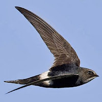
The white-throated needletail is a sleekly designed
bird, with swept back wings and a large chest. The
chest is large to accommodate huge breast muscles
and a larger than normal heart a lungs. Its tail can be
splayed out for turns, or turned into a needle shape
to reduce drag.


If youwant top speed in flapping flight from a bird, bet on the white-throated needletail. It used to be considered a member of the swift family (appropriately named), but is now in its own genus. With a tail wind and the proper motivation, these small birds can reach speeds of 100 mph. Living on the northern coasts of Australia, it is bigger than you might expect for a fast bird, especially one that used to be considered a swift. It has long, swept back wings that help it pick up speed and still be able to maneuver.

Most airplanes have this swept wing design to improve flight characteristics; and as with many of humans so called ideas, we stole it from nature. However, we are getting better at stealing. New aircraft wing designs are based on the swifts’ ability to wing morph, changing the shape of its wings to take better advantage of the flying conditions at the time.

If we drop down into the seas, we can look at speed in the fishes. The sailfish is considered to be fastest. It is of course built in a streamlined fashion, meant to build the speed needed to catch the fish and octopuses it eats. It has been clocked at 68 mph, which in my book makes it faster than the cheetah, since it is moving through water, a much more dense medium as compared to air.

The long upper jaw of the sailfish isn’t just for cutting
through the water. It can skewer large fish with it while
hunting, although this isn’t its normal hunting behavior.  
Sailfish have been seen to cut mackerel in half with a
flick of the bill. Its cousin the marlin has a bill that can
cut through the hull of small boats.

Sohow do bacteria stack up against these speeds? Not too well, despite what we saw in the first video. The fastest bacteria, members of the Vibrio family, move about 200 µm/sec – this is about 0.00045 mph (0.00072 kph). Even my teenage son on his way to clean his room moves faster than that! However, when you sneeze, you send bacteria (and mucus) out of your nose and mouth at over 100 mph – you can almost hear the little bugs screaming.

Because we are looking at a very small area under the microscope, it appears that the bacteria are covering a good distance. But at 1000x magnification, the least magnification you would need to observe bacteria, the field is usually just 500-800 µm across (0.02-0.03 inches). This makes the bacteria appear to be moving quickly.

Vibro cholerae is a gram negative bacteria with a single
polar (meaning at one end) flagellum. This long
appendage rotates and provides the locomotive force for
this fastest species of bacteria. The flagellar motor can
spin in either direction and is driven by a sodium ion
gradient. Too much salt and it slows down; not enough
salt, it slows down. It’s like a microbial Goldilocks.

Like the fish, bacteria are moving through an aqueous (water) medium, so the density is much greater. But it is even worse for them because of their small size. The effects of density are much larger on small organisms, sort of like us trying to walk through a pool filled with caramel (not a bad idea).

Butwhat if we measured speed in a different manner, say….. bodies lengths per second. Vibrio are approximately 2 µm (0.00008 inches) in length and they move about 200 µm/sec. This is about 100 body lengths per second. Now that seems pretty fast, especially for swimming through something thick. 


How does that compare to our other candidates:

                                                Avg. Length            Top speed (kph)              Body lengths/sec
Cheetah                                     125 cm                         112.7                                     25.0
White throated needle tail           25 cm                         160.9                                    178.7
Sailfish                                      340 cm                         109.4                                      8.9
Vibro choleraebacteria            0.0002 cm                   0.00072                                   100
S. Giant Darner dragonfly          12.7 cm                       57.9                                       126
Australian tiger beetle                0.10 cm                        9.01                                    2502.8


So the bacteria are pretty fast, it just depends on how you measure it. But the needle tail still holds its own, even though it is only traveling through air. Comparatively, Usain Bolt moves at a top speed of about 6.2 body lengths/sec. Since humans walk upright, we could measure him at body depths/sec, which makes him sound faster, about 30 body depths/sec (assume 15 inch body depth). But we don’t all run like Usain Bolt.

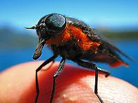
Black horseflies can measure up to 1 inch in length. They
pester people and domestic animals, but worse, can carry
mosquitoes, only the females feed on blood, the gentler
males prefer nectar and help pollinate many flower species.
I could make an analogy between horseflies and humans,
but I won’
In the table above I gave you a couple more examples so that we can find an overall winner.  As always, nothing seems to top the insect world, the Australian tiger beetle can move at over 2500 body lengths/sec, while the darner dragonfly shown above is merely the scientifically confirmed fastest flying insect. However, if you want to go with the most recent estimate for the male horsefly (Hybomitra hinei wrighti), we are talking about speeds of 145 kph when he's in pursuit of a female- typical male behavior. That works out to roughly 4000 body lengths/second!

Next week we can look at plants lifting weights; they have to be in shape in order to photosynthesize!


Penny E. Hudson, Sandra A. Corr and Alan M. Wilson (2012). High speed galloping in the cheetah (Acinonyx jubatus) and the racing greyhound (Canis familiaris): spatio-temporal and kinetic characteristics J Exp Biol DOI: 10.1242/​jeb.066720

Wilson RP, Griffiths IW, Mills MG, Carbone C, Wilson JW, & Scantlebury DM (2015). Mass enhances speed but diminishes turn capacity in terrestrial pursuit predators. eLife, 4 PMID: 26252515

Leaves Suck!

$
0
0
We have talked about the reactions of photosynthesis
before. Basically, the plant uses the energy of the sun to
fix carbon (change it from gas to solid) by adding water
to it chemically. Then it splits them again to make energy.
In previous posts we have talked about photosynthesis – carbohydrates and made from carbon (carbo-) with water added (-hydrate, as in- when you are very thirsty, you are dehydrated). Therefore, the leaves must have a constant and reliable source of carbon (from carbon dioxide in the air) and water.

Question of the day:
How do trees move the water up into their leaves, against the force of gravity, in order to carry out photosynthesis?

Water is quite massive (1kg/L or 8.3 pounds/gallon), and a mature oak tree needs 40-60 gallons of water every day. So how does this huge amount of water get to the top of the tree? Does it travel there from someplace else? Could it be absorbed by the leaf from the air in the same way carbon dioxide is brought in? Or maybe plants don’t have to drink and they use the water they make during metabolism, like the kangaroo rat we talked about last year -they don’t drink at all and seem to get along just fine.

We might be able to eliminate one possible explanation right away – what happens when you don’t water your houseplants? Do they grow or do they die? So do you think most plants need a source of external water or could get along on the water they make during aerobic respiration? Right… I think we are down to absorption or movement from some other place on the plant, namely the roots.

Keep in mind that not every plant has to move water from its roots to its leaves, take the bromeliads for instance. Many of these plants don’t have roots, we have discussed how they have special structures that help them absorb water at the base of their leaves.

You could test other types of plants to see if water on just the leaves is enough to keep them alive. How might you do that? Cover the dirt with something that repels water and then just mist the leaves – that might do it. Try it for a while and see how the plants do.

I think that you will find that they do not thrive after the moisture in the dirt is used up. For most plants, 99% or more of the water they use must be absorbed by the roots and transported up the stem (trunk if it is a tree) to the leaves.

Celery stalks and carnations are good to show the flow
of water against gravity. Dark colors show up better.
Maybe you could have races between the two plants and
then cut them crosswise to look at the size of the vessels.
I bet the smaller ones move water faster.
To modelthe answer to our question of the day all you need is a straw. But that isn’t very illustrative or fancy – so how about cut carnation stems or celery stalks (with the leaves) in a glass of colored water. Lighter colored flowers and darker colored water works best (I use blue food coloring), but I have had students who have really gotten into this and tried to measure the time by adding one color, then switching to another and seeing how long it takes the color to change in the flower and if all the color is lost along with the water.

Over a couple of days, the color will indeed be drawn into the petals of the flower. How does the color get there? Is the water level the same? Water is moving up and taking dye with it. So you can see that it does happen – but this still doesn’t explain HOW it happens. Hint - it isn't capillary action. Even in a very thin capillary tube, water will only move up a few centimeters. How could it possible move from the roots to the top of a redwood tree?

To answer this, you might ask what happens to the water that is being drawn up into the leaves (and flowers of the carnation model). Try putting a baggie over the end of a tree branch and tying it tight.  You will see condensation develop over a day or so. Where did this water come from?

Here are the vessels in a tree. 1) pith– it gets crushed as the tree grows 
2&3) annual growth ringsmade of water carrying xylem. Why 
do you see different rings if they are all xylem? Because spring 
xylem vessels are big, and summer xylem (less water available, so
less growth) vessels are smaller. The line is the change from 
small to big. 6) phloem– this is what carries the carbohydrate to 
the roots and other parts of the tree.
The answer is a process called transpiration (or evapotranspiration). The water evaporates from the leaves, out of pores called stomates, and this creates a negative pressure – like the negative pressure in your mouth when you suck on a straw. This negative pressure actually pulls water up from the roots through the xylem of the plant, to the leaves. In the case of the carnation flower or celery, it also pulled up the very small dye molecules in the water. This evaporative force is quite strong, but not strong enough on its own to lift that 350-500 lb.s (40-60 gallons) needed for an oak tree each day.

The water itself helps in the process. Water is a social molecule, it likes to stick to itself and to other things. It will climb up the sides of container, just look at the meniscusformed in a narrow graduated cylinder when water is added, or note how water travels up a thin capillary tube.

The capillary action comes from the water’s cohesive force, and helps the tree stay hydrated. Evapotranspiration’s negative pressure pulling water up is combined with water’s ability to climb up, and together this is enough to keep the tree’s leaves in the pink, no matter how tall it grows.

But like everything else, there are exceptions, like the plants that don’t have xylem. The non-vascular plants (like mosses and hornworts) only survive based on water absorption and capillary movement from cell to cell. Therefore, they cannot be very tall; you need vessels (xylem) to allow water movement and tall growth. The tallest of the non-vascular plants, the Polytrichummosses, may get to be two feet tall, but that’s it.

Evapotranspiration via vasculature and leaf stomates leads to another question – if water is being lost through the leaves all the time, doesn’t this hurt the plant in times of drought. Well… yes. But plants have evolved some pretty neat tricks to help out.

Some plants don’t use the most forward strategy of
photosynthesis because it would drain them of all their
water during the hottest weather. CAM plants can close
their stomates during the day and only fix carbon dioxide
at night when it is cooler. We should probably talk about
these plants in more detail later this year, they have some
mighty cool adaptations.
1) Stomatescan open and close to regulate water loss. Some plants can close their stomata completely during the hot day, and save their built up radiate energy to convert carbon dioxide and water into carbohydrates only at night, when they will lose less water. Cacti are a good example of this.

2) Leaves, especially the sun-exposed sides of leaves, are covered with a waxy substance called cuticle that greatly reduces the loss of water by diffusion through the cell wall. If water were allowed to travel through the cell membrane and wall, then it would evaporate and set up a negative osmotic and evaporative pressure that would quickly dehydrate every surface cell.

3) Here's a trick many people don’t really consider – many plants have two types of leaves! You might be able to find a tree or two with which to investigate this.

Big leaves have large surface area, so more water will be lost as compared to smaller leaves. Leaves in the direct sunlight should be structured in order to carry out the most photosynthesis, but if they are small, how can this be maximized?

Many trees have sun leaves and shade leaves. Sun leaves are smaller, thicker, have more stomata, and are located where the direct sunlight hits the tree during a good portion of the day.  Shade leaves are bigger, thinner, and have fewer stomates to reduce water loss.

Sun leaves have more layers of pallisade cells, the cells that have the most chlorophyll and do most of the photosynthesis. They are located at the ends of branches, especially on the north side, and on the crown (top) of the tree.

Sun leaves are a smaller and thicker, and they often have
fewer in and outs in their shapes. The smaller shape
reduces water loss, the thicker body provides extra layers
of cells for photosynthesis, the reduced number of cuts
and points…. I have no idea. It is a continuum, leaves that
get a good amount of light land somewhere in the middle.
Shadeleaves have to rely on lower levels of sunlight (they are in the shade), so they have even higher concentrations of chlorophyll than sun leaves, although they are thinner. They can process light more efficiently than sun leaves, so they are actually very important to the plant despite their little time in the sun.

Look at the trees around you, do some have large leave on inner branches and lower on the canopy, while having smaller leaves on top or on the ends? These are probably shade tolerant trees. They have developed the ability to still do enough photosynthesis despite low levels of light.

On the other hand, do you see a tree that has just one size of leaf (not including newly formed leaves) and only has leaves on the ends of the branches? This is probably a shade intolerant tree.

The conifers are an interesting exception, some are shade tolerant, usually the firs, while others are shade intolerant, mostly the pines. However, neither type has sun leaves and shade leaves. Their shade tolerance has more to do with their branch geometry and ability to allow just about all their leaves (needles) see the same amount of sunlight.

Next  week, we will ask if there is any limit to interspecies mating, can you cross a cat with a dog? 


von Caemmerer, S., & Baker, N. (2006). The Biology of Transpiration. From Guard Cells to Globe PLANT PHYSIOLOGY, 143 (1), 3-3 DOI: 10.1104/pp.104.900213

Terashima, I. (2005). Irradiance and phenotype: comparative eco-development of sun and shade leaves in relation to photosynthetic CO2 diffusion Journal of Experimental Botany, 57 (2), 343-354 DOI: 10.1093/jxb/erj014

Lions And Tigers and Ligers, Oh My!

$
0
0
A full grown liger is a biiggg cat! A male lion and
a female tiger get to know each well, and their
love child is huge. It doesn’t happen in the wild
because the ranges of lions and tigers don’t
overlap, and they don’t have computer dating
services. The liger is bigger than either parent,
and this is a problem during birthing. They have
many birth defects and often die young.

Whenputting organisms into categories (taxonomy), we go from bigger, more general categories to smaller more specific ones. The more similar two organisms are, evolutionarily and genetically, the more levels of their taxonomic classification they will have in common.

Question of the day:
A tigon is the result of a cross between male tiger and a female lion, while a liger is the offspring of a male lion and a tigress. But are these new species? And just how far can you go when you cross-breed?

There are instances when different species mate, but the outcomes, while interesting, may or may not be new species. It helps to know the classification levels.

Kingdom (domain) - Insects, dogs and people are all very different, but they are all animals.

Phylum (Division for plants) – usually these group things by a common body plan or some other morphologic character, or a certain degree of genetic relatedness. Arthropods are all related by a chitin exoskeleton, so flies and lobsters are both arthropods, but flowering plants have fruits and conifers have cones, so they are in different divisions.

Class– these groups have more in common, either physiologically or genetically. Cows and dogs both have hair and give birth to live young, so they are both in the class Mammalia. However, flowering plants are divided into two classes, monocots and dicots, based on seed and vascular tissue differences.

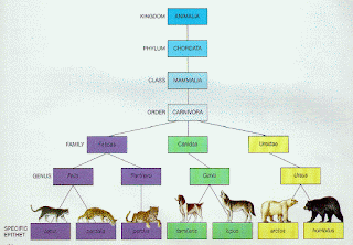
Here is the classification scheme for several familiar
mammals. Cats are all in the same family, but there are
several genera, while all dogs fit in one genus and all
wolves in another. Black bears and Kodiak bears are a
different genus than polar bears, but hikers and bigfoot
monsters have reported sightings of polar and Kodiak
bear hybrids…. well hikers have been reporting them.
Order– Even more specific, this level of classification has been altered greatly by molecular biology. Armadilloes and anteaters are both in the Order Edentata– as toothless mammals. However, roses and magnolia trees are both dicots, but they belong to different orders (Magnoliidae and Rosidae).

Family– Now we are getting down to smaller differences, but still just as important.  All the big-eared bats and all the thick thumbed bats are both included in vespertine family, because they come out to feed in the evenings. On the other hand, maples and mahogany trees are both in the order of Sapindalae, but they belong to different families based on their leaf and flower anatomies.

Genus–comes from the Greek for “kin,” so these organisms in the same genus are closely related. In animals, both moths and butterflies are in the same order, but they are broken into 124 different families, and the family Nyphalidae, which contains the Monarch butterfly, has over 600 genera (the plural of genus).

Species– These are the individual distinct group of organisms. Usually, the distinction is made based on whether their breeding can produce fertile offspring. So bulldogs and St. Bernards are both species of dog, since they can make mutts.

The africanized honeybee is more likely to swarm and
migrate when food supplies are low, so they can be seen
in masses like the one above. For hives, they usually
invade an existing hive, quit out the queen and install their
own. The danger in these bees is that they are more likely
to swarm when agitated, and they will chase the agitator
for a much longer distance (a mile or more) than regular
honeybees (100 yards or so).
Sub-Species - this is like the different breeds of cats we keep or that unfortunate incident where African bees were crossed with South American honeybees and created killer bees!

Now that we have that information – let’s rephrase our question of the day. Can you breed (hybridize) different species and create a new species?

Cross-breeds are common within species (intraspecies hybridization), like with cats or dogs – not cats with dogs, you’d never want to do that! And we know they are fertile, so you end up with some dogs that are ¼ this, 1/8 that, and ¼ the other. What about between species?

Interspecies hybridsusually don’t give you fertile offspring. Since the definition of a species is a group of animals that can mate to give fertile offspring, then you would be hard pressed to create a new species by breeding different species together.

For example, the liger and tigon males are always sterile, so even though the females are sometimes fertile, they still can’t mate a tigon to a tigon. This would be necessary to make a stable species. So the chances are low on the interspecies level.

That would mean that new species coming from breeding of animals from different genera would be even less likely to produce new species. However, individuals can be hybridized. Intergeneric hybridization is easier to do in plants; orchid growers have made many different intergeneric crosses, like little Dr. Frankensteins with green thumbs.

Compare the two marine mammals that are jumping. One
is bigger, darker colored, and apparently can jump
higher. That one is the wolphin. Her name is Kekaimalu,
the offspring of a bottlenose dolphin and a false killer
whale. Her offspring, Kawili Kai, is bigger than a dolphin
as well, but is lighter colored than mama.
Butthey can occur in animals. A wolphinwas born at Hawaii Sea Park in 1985, the result of a mating between a bottlenose dolphin and a false killer whale. These species are in the same family (Delphinidae) but different genera. Named Kekaimalu, this female is fertile and has mated with male bottlenose dolphins. The first two offspring did not live very long, but her third calf is still alive and well, at ¾ dolphin and ¼ false killer whale. However, this wouldn’t be a new species unless wolphins mated with wolphins and produced fertile wolphins.

The rarest hybridization is the interfamilial hybrid. Most examples have occurred in birds, where game fowl are housed together. The Pea-guinea is a hybrid between a peacock and a guinea fowl hen. They look weird and don’t survive beyond a year or two, so there is no way that these could form a stable species.

It would take a bunch of posts to talk about why certain hybrids will work and others won’t, and why new species are not generally produced in this way. But for now - how about two exceptions?

What do you get when you cross a blueberry with a
snowberry (maggots, that is)? You get a Lornicera fly –
O.K. not a funny joke, but a pretty cool twist in
evolution. As fruitflies go, this is a pretty cool looking
one; you don’t have the ghoulish red eyes to deal with
and three stripes make it look a little like a lightbulb!
The Lonicera fly is a new species produced by natural interspecies hybridization! Just when you think you understand nature, there it goes again, kicking you in the seat of your pants.

The creation of this new fruit fly species did have a little help from humans. For about 250 years, honeysuckle plants have been imported to the North America from Europe. In the 1990’s scientists found the Lonicera fly and tried to see what other flies it was related to. Low and behold it was a hybrid of the snowberry maggot and the blueberry maggot. But why didn’t the hybrids breed with the parent species and dilute the hybrid genome back into the two stable species? How did the hybrid become a new, stable species?

These hybrid flies preferred to feed on the honeysuckle, so they lived on the imported plants, while the parent species lived on their favorites (snowberry bush or blueberry bush).  This is a kind of geographic isolation; the Lonicera hybrids find only Lonicera hybrids when it comes time to mate and they end up mating hybrid to hybrid for many generations. This resulted in a stable species, the process is called hybrid speciation.

The Heliconius heurippa butterfly has an unusually
large black bar that crosses its body. This must be
fairly obvious to other butterflies of the same
hybridization and it must also be pretty attractive.
Both male and female hybrids search out the wide
black bands. I would love to know the molecular
biology of that specificity of attraction.
The second exception is the Heliconius heurippa butterfly in South America. An interesting 2006 study in which hybridization was repeated in the laboratory showed that H. heurippa in nature is the result of breeding of two other species of butterflies. The hybrid does produce fertile offspring, both male and female, but that isn’t the end of the story. In this case, there isn’t any geographic isolation forcing hybrid-hybrid mating - they chooseto mate together. Their choice is related to the fact that the hybrids have bold black stripes on its wings, while neither parent species does.

The hybrids preferentially mate with other butterflies with the bold stripes, so they are mating hybrid to hybrid and are stabilizing the new species. Darwin would blow his top – or maybe not. He never said this couldn’t happen, just that it was less likely.

Stay tuned, molecular techniques are beginning to show us that this may not be such an exception – a 2011 study identified another butterfly species created by hybrid speciation and it happens all the time in plants, like sunflowers. Three younger species, the desert, the puzzle, and the sand – seem to live where their parent species cannot, so they tend to pollinate with their similar hybrid brethren and make new species.

Next week we will ask why some birds migrate while others stay put year round.


Jesús Mavárez1, Camilo A. Salazar, Eldredge Bermingham1, Christian Salcedo, Chris D. Jiggins & Mauricio Linares (2006). Speciation by hybridization in Heliconius butterflies Nature, 41, 868-871 DOI: 10.1038/nature04738

I’ll Fly Home—Or Not

$
0
0
The snowy owl is sedentary, meaning it does not
migrate. The males are almost perfectly white,
while the females and juveniles can have black
barring. They sit, look, and listen for their prey,
which includes small rodents and even other birds.
Their hearing is good enough to let them target a
mouse under the snow from hundreds of yards away.

The Arctic tern travels from north of the Arctic Circle to Antarctica and back again every year. On the other hand, the Snowy Owl lives in the arctic region year-round; it doesn't migrate at all.

The Question of the Day:
Why do some birds migrate while other birds stay in one place?

The possible explanations are many. Maybe the type of food they eat is present only part of the year, or maybe they can’t stand the cold temperature. They might need to have their babies in a place away from predators, or perhaps migration is an evolutionary holdover that had a reason in the past, but no longer isnecessary.

How could you start to determine the reason for migration in only some bird species? I would start by looking at how closely related the migratory and non-migratory species are. Maybe all the birds that migrate are more closely related to one another than to the birds that don’t move around during the year. This would suggest that there is a genetic basis for why only some species migrate.

One research group did just this in 2007. The looked at 379 species of flycatchers, a closely related group of birds. They found that almost equal numbers of species were migratory or resident, so it doesn’t appear that genetic relatedness is the answer.

The painted bunting is among the most colorful
birds in North America. This male has several colors,
while the female is a brilliant green. They breed in
south central US or on the southern east coast, but
winter in Central America; therefore, they are
seasonally migratory.
Maybethe need to migrate has to do with the geographic region. About 90% of birds in the arctic migrate, some are present there only in the mid-summer months. The arctic tern is a good example. Arctic terns move with the summer, breeding in the arctic in May-July, moving down along continental coasts to arrive in Antarctica for the months of December to February. The entire distance traveled could be as much as 32,000 km (20,000 miles) in a single year.

Similar to the arctic region, the east coast of North America has species that migrate and species that are sedentary. About 80% of the birds species of the east coast move south during the colder months, but on the Pacific coast, almost all the bird species are non-migratory.

So migration is not due to the type of geography around the birds. However, the east coast of North America does have larger temperature swings than the west coast, so maybe it is just that some birds can’t deal with the cold. 


Some non-migratory birds can control the amount
of blood that travels to the legs in order to conserve
body heat. This works even better if they can reduce
the amount of contact with the cold surfaces, so some
of these birds perch on one leg at a time.
Manybirds that do not migrate have special adaptations to deal with the cold. Trying to keep a constant body temperature (endothermy) takes a lot of energy, and birds live right on the edge of having enough energy anyway. Flying requires a huge amount of energy and they must eat almost constantly just to keep enough carbohydrates in their system to be able to move around to find more food.

Burning more energy to keep warm might tip them over the edge into starvation. To alleviate this problem, many birds can allow parts of their bodies cool down to freezing or near freezing, while keeping their internal organs at a temperature that will preserve their function. Blood flow is a major way to keep parts of the body warm, a duck standing on the ice can reduce the blood flow to its feet and reduce the amount of heat lost to the cold ice. The duck’s chest may be 40˚C, but its feet could be just one degree above freezing.

But let us look again at the arctic tern. It migrates from the north polar region to the Antarctic region in such a way that it sees two summers each year. But these are summers in name only. The arctic summer has an average temperature from -10˚C to 10˚C, so much of the time the tern is there, the temperatures are near zero.

The arctic tern has a ghastly commute each year.
The trip is even more amazing when you consider
that during its yearly molting, the tern flies very
little.  So, all that distance must be fit in to just a part
of the year, not the entire 365 days. I guess they
vacation by NOT traveling.
Thenwhen they reach the Antarctic, the summer there has an average temperature of -2˚C to 2˚C. This is hardly a balmy vacation destination for the tern. The temperatures in both its breeding grounds and wintering grounds would require it to have elaborate temperature control and energy-saving adaptations. Therefore, inability to tolerate cold temperatures is not the reason for migration, at least not for many birds.

The group who carried out the 2007 study concluded that the main reason that only some flycatcher species migrate is not due to what they eat, or when they breed, or what is trying to et them, but to how available their food source is. Whether they are fruit eaters, or insect eaters, or seed eaters, how easy it is to find their food is the most common reason that migration has evolved for a specific species in a specific location.

The food availability hypothesis is supported by certain types of migration that are common in North America. Irruptive migration is characterized by a population moving to another place, but there is no yearly, seasonal, or geographic pattern. The birds may migrate one year, then not again for a dozen years, or they might go for several years in a row. North American seed-eating birds are famous for these migrations. The distance and number of individuals that migrate are also not very predictable, and this all makes it sound like the movement is linked to food availability. However, it could also be to escape some population explosion in a predator species or for some other reason.

It isn’t only birds that might undergo partial migration.
Some crab species will migrate for breeding purposes,
like these Christmas Island Red Crabs.  Individuals
that won't breed just don’t make the trip. They may be
too young, too old, too lazy. In other cases, when
populations migrate away from the breeding grounds,
some individuals may remain there the year round.
Anothertype of migration is partial migration, a pattern wherein not all birds of a species in a certain location will migrate, only some of them leave in non-breeding times, while others stick around year-round. Food may be available for some, but not all, or the environment may be unsuitable for some weaker individuals to have enough time to forage for a sufficient amount of food. These (and other reasons) might explain why partial migration exists, but one question remains, who stays and who goes? The choices could be based on age, altruism, suitability, dominance… laziness?

As an aside, it isn’t just birds that migrate. Mammals move from place to place, sometimes with a defined pattern during the year, but sometimes they just follow the food, a process called random migration. And some insects migrate as well.

For many years, the migration of the Monarch butterfly was believed to be the longest insect migration. But this is not the typical migration we think of, where an individual moves from one place to another and then back again. The migration of the monarch butterfly takes four generations to complete. Some generations are born and fly a long distance to lay their eggs, while others are born, live, and reproduce in a small area. But altogether, this butterfly moves from as far north as Canada to the high mountains of Mexico and back each year, about 7000 km (4400 miles).

Globe skimmer dragonflies breed in freshwater pools,
so they migrate from India’s monsoon season to the rainy
season in East Africa, all in search of a place to lay
eggs. They make stopovers on the Indian Ocean islands,
but only to rest, because there are very few
pools of freshwater on these coral cay islands.
A few years ago, a biologist in the Maldive Islands started to wonder about the movement of globe skimmer dragonflies where he lived. They seemed to be plentiful in some periods and absent in others. He started to track them, and found that they have an even larger migration pattern than the monarch butterfly. What is more, they fly long distances over the ocean with no place to stop and rest.

Over a series of generations, the dragonflies move from India to the Maldives, some 600-800 km across the open sea. Then they move to east Africa, from Uganda to Kenya and Mozambique. In January, they start back toward India, and complete their migration of more than 18,000 km (>11,000 miles).

So birds may get most of the publicity, but insects hold their own in the migration game. Of course, it does take four generations of dragonflies or butterflies to make their complete journey, where a single arctic tern may make its entire 20,000 mile trip thirty times in its lifetime. O.K., they are both pretty impressive when you consider most people need a car to go down the street to the grocery store and back.


Davenport LC, Goodenough KS, & Haugaasen T (2016). Birds of Two Oceans? Trans-Andean and Divergent Migration of Black Skimmers (Rynchops niger cinerascens) from the Peruvian Amazon. PloS one, 11 (1) PMID: 26760301

Ahola MP, Laaksonen T, Eeva T, & Lehikoinen E (2007). Climate change can alter competitive relationships between resident and migratory birds. The Journal of animal ecology, 76 (6), 1045-52 PMID: 17922701

Hobson KA, Anderson RC, Soto DX, & Wassenaar LI (2012). Isotopic evidence that dragonflies (Pantala flavescens) migrating through the Maldives come from the northern Indian subcontinent. PloS one, 7 (12) PMID: 23285106

Ivy League Climber

$
0
0
Wrigley Field was originally called Weeghman Park,
after a local lunchroom owner. The first team to call
the park home was the Chicago Whales - strange name
for a city on a freshwater lake. The ivy on the outfield
wall is actually Boston Ivy and Japanese Bittersweet,
since English Ivy would have a tough time with Chicago
winters- just like everyone else.
Wrigley Field is the venerable 1914 baseball stadium on Chicago’s north side. One of its most characteristic features is the ivy covered outfield wall that occasionally swallows a hit ball, never to be seen again – a ground rule double.

The question of the day:
Does ivy stick to a wall or grab it, and will ivy have enough strength to destroy the wall over time?

English ivy (Hedera helix) is of the Araliacae family, but doesn’t have spines like some other species in the family, like the aptly named devil’s walking stick (Aralia spinosa). I can’t imagine a Chicago Cub outfielder diving into a wall covered with devil’s walking stick to make a catch; although being a Cubs fan can feel like that.

English ivy is an evergreen climbing vine, but it will grow along the ground just fine if there is nothing available to climb. Not unlike Kudzu in the US south, ivy can become invasive and choke out other plants, creating “ivy deserts.”

As English ivy grows along the ground, it shows its juvenile form. It has light colored leaves with lobes and points, no flowers, and can form roots very easily. When the ivy finds something on which to grow vertically, it transitions to the adult stage, with leaves that are less lobed or pointy, less root growth and can produce flowers and berries.

The stem of ivy is not capable of supporting the weight of the vine – it can’t stand up on its own, but yet it easily grows 30 m (98 ft) against the force of gravity and can reach heights of more than 90 m (300 ft) in conifer trees with seemingly no problem whatsoever. The mechanism by which it accomplishes this was investigated by none other than Charles Darwin, but much more recent work is showing the ivy plant to be quite a surprise.

English ivy sends out thousands of adventitious roots
per foot. These roots are responsible for the ivy’s
adherence to the substrate. They are aerial roots, but
can also grow into the ground and act as regular roots
as well.

Darwinnoted that ivy sends out adventitious roots from its stem. This is where the devil’s club or walking stick and the ivy are similar, but in the case of ivy, they are induced by a vertical substrate and don’t cause pain.

Adventitious roots are those that arise from someplace other there where you would expect them, like directly from the sides of stems, or off leaves, or off old woody roots. In the case of ivy, they are aerial, adventitious roots, since they do not get buried in dirt. They can still collect water, but are protected from dehydration by having a thicker, waxier surface.

Darwin also noted that ivy was not wrapping the adventitious roots around some protruberance on the vertical surface to allow the vine to cling. Those that do wrap around and grab are called tendril climbers, andinclude clematis, grapes, and sweet peas. In some cases, the clinging apparatus will have only that function, in other plants they will grasp, but can leaf or fruit as well.

Other vines use their stems to wrap around a vertical substrate, the stem twiners and tendril climbers are both examples of thigmotropism (thigmo = to touch, and trope = turn). Interestingly, honeysuckle always coils clockwise while wisteria always turns counterclockwise.

Pea plants grab hold of vertical surfaces using tendrils
that coil upon contact with a surface. The tendrils are
modified leaves, stems, or shoots. Supposedly they taste
good and are a vogue ingredient in cooking nowadays.

English ivy doesn’t twine, it doesn’t tendril wrap, and it doesn’t burrow into a flat surface to gain an anchor, although it will exploit a crack and grow through or along it. Neither does it just grow up until it touches something and then use its growth to ramble through and around the substrate. Climbing rose is an example of a rambler, it will use its hook shaped thorns to help it stand up as it grows through and around another plant.

No, English ivy uses a chemical adhesive secreted by it adventitious rootlet ends in order to stick to a vertical surface – it can even cling to something as smooth as glass. The secretion is yellowish and forms circular dots on the vertical surface. It is very sticky, and becomes stickier as it dehydrates.

The compound contains polysaccharides that act as a carrying agent for discrete nanoparticles (70 nm diameter) that are responsible for the adhesion to the wall. Amazingly, the way ivy clings to a wall is very similar to how a gecko walks up a wall or hang upside down.

This is an electron micrograph of the nanoparticles of
ivy adhesive. The particles have an average diameter
of about 65 nm and can get so close to the substrate
that the electrons and nuclei of each will interact and
attract one another.

The nanoparticles are like the nanohairs on a gecko’s (or fly’s) foot. They increase the surface area of the material greatly and are so small that they can make very intimate contact with the surface. They get so close to one other that they can use van der Waal’s forces on the atomic level to attract one surface to the other. Studies from 2010 showed that the interactions of the nanoparticles in the yellowish ivy secretion were enough to create the bond, and mimics using polystyrene nanoparticles have become excellent adhesives.

But the amazing abilities of the ivy nanoparticles don’t stop there. They seem to disperse and absorb light energy much better than the metal nanoparticles that we currently use in our sunscreens. Titanium oxide and zinc oxide are the current state of the art in terms of reflecting, dispersing and absorbing ultraviolet rays, but it seems that ivy nanoparticles are 70X better at these jobs than are the metal oxide particles. Our next generation of sunblock may come from ivy – talk about green technologies!

Ivy can help with sun damage in another way as well. By covering the walls of a building, ivy keeps the heat in during the winter by acting as insulation and reflects the sunlight away in the summer, keeping the building warmer or cooler as the case may be. Ivy also deflects much of the rain from getting to the surface that is covered, so it can protect against acid rain damage or other weathering.

But ivy can do damage as well. Any surface that has gaps, like shutters against a wall or wood siding will allow ivy to grow in the cracks and pull them from the wall over time.  It may not create holes in mortar or brick, but it will grow into them and then expand when the stem fills with water. This hydraulic action can break down stone over time and bring a building down if given enough time and opportunity.

The mass of an ivy vine can also cause damage. It can cover an entire plant and keep it from getting enough sunlight to live, but it can also make it top heavy and cause it to fall in a strong wind. I have wondered about this in terms of ivy growing on a building. How much weight does it add to the wall, and would it ever be enough to pull the wall down?

The quintessential ivy covered cottage. How much weight
must this add to the house? The roof could easily collapse,
and who knows what is living in there. But there is no
arguing that it looks great.

Look atan ivy-covered wall. How much must all that vine weigh? Forestry workers pulling ivy off of conifers say it is not unusual for there to be over 2000 lb.s (907 kg) of ivy on a single tree.

I have been wondering how to estimate the mass of ivy that is clinging to a wall. You might estimate the square footage covered, then cut out one square foot and find its mass, and then do the math to find the total. But if you cut from the bottom, then everything above it will die – not the best experimental design. If you cut from the top or edge, the vine will be immature and have less mass per sq/ft than the average along the entire wall.

Maybe you could advertise free ivy removal, find a client, measure the square footage and the find the mass of everything you take down. But remember, that is one great adhesive; you will probably leave a decent amount behind, leading to a low estimate. Or, you will bring parts of the wall with it, leading to an overestimation.  Any ideas?

Next week we will begin a series of posts on getting sick - the exceptional thing is that sometimes it is good for you to get sick.

Lijin Xia, Scott C Lenaghan, Mingjun Zhang, Zhili Zhang and Quanshui Li (2010). Naturally occurring nanoparticles from English ivy: an alternative to metal-based nanoparticles for UV protection Journal of Nanbiotechnology DOI: 10.1186/1477-3155-8-12

Lucky For Me, I’m Diseased

$
0
0
Biology Concepts – disease, vaccination, single nucleotide polymorphisms

Jill Bolte Taylor is an Indiana University neuroscience
professor who suffered a massive stroke. She recognized
what was happening and has translated her thoughts
and feelings into a narrative to help us understand. She
is eloquent in describing how her stroke has affected her
in a positive way. --- Soon to be a major motion picture!

Yourarely hear someone say how glad they are to be sick – unless a business meeting, unit test, or visit to the in-laws is involved. Robust health is a sign of good genes, and animals (including humans) instinctually seek out good genes when selecting mates. We don’t like to be sick, and we don’t want others (potential mates) to see us being sick.

True, there is that one person in a thousand who argues quite eloquently that an illness showed them another side of life, expanding their world-view and making them a better person. I applaud this attitude, but did you ever notice that it’s only the survivors that can gain this insight?

Our entire health care system is based on the idea that it is preferable to not be sick. The best way to bring this about is to reduce the chances that we will encounter anything that might provoke a response from our body, including pathogens (disease causing organisms, from pathos = disease and genique = to produce) and allergens (living or non-living molecules that can induce an allergic response).

But what does it mean to be “sick?” If you are infected by a pathogen, are you necessarily sick? There are infections that are subclinical or asymptomatic (without signs of disease), and there are carrier states, when a person is infected and can transmit the disease, but does not have symptoms. Are these people still sick?

You can be in a social situation where you feel empathy or regret, “I feel just sick about how I treated her.” Is this true sickness? Your mental state of mind is important in your health; if you talk yourself into being sick, are you really sick?

Single nucleotide polymorphisms (SNPs) are one base
changes in a gene sequence. “Polymorphism” means
that the population will show different sequences at this
point. SNPs may produce no change in the protein, but in
some cases they may change the shape or function of the
protein just slightly. This may not cause disease, but may
affect the course of a disease, or how drugs will work in
that individual. SNPs may one day lead to personalized
medicines in a new science called pharmacogenomics.
Drugs will be designed to work best for your particular
DNA sequence.

What about genetic mutations? Can everyone with a genetic mutation be considered sick? If yes, then we are all sick, because everyone one of us has thousands, perhaps millions of single nucleotide polymorphisms (differences in a single base of DNA that might lead to change in function of a protein). I would suspect that most of us have larger mutations as well; the older we are, the more mutations we have. Some mutations render a person predisposed (more likely) to develop a disease – is this person sick even before he/she acquires the illness?

Osteoarthritis is a disease that can wear away joint surfaces and necessitate hip or knee replacement. My father has two artificial hips due to osteoarthritis, but does that make him sick or ill?

You see someone coughing, sneezing and blowing his/her nose. It could be due to respiratory allergies or a bacterial or viral infection. Are they sick in one instance, but not the other? I have seen TV ads that try to convince allergy sufferers that they are a menace to society, and should be embarrassed about their condition (unless they use their wonderful product). The entirety of the message in our society is that any illness or condition is a deficit.

To summarize our man-made rule: diseases are bad, and being exposed to diseases is bad, so keep your environment clean and antiseptic. Don’t get me wrong – I am not mocking the rule. I would rather not be sick - so much so that I am careful where I go and what I touch – in some places I simply choose not to breathe, just to be on the safe side. Disease prevention is an important part of life expectancy.

But are there exceptions? Is it sometimes good to get sick, either in general or with some specific disease? I think you know there must be exceptions, otherwise we would just be left with an interesting discussion of what it means to be sick. I bet you can even come up with at least example on your own. There are in fact boatloads of general and specific exceptions to this rule. Let’s take a few weeks and cover a few examples that are exceptions to "disease is bad" rule.

Our first exception is one that you may have already thought of – vaccines. With many vaccines, getting the disease is the key to not getting the disease – counterintuitive, isn’t it? I will use smallpox as an example of the idea that sickness prevents sickness, but there are many others.

Smallpox survivors had a very distinct look. It was
unfortunate that the lesions showed up most heavily on
the face and arms. Thankfully, the disease has been
eradicated, and the virus only exists now in two
laboratories, at the Centers for Disease Control in Atlanta
and the “vector” lab in Siberia. Whether these stocks
should be destroyed is a matter of some debate.
Smallpox, until recently, had been a scourge on mankind for thousands of years. The infection is caused by a virus (Variola major or minor) and may present in several different forms. It was a very dangerous disease, the hemorrhagic form was almost universally lethal. Those that survived smallpox were marked for life (see picture).

In the 1790’s, Edward Jenner of Gloucestershire, England noticed that milk hands and milkmaids seemed to be immune (from Latin, immunis = exempt) to smallpox and he wondered why that might be. The milking workers told him they felt protected because they worked with diseased cows, those that had a mild disease called cowpox. For some reason, having had cowpox kept the milkmaids from catching smallpox.

It turns out that cowpox and smallpox are enough alike that having one will prevent you from having the other. It was on this basis that Jenner developed the first vaccine (Latin from vaccinus = from cows, coined by Louis Pasteur as a tribute to Jenner). By pricking the skin of a young boy with a needle contaminated with the pus from a young milkmaid with cowpox, Jenner showed that this could prevent infection with smallpox (Jenner wasn’t the first to vaccinate with cowpox, just the first to prove it prevented smallpox).

Contracting cowpox, a mild disease that did not kill or scar, could prevent one from catching smallpox, a terrible disease that often killed and left survivors with permanent reminders of their ordeal. Maybe getting sick ain’t always so bad. We will talk more next week about just how vaccination works to produce a protective immune response.

Cowpox vaccination is an example of using one disease to prevent another, but even 100 years before Jenner it was recognized that you could prevent smallpox by giving people smallpox. Strange, isn’t it? Variolation was performed by blowing ground smallpox scabs up the nose of another person, or by pricking them to place the material under the skin.

The virus in the olds scabs was definitely variola, it was just weakened (attenuated) by its age and its time outside of healthy cells. The virus was recognized by the body and an immune response is mounted, but the virus was too weak to produce a fulminant infection was eliminated by the body. But not before it helped the vicitim become immune to subsequent smallpox infection.

Poliomyelitis infection led to a paralysis of the muscles.
This could include the respiratory muscles, so iron lungs
were used to force air in and out of the patients’ lungs.
Before a vaccine was developed, a treatment was
developed by an unaccredited nurse from Australia.
Sister Elizabeth Kenny overcame much professional and
gender prejudice to show that heat and passive exercise
to retrain muscles was better than the then used
immobilization therapy. Try to see the biopic “Sister
Kenny” on TCM some time.

Attenuatedvaccines do carry some risk. Paralytic poliomyelitis has almost been eradicated thanks to Jonas Salk’s inactivated (dead) vaccine injections and Sabin’s orallly taken, attenuated vaccine. The attenuated vaccine is better at preventing a natural infection, but in rare cases the vaccine virus can revert back to a wild form and result in iatrogenic (iatro = doctor and genique = to cause) polio, also called vaccine associated paralytic poliomyelitis (VAPP). Thankfully, widespread use of the Salk and Sabin vaccines in the 1950’s has made vaccination in the US (as of 2000) and UK (2004) unnecessary.

Many of the vaccines used today are engineered in a laboratory from just a portion of the organism. By using only the antigenic portion (that part that elicits an immune response) of the virus, there is no risk of iatrogenic disease. If the viral portion is produced in a laboratory using DNA technologies, it is called a recombinant vaccine. In some cases, the antigenicpart of the virus is weak on its own, so these subunit vaccines may be conjugated (joined to) some other molecule that will elicit a stronger immune response.

Unfortunately, there is a growing number of people ignoring history and putting are their children and the population at large at risk. Some parents’ reluctance to vaccinate is based on a single 1998 study in which vaccination was linked to autism, even though the author of the paper, Andrew Wakefield, has been convicted of scientific fraud and banned from the practice of medicine. Wakefield was an investor in a company that was going to offer medical testing for vaccine-associated autism and as well as assist in autism/vaccine lawsuits, so he falsified his data in an effort to make his company profitable.  

As a result of the vaccine scare, the UK has seen a rise in the number of measles, mumps, and rubella cases in the last decade. These are diseases associated with childhood, but can cause severe disease or death in many victims, especially adults.

Pertussis, also called whooping cough, is transmitted only
from person to person. If no around you has it, you can’t
get it. However, symptoms may not show for 6 weeks after
infection, so everyone should be vaccinated. The coughing
can be so violent that it breaks blood vessels around the
eyes and nose – and it can kill young children.

Manyin the US are also selecting to apply for vaccination exemption due to medical, religious, or personal beliefs; therefore, disease incidence is rising in America as well. In July, 2012, the CDC reported that the US had 18,000 cases of pertussis (whooping cough) in the past year, including an epidemic of more than 2500 cases in Washington state from January to June. This points out the need for vigilance in monitoring, as some of these patients had been vaccinated. This suggests that that the protection may not be lifelong; a booster vaccination may be necessary, although it is also telling that Washington state has one of the highest vaccination exemption rates in the country.

This also brings up the idea of herd immunity. There are some people who have been vaccinated, but protection is not complete. The elderly may not be able to react completely even if vaccinated, as might the very young. Some vaccinations may not take - how many time have you had an antibody titer test to make sure your vaccine worked? It is very rare to get titers unless something is suspected and you are already sick. Therefore, many people must count on the vaccination of the herd - a critical percentage of population needs to be protected in order to keep the incidence of the disease below a crucial level. If the level rises - as with too many people choosing not to vaccinate - then the incidence will sky rocket because it will affect those people who don't happen to know they are not protected. Un vaccinated people affect everyone, not just themselves.

Next week we will look at vaccine driven immune responses in a bit more depth, in an effort to understand why we have to get a flu vaccination every year.

Centers for Disease Control and Prevention (CDC) (2012). Pertussis epidemic - washington, 2012. MMWR. Morbidity and mortality weekly report, 61, 517-22 PMID: 22810264

For more information on these subjects, or classroom activities, see:

Sick/diseased/ill:

Single nucleotide polymorphisms and pharmacogenomics:

Vaccines:

Lack of vaccination:

Your Body Has A Photographic Memory

$
0
0
Biology Concepts – innate immunity, acquired immunity, memory response, influenza

Your body is exposed to tens of thousands of foreign molecules every day. Some can do you harm, some can’t. Your immune system sorts them by matching receptors on immune cells to molecules on the foreign objects.

Legos and biology are a good fit. They can be used to analogize the 
rearrangement T cell receptor genes or hypervariable regions 
of antibody genes, or they can be used to model the entire 
body. One scientist uses them to model building 
complex systems from repetitive units. And they’re fun.
Thinkof the receptors as Legos; your DNA provides for several different types of Lego blocks to be made, and your immune cells can rearrange the different types and put them together as a receptor, so there can be millions of different receptors. Each immune cell has just one type of Lego receptor, although it may have many copies of that one form. Each different Lego receptor will fit, key in lock style, with a specific foreign molecule.

The receptors exist on many types of cells, and antibodies sometimes function as receptors when attached to the surface of specialized immune cells. Even circulating antibodies (Ab) in the blood take the form of key and lock systems, whether as single Ab, dimers (2) or pentamer (5) complexes.

The immune system of higher animals can be described as several sets of pairs. Each member of a pair attacks a problem in a certain way, and has independent pathways, but each pair also has overlap and must work together in an overall response. We could spend weeks just on this system, but lets look at the major parts by describing each pair, from largest to smallest.

Innate immunityvs. adaptive immunity– the innate immune responses are fast but short. They don’t depend on your immune system recognizing the specific foreign molecule (antigen) with a specific receptor, but respond with the same types of reactions no matter what it is. Almost all plants and animals have some form of innate immune system.

Vertebrates take the immune system further. They have developed an adaptive immune system that does depend on your immune system recognizing the specific foreign invader. It then generates a tailored response to that one foreign organism or molecule. The faster, but more general, innate response helps the slower, but longer lasting and more specific, adaptive response to kick in.

These are cartoons of an antibody. The model on the left is a much 
more realistic image. The Fc portion is the same through most 
antibodies (c= constant), while the gene rearrangement takes place 
in the light chain and heavy chain variable regions. The 
different variable regions are the Lego blocks that can be put 
together differently to make the millions of different antigen 
binding sites.
Humoral immunityvs. cellular immunity– when an antigen is recognized by an adaptive immune cell (often through antigen presentation by the innate system), an early response is for the cell to divide and make more of itself. You don’t get sick from one bacterium infecting you; many infect you at once and then divide to become many more. You need many copies of that specific immune cell in order to battle the invading horde of bacteria.

The immune cells can generate an antibody response (humoral immunity) and/or trigger specific killing and directing cells to be produced (cellular immunity). The antibody (produced by B lymphocytes) is a protein that recognizes the specific antigen. The cellular immune response is mediated primarily by T lymphocytes.

However, B cell-produced antibodies are important for T cells to do their work, and antibodies also help the innate immune response to keep working after specific recognition has been made. In addition, the cellular immune response can control and ramp-up the humoral response. You see what I mean about each pair being separate but connected.

Effector T cellsvs. regulatory T cells– There are pairs of T cells as well. I use the term “effector T” cells to lump CD8+and CD4+ lymphocytes together (CD = cluster of differentiation markers on the cell surfaces). Effector T lymphocytes are either directly cytotoxic (CD8+, cyto = cell and toxic = damaging) or command (CD4+) the many adaptive responses. Effector cells are contrasted with regulatory cells, which include regulatory and suppressor T lymphocytes. The purpose of these cells is to stem the effector response so it doesn’t get out of hand; parts of the immune response are inflammation and non-specific cell killing – too much of that and you die too.

Memory Immune System– This last part of the immune response is not a member of a pair. When your innate immune system is activated, it ramps up, does its job, and hopefully is turned back off. The adaptive immune system responds to the antigen by producing more cells, antibodies and chemical signals (cytokines), and after the invader is vanquished you want this response to diminish as well. The innate system always starts over from zero, but the adaptive system remembers the infection you had.

The dendritc cell on the left is an innate immune cell that works 
to present the antigen to the adaptive immune cells (Th1, 
Th2, and B cells). The adaptive cells reproduce and make 
cytokines to stimulate other immune cells. They also generate 
some memory cells that recognize the same antigen, but stay 
around for a long time and can react strongly and quickly.
Duringthe adaptive response, some of the produced immune cells become “memory cells,” they still recognize the antigen from the initial infection, but hang around in larger numbers; in many cases they circulate in your body for the rest of your life. If your body sees that specific antigen again, the memory response can be re-initaited very quickly and very aggressively. You might be infected again, but your memory response is so fast and effective that you never know it.

In a world without vaccines, you are infected, get the disease, recover (hopefully), and then have a memory immune system for that antigen. Vaccines take the initial infection and disease out of the equation; you get to develop a memory without having had the experience!

As we discussed last week with smallpox, vaccines present your immune system with the antigen in the form of a dead or weakened pathogen, or just the antigen molecule itself. Your body doesn’t know the difference, it develops an adaptive and memory response just as if it were the real infection.

In the majority of cases, you develop memory B and T lymphocytes when infected or vaccinated. However, there are exceptions. Most antigens cannot fully activate B cells to make antibody, they have to be helped along by antigen-activated T cells. But there are T cell-independent antigens that can fully activate B cells on their own. In these infections, you can develop a B cell memory without a T cell memory.

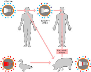
On the other hand, there are other infections that develop a full memory response, but it is not useful. Influenza is an example of this. Influenza has been around for thousands of years; some years we have severe epidemics or even world-wide pandemics. The 1918-1919 Spanish flu pandemic killed over 50 million people, many more than the contemporaneous WWI (16 million deaths).

Flu is difficult to vaccinate against because it keeps changing. Influenza virus has two antigens, called H (hemagglutinin) and N (neuraminidase). These are the molecules on the virus particle that your body mounts an immune response against.

The H molecule on the viral coat binds to sialic acid receptors on respiratory cells and allows the virus to enter. When the newly produced viruses bud off of the cell, they place H on the cell surface, but there are still host sialic acid receptors there as well. These receptors would bind up the H and prevent the new viral particles from attaching to and infecting other cells, so the N molecule cleaves the sialic acid receptors from the new viral particles.

Influenza virus can mutate by antigenic drift or antigenic
shift. The top line shows that by passing from person to
person, the antigens (and virulence) shift slightly. The lower
line shows that by passing through other animals and
recombining, the antigens can have small or big changes. When
shifted virus moves into humans, it’s a recipe for a pandemic.
Theproblem arises when the H and N antigens mutate.... and they do. Scientists have identified 16 different classes of H’s and 9 different N’s, and they can be paired up in many combinations. Small changes (antigenic drift) usually mean that memory might have a slight protective effect, and major epidemics do not occur. But major changes in H and N (antigenic shift) mean that previously infected people have no memory protection.

Different strains of influenza virus can infect the same animal (often pigs and ducks – thus avian flus and swine flus) and can mix their H’s and N’s. What emerges and might be transmitted to humans can be a virus with H’s and N’s similar to years past, or with new H’s or N’s. That is why a new vaccine must be produced each year, after scientists see which H’s and N’s the new virus has and how much they have drifted. Avian flu is H5N1, while swine flu is H1N1. However, antigenic drift means that each H1N1 will not be exactly like the previous H1N1 to emerge. The 1918 pandemic was caused by an antigenically shifted H1N1 sub-strain.

Like flu, other infections may not provide life-long memory. If the memory response is weak or the initial response was not strong, then memory may fade over time. This is why some vaccinations require boosters in later years. A fading of the memory response to influenza is also implicated in the need for yearly vaccinations.

Here's a great book that discusses both the biology
and sociology of influenza. There are great personal
stories as well as medical detective work. This
pandemic was a jolt that brought infectious
disease research into a new century. I highly
recommend it.
Now for the exception to the exception. Influenza changes each year, so memory does not help much, but a 2010 report from scientists in Hong Kong suggests that prior exposure to any seasonal influenza (either by infection or vaccination) might have been a contributing factor as to why the 2009 pandemic of antigenically shifted swine flu (H1N1) was much milder than expected.

The 2009 seasonal flu vaccine did not have any cross-reactivity with pandemic H1N1, so the scientists suggest that previous years seasonal influenzas did generate some memory response that was partially effective against 2009’s H1N1 swine flu. Cross-reactivity means that the H and N antigens were not identical to previous version; the Legos don’t fit together exactly, but they were similar enough to fit together and initiate a partial response. Once again, we see that getting sick may save your life down the line.

Next week will look at examples wherein having one disease can protect you from catching another.


Mathews, J., McBryde, E., McVernon, J., Pallaghy, P., & McCaw, J. (2010). Prior immunity helps to explain wave-like behaviour of pandemic influenza in 1918-9 BMC Infectious Diseases, 10 (1) DOI: 10.1186/1471-2334-10-128

Kash, J., Qi, L., Dugan, V., Jagger, B., Hrabal, R., Memoli, M., Morens, D., & Taubenberger, J. (2010). Prior infection with classical swine H1N1 influenza viruses is associated with protective immunity to the 2009 pandemic H1N1 virus Influenza and Other Respiratory Viruses, 4 (3), 121-127 DOI: 10.1111/j.1750-2659.2010.00132.x

Cowling, B., Ng, S., Ma, E., Cheng, C., Wai, W., Fang, V., Chan, K., Ip, D., Chiu, S., Peiris, J., & Leung, G. (2010). Protective Efficacy of Seasonal Influenza Vaccination against Seasonal and Pandemic Influenza Virus Infection during 2009 in Hong Kong Clinical Infectious Diseases, 51 (12), 1370-1379 DOI: 10.1086/657311


For more information or classroom activities, see:

innate immunity:

adaptive immunity:

memory immune response:

influenza virus:
http://www.xvivo.net/zirus-antivirotics-condensed/

Feelin' Hot Hot Hot!

$
0
0
Biology concepts –  fever, infectious disease, sexually transmitted disease, innate immune system

Would you be willing to be a human guinea pig, to
see if one disease might stop another? The term
“human guinea pig” refers to the fact that from the 1890’s
to the 1920’s guinea pigs were a major model for medical
research. Later replaced by rats and mice that could be
bred faster, guinea pigs were used to develop the first
diphtheria antitoxins, which subsequently saved
millions of lives.
Wouldyou be willing to let a doctor give you a disease? What if that might save you from another disease? You suppose this might be O.K., if the disease you were being given on purpose wasn’t too nasty.

What if the disease you're to be given as treatment is a form of the infection that kills a million people each year, the second most of any infection? Now you're thinking the disease you already have must be pretty horrible if this is the best idea for a cure. Let’s investigate and see if it might be worth it to save you from.... neurosyphilis.

Syphilis is a sexually transmitted disease that has distinct stages. The primary infection is marked by a lesion on those parts of your body that are most at play in the contracting of a sexually transmitted disease. It is amazing that some scientists believe that sexually transmitted diseases such as syphilis, in their primary stages, actually make sexual relations feel better. The organism (Treponema pallidum) benefits from this because the infected individuals might be more likely to have sex more often and this is an opportunity for the organism to be transmitted to additional hosts. Called “host manipulation” this is an evolutionary process that is just now gaining more attention, and will be something we will talk a lot more about in this blog in the near future.

The gumma lesions (left side) of secondary syphilis are not
meant for polite society. It's no wonder the Elizabethans
opted for the ruff collar (right side).
Thesecond stage of syphilis is marked by lesions, called gumma, on many parts of your body. You know those huge collars that the Elizabethans wore in 1500-1600 England? (see picture) As the story goes, the collars actually came into fashion as an attempt to keep syphilitic gummas out of sight. Syphilis ran roughshod through the English royal families at the time. Whatever the royals did everyone else wanted to do, so the collars became a fashion hit.

The tertiary (3rd) stage of syphilis is much more likely to be fatal. Appearing anywhere from 3-15 years after the primary lesion, tertiary syphilis attacks the brain, heart, liver, or bone tissues of the victim. Neurosyphilis can bring dementia, hallucinations, psychosis, as well as unsteady gait and movements (ataxia or paresis). While only a quarter of the patients reach this stage, it is a nasty way to go.

Do you agree that being purposefully infected with one disease to avoid the ravages of neurosyphilis might be worth considering? Even if the doctors were going to give you....... malaria?

This is the spirochete bacterium Treponema pallidum,
the causative organism of syphilis. Recent evidence
suggests that the bacterium is flatter and less like a
corkscrew than previously thought. They don’t look like
they have a flagella to move around, but they do. It is
located INSIDE the cell, which makes the whole cell
whip back and forth, not just the tail.
In the modern day, the treatment for syphilis is antibiotics; penicillin G can easily kill T. pallidum in the primary and secondary stages. However, antibiotics do not cross the blood brain barrier very easily (this barrier is made by very tight junctions between the cells and reduced movement of molecules through the cells, in order to protect your brain from toxins and infectious agents). Very high doses of drugs must be used to treat neurosyphilis. They may not work at all and might bring side effects.

But in the days before antibiotics, other treatments had to be sought. In the state of Indiana, USA, just as in all states and countries at the turn of the 20th century, syphilis was rampant in mental hospitals. This was both the cause and effect for some of the incarcerations, and was a source of constant battle in the institutions.

For better or worse, these patients were a stable population for the testing of different therapies for neurosyphilis, and Walter Bruetsch at the Central State Hospital in Indiana was a leading American researcher on the use of malaria to combat neurosyphilis.

Originally developed by Professor Julius Wagner-Jauregg of Vienna, Austria, the “malaria cure” was used to originally to treat paresis (very unsteady) and general paralysis patients; he suggested that fevers were helpful in paresis and tertiary syphilis.

Wagner-Jauregg had noted as early as 1887 that in the tropics, both malaria and syphilis were common, but those with syphilis rarely progressed to the tertiary stage, with the paresis that if often brought. In 1917, he treated nine paretic patients with good results, so other institutions expanded the study of this treatment. In Indiana, several decades of work were summarized in a series of papers in the 1940’s, making Indiana the prime American spot for “malaria cure work.”

The female Anopheles mosquito can take in quite a bit
of blood in just a short time. Take too long and they
could get squished.....but take hot blood in too fast
and they roast. That drop of fluid at the end of their
abdomen evaporates and helps cool their body as they
suck up the 37˚C blood, according to a 2011 study.
So how might malaria help in the treatment of syphilis? To discuss this, we have to know a few things about malaria. It is an infectious disease caused by an apicomplexan parasite called Plasmodium falciparum, although early hypotheses implicated bad air in the disease – hence the name; mal= bad, and airia = air. This organism has a complex life cycle, part of which occurs in the gut and salivary glands of the Anopheles mosquito and part of which occurs in human liver and then red blood cells (erythrocytes, RBCs).

There are five species of malaria parasites; P. falciparum is the one that causes the most severe disease. Other species include P. vivax  and P. malariae, which are dangerous but do not cause as many deaths. They are also the prevalent species outside of Africa.

The merozoite (meros= portion, and zoo = animal, so like half an animal) stage of the organism invades the RBC’s and reproduces asexually. Periodically the merozoites burst out of the depleted erythrocytes and look for new blood cells to infect. These periodic bursts are timed differently in the different species, from every 48 hours for P. falciparum, or every 36 hours for P. vivax. When they break the RBCs and escape into the bloodstream, an immune reaction is stimulated by the broken cells, including a very high fever, from 103-110˚F!

The fever itself may be lethal, but there other factors, such as the fact that infected cells have parasite proteins on their surface that makes them sticky. The infected RBC's don't pass through the entire circulation and can block circulation in the brain or spleen and cause other problems. So which part of the infection was helpful in tertiary syphilis?

The consensus idea was that the malarial fever killed the T. pallidum of syphilis. Microorganisms like to live inside us because we provide them with something they need, and they have evolved to live best at our temperature. A fever is one way your body tries to make you a bad host for the organism. A high fever, induced by malaria, would make you a very inhospitable host for T. pallidum, and could be lethal to the organism.

Think about this the next time you want to take an Advil or Tylenol for that low grade fever. By medicating yourself, you are preventing your body from using one of its natural defenses against infectious agents. But high fevers cause damage on their own, so declining an anti-febrile (anti-fever) drug when your temperature is 100˚F is much different that counting on your body alone when the fever is 105˚F and you're having convulsions.

Here is a macrophage (false color image) ingesting
bacteria. The macrophage is part of the innate
immune system, it can phagocytose (eat) many
different foreign invaders. One macrophage can
take up and destroy hundreds of bacteria. They
stick to tissue culture plates not because they are
sticky, but because they're trying to eat the plate!
Work done by Dr. Walter Bruetsch at Central State Hospital during the 1940’s questioned whether it was the high temperature of the fever that stimulated T. pallidum destruction. Artificial fevers were not as effective as malarial fever in treating neurosyphilis; Bruetsch suggested that malarial fever and the RBC destruction it brought stimulated innate immune macrophage activity, while artificial fever stimulated only adaptive immune lymphocytes and resulted in lowered Ab concentrations (called titers) at the same time, making the adaptive response less effective. Bruetsch concluded that it was the activation of the innate system that produced results in treating general paralysis and neurosyphilitic paresis. The obvious answer isn't always the complete answer.

In later years, antibiotics took over as the major treatment for syphilis, and only rarely does the infection progress to the tertiary stage. However, proponents of fever therapy have, over the years, suggested that malaria as a treatment could be used for a variety of infections, from lyme disease to HIV.

The primary cheerleader for using malaria to treat HIV infection was none other that Henry Heimlich, inventor of the Heimlich maneuver. In the late 1990’s and early 2000’s Heimlich carried out a series of highly questionablestudies on malaria fever in HIV infection. It is not altogether clear whether proper informed consent was used, and the results of the studies have been universally discounted. But that is not where HIV and malaria part company.

A schematic cartoon shows how HIV replicates. It
first attaches and uncoats. The RNA is reverse transcribed
and then transcribed and translated into protein.
When the new virus assembles itself, the coat proteins
have to be chopped up into usable pieces. This is the
job of the HIV protease. Protease inhibitors stop this
and prevent virus maturation.
It turns out that protease inhibitors used to treat HIV infection may be potent inhibitors of P. falciparum as well. HIV takes over a cell and forces it to produce the proteins and RNA to form new HIV particles. Many of the proteins must have portions cut off to make them functional; this is the job of the protease (prote= protein, and ase = cut). Protease inhibitors prevent this cleavage and therefore stop the formation of new viral particles.

It turns out that malaria parasites use proteases very similar to those of HIV, and preliminary studies indicate that these drugs can prevent reproduction of the organisms. As hard as it has been to come up with useful malaria drugs, here’s hoping that human studies are successful.

Finally, there is some speculation that malaria and HIV are linked. The dangerous P. falciparum was not used to induce fevers in syphilis patients; doctors used less virulent Plasmodium species, such as P. malariae or P. vivax. Charles Gilks, in a 2001 paper in Philosophical Transactions of the Royal Society, suggests that some primate strains of malaria were also used, wherein infected monkey blood was injected directly into the syphilis patients. Gilks wonders if this is where a simian immunodeficiency virus made the jump to mankind. I think that is an extremely long leap.

Next week let’s work the other side of the street; do some diseases keep you from getting malaria? Yes, and there are more than you might have guessed.


C. Gilks (2001). Man, monkeys, and malaria Philos Trans R Soc Lond B Biol Sci DOI: 10.1098/rstb.2001.0880


For more information and classroom activities, see:

Syphilis –


Malariotherapy in syphilis and other infectious diseases –


Malariotherapy in HIV –


Protease inhibitors-
http://www.thebody.com/content/art12606.html

Viva La Evolution

$
0
0
Biology concepts – evolution, reproductive advantage, natural selection, co-dominance, X-linked genes

Last week we learned how less aggressive strains of malaria were used to treat neurosyphilis and how they may be useful in treating HIV infection. This week, we will turn 180˚ and see if other diseases can help prevent or lessen the effects of malaria. In the process, much can be learned about natural selection and reproductive advantage.

Plasmodium-infected red blood cells develop knobs,
the surface protrusions seen on the left erythrocyte.
These knobs are covered in a certain protein that
inhibits the immune system’s ability to recognize this
cell as infected and respond to it. The cell on the right
is also infected with P. falciparum, but has a mutation
that prevents knob formation. Image credit: Ross
Waller and Alan Cowman.
As youundoubtedly remember from last week, malaria is a parasite-caused infectious disease that is transmitted from human to human by mosquitoes. The parasite, Plasmodium falciparum, takes up residence in the red blood cells (RBC) to reproduce. The red cells burst to release the organisms, and this brings fever and weakness.

As far back as the 15th and 16thcenturies, quinine, made from the bark of the cinchona tree, was being used in Peru to treat malaria. Chloroquine, mefloquine, and quinine all work against malaria in similar fashion. Because of their neutral pH, they move across membranes easily including the lysosomemembrane. Once inside the lysosome, they become charged and can’t get out. This includes the trophozoite-containing lysosomes. In the RBC, trophozoites consume hemoglobin to obtain amino acids, and the heme is digested in the lysosomes to form a black malaria pigment. The quinine drugs in the lysosome bind up the heme and produce a toxic product (cytotoxic heme) that kills the parasite.

There are other classes of drugs that are useful against P. falciparum. Primaquine and the artemisinin drug, artesunate, act by a completely different mechanism from that the quinine drugs. Artesunate is excellent for treating P. falciparum malaria, while primaquine is often used in conjunction with quinine to treat P. vivax or P. ovale forms of the disease.

These drugs work by breaking down – weird, but this is how many drugs work. It isn’t what you swallow that kills the organism, it's the metabolites (the products made by your biochemistry breaking down the drug) that are active. In the case of artesunate and primaquine, the heme molecule in the red blood cells releases peroxide from the parent compound (the drug you take). This is just like the peroxide you use to wipe out cut in order to prevent infection.

Artusenate comes from the sweet wormwood
plant. Chinese herbal medicine has used it for
thousands of years. A recipe for an Artemisia
based malaria medicine was found on a tablet
from the Han Dynasty (206 BCE to 20 CE). It is
now being investigated as a treatment for breast
cancer, also based on its ability to form radicals.
Oxygenis crucial for cellular function because it can gain electrons and can react with many other atoms. Unfortunately, this also makes it harmful to your cells as well. Without proper supervision, forms of oxygen that have picked up an extra electron or two (peroxide, superoxide, nitric oxide) can react with many important molecules in your cells and leave the cell impossibly damaged.

The cell has defenses against free radical damage, but higher than normal concentrations render the RBC fragile; on the tipping point of destruction. Treatment with primaquine or artesunate makes the cell inhospitable for the parasite, the red blood cells become flop houses instead of five star hotels. The parasite’s operating instructions are to survive and reproduce, but these drugs pull up the erythrocyte welcome mat and the parasite seeks moves on to seek friendlier accommodations.

Unfortunately, some strains of P. falciparum have become resistant to some quinine drugs, especially chloroquine. The free radical generating drugs are still useful, but scientists in Western Cambodia recently reported artesunate drug resistance there. The parasite has evolved – evolutionary pressure is everywhere. The actions of humans have put pressure on the organism to evolve; those parasites with mutations to resist the drugs have a reproductive advantage, and those mutations get passed on. We had better have something else on our plate to combat malaria – we're working on it, but nature has provided some help as well.

There are natural defenses against malaria. We have seen that a fragile red blood cell helps in preventing are lessening the disease course of malaria. What else might do that? This is where human genes come into play.

Sickle cell diseasecreates a very fragile RBC. The mutation is just a single DNA base change in the hemoglobin beta chain peptide, but the result is a hemoglobin molecule that becomes pointy and can tear the red blood cell apart, or can get stuck in small blood vessels and prevent good blood flow. Reduced blood flow starves the downstream tissues of oxygen.

You get one gene for hemoglobin beta chain from each parent. The disease comes when an individual receives mutated genes from both parents. But that doesn’t mean that sickle cell anemia is a recessive trait. If you have one copy of the mutated gene, then you will have sickling problems when oxygen concentrations are low, like during exercise or at high altitude.
Sickle cell disease or a sickle cell trait episode can result in red blood
cells clogging up vessels and organs. On the left is an absolutely
HUGE spleen from a sickle cell patient. On the right is a normal sized
spleen, about 20% the size of the injured spleen on the left. A normal 
spleen is about the size of your hand, maybe a little skinnier.

If sickle cell anemia was a recessive disease, then a single wild type (normal) gene would be dominant, and you would show no disease. Instead, sickle cell anemia is co-dominant, one mutated allele (copy of the gene) is like having half the disease; it only shows up in certain circumstances.

This can still be a pebble in your shoe, just ask Ryan Clark, the Pro-Bowl safety for the Pittsburgh Steelers. In a 2007 game in Denver (altitude 5300 ft, 1616 m), Ryan almost died from a sickling attack during the game, and ended up having his spleen and gall bladder removed (remember that sickled RBCs can clog blood vessels, especially in blood rich organs like the spleen).

When Pittsburgh next played Denver, Clark didn’t even make the trip. This just happened to be the 2011 playoff game in which Tim Tebow threw a long touchdown pass in overtime to the receiver being covered by Clark’s replacement. Sometimes disease can change how sports evolve as well.

Thalassemia is another example. This is a group of inherited disorders wherein there is reduced production of one of the subunits of hemoglobin (hemoglobin is made from 2 alpha and 2 beta subunits). Alpha-thalassemias have mutations in the alpha subunit; likewise for beta-thalassemia.

Reduced subunit number means reduced hemoglobin number; the blood won’t carry enough oxygen, and the patient is constantly oxygen-poor in his/her tissues. Having two mutated alpha genes is lethal in the very young (called hydrops fetalis), but you can live with one mutated alpha gene, one mutated beta gene, or even two mutated beta genes.

This the broad bean, or fava bean in opened pod
and out of the pod in a bowl. The ancient Greeks
used to vote with fava beans, a young white bean
meant yes, and old black one meant no.
Sickle cell trait (one mutated allele), and thalassemias result in fragile erythrocytes. This makes them poor hosts for malaria, and confer a resistance to the disease - bad genes aren’t bad in every case. And just for good measure here is another example.

Favism, better called glucose-6 phosphate dehydrogenase deficiency (G6PDH), is an X-linked genetic disease; the gene is on the X chromosome. A female (XX) has two copies, so having one mutant copy is no problem, but a male (XY) has only one, so getting a mutated copy from your mother means that you ONLY have the mutated gene – this brings the disease.

The enzyme G6PDH works in several pathways; in your red blood cells, it is the only source of reduced glutathione, an important antioxidant. This means that things that trigger free radical formation in your red blood cells will trigger the disease – lots of weakness and lack of energy. If there is enough erythrocyte destruction, one could die.

Triggers include broad beans (fava beans), hence the name favism. Other triggers include many drugs, including primaquine and artesunate, the anti-malaria drugs that induce free radicals. Having G6PDH-deficiency is like having your own artesunate pharmacy right in your cells - you naturally have higher oxygen radical levels in your RBCs, so the malarial parasite can't live there.

Not by accident, sickle cell mutation is more prevalent in people of Sub-Saharan African descent, thalassemia mutation is more common in people from the warm, moist Mediterranean, and G6PDH-deficiency is found most commonly in the Mediterranean and Southeast Asia. These just happen to be the areas where malaria-carrying mosquitoes are most abundant. Evolutionary biologists make the argument that natural selection has maintained these genes in the populations because they provide a reproductive advantage to the species.

Left image: dark green is where there is thalassemia and yellow and red are where there is sickle cell. Right image, light green is where there is favism, and inside the light blue outline is duffy antigen mutation. It is
interesting that these areas are also where malaria is endemic.


Youmight die from sickle cell disease, but probably not from sickle cell trait or beta-thalassemia. Learning not to eat fava beans makes the G6PDH mutation less lethal. One might very well live to an age where one could mate and pass on his/her genes. The diseases might still kill the patient, just not as soon as malaria would.

Malaria is a killer, and significantly, a killer of the young. In East Africa, children are bitten by the anopheles mosquito on average 50-80 times each month. They very well might not reach an age to reproduce. Therefore, having sickle cell trait, thalassemia, or favism provides a reproductive advantage in these environments and natural selection has resulted in these alleles remaining in the populations in these areas.

The Duffy antigen (DARC) is important for P. vivax
entrance into the red blood cell. The Duffy binding
protein (DBP) interacts with DARC, the yellow parts
of the DBP are variable, and can be used to bind an
antibody. These variable areas overlap the binding
site, and can be used to make a vaccine for P. vivax.
Evolution maintains some diseases in order to combat others. It isn’t by design, it's by biology; no big plan is involved, as exemplified by the Duffy antigen. All your cells have proteins on their surfaces. One, called DARC (Duffy Antigen Receptor for Chemokines, or Duffy antigen) helps your cells receive signals from your immune system. In those people with a specific single nucleotide polymorphism(SNP) for Duffy Ag, the antigen is not present on red blood cells (though it is still present on all other cells).

SinceP. vivax uses Duffy Ag as a way to enter the red blood cells, those with the Duffy SNP are resistant to P. vivax malaria – they don’t even have to suffer with some other disease; just a simple case of chance.  And the prepared mind exploits chance – the Duffy antigen binding protein is now a candidate for use as a P. vivax vaccine.

Next week, how the plague was defeated by a genetic disease.


Chootong P, Panichakul T, Permmongkol C, Barnes SJ, Udomsangpetch R, et al. (2012). Characterization of Inhibitory Anti-Duffy Binding Protein II Immunity: Approach to Plasmodium vivax Vaccine Development in Thailand. PLoS ONE , 7 (4) DOI: 10.1371/journal.pone.0035769

For more information or classroom activities, see:

Malaria –

sickle cell mutation –

thalassemia –

favism –

duffy antigen –

Ironing Out The Black Death

$
0
0
Biology concepts – iron, genetic disease, infectious disease, immune evasion

It is strange to think of people as rusting, but there are 
days when I get up and swear that my joints have 
frozen – my age makes me assume it is rust. 
In truth the molecules of rust are very much like 
some molecules in your body; too many of these in 
the wrong places, and maybe you are rusting.

Believeit or not, someone you know is rusting - and it probably saved his/her ancestor’s life.

Animals require iron to survive; normal adult humans carry about 3.5-4 grams of iron in their bodies. It’s vital for every cell. Red blood cells use iron as part of the hemoglobin molecule that carries oxygen, But all other cells use iron in part of electron transport chain that makes ATP, and in the synthesis of DNA.

In plants, iron is used in chlororphyll production, in nitrogen fixation, and in regulation of transpiration (moving water and nutrients up to the leaves). Plants are a decent source of dietary iron, but heme iron (from meat) is much more easily absorbed.

In both plants and animals, the amount of iron is highly regulated. Iron is most often bound to proteins; one type in cells, another in the blood, and they lock it up tight. When you need more, your gut cells (enterocytes) release some of their stored iron and then take in more from the food you eat.

People who absorb too little iron (from poor diet or absorption defects) have a hard time carrying oxygen to their tissues because they don’t have enough hemoglobin. They are fatigued, dizzy, lose their hair, and less able to fight off infections. Weirdly, they may demonstrate pagophagia; a compulsion to eat ice! The reason for this is open for discussion, but one hypothesis says there is an ancient crunching desire, related to chewing on bones to get at the iron-rich marrow.

Pagophagia (eating ice) is one type of pica. In pica, a
person craves to eat something that is not a food source.
Some people with pica will eat hair (trichophagia)
or dirt (geophagy). I guess if you have to have pica,
ice craving isn’t so bad. And yes, some people crave
plastic, like parts of your keyboard.

Too little iron keeps you sick - and apparently always refilling the ice tray. But too much iron is just as bad; both ends of the scale can kill you.

Hereditary hemochromatosis (HH) is an autosomal recessive (need two mutated copies) disease of iron storage and transport.  Patients with this disease may have as much as 20-40 grams of iron in their bodies; they can even set off metal detectors at airports!

All this iron causes medical problems too. People with HH will accumulate iron in their liver, heart, skin and other tissues. Excess iron plus fats can produce free radicals and oxygen radicals. The radicals can react with many molecules, including those you need in order to keep your cells functioning properly.

Radicals can break down enzymes, destroy mitochondria, and even react with the iron itself to produce iron oxide – rust; biological rust being called hemosiderin. Could HH patients be like the frozen Tin Man that Dorothy finds in the Wizard of Oz? Of course not, tin doesn’t rust – it’s a good thing L. Frank Baum was a writer and not a metallurgist!

The brown color is hemosiderin pigment that has been
deposited in the tissues.  Most times, your body will
resorb this colored material, like when a bruise goes
away over time. In hemochromatosis, there is too
much hemosiderin to be completely removed.

Over time, the damage from free radicals and from hemosiderin buildup causes systems to shut down. Without treatment HH is lethal - so it is important to know how all that iron gets there.

We said above that enterocytes are the storage area for iron absorbed from your diet. In HH, the export signal is broken and they keep dumping their stored iron into the bloodstream. Even worse, the enterocytes lose the ability to sense if the body needs more iron. As a result of HH, gut cells keep absorbing more iron and releasing it into the bloodstream.

It’s a bad thing to inherit hemochromatosis…..EXCEPT if Yersinia pestis is lurking in the environment. Y. pestis is the bacterium that causes the plague. The organism can be passed from person to person, but also from fleas to people, and from fleas to animals to people.

You can read about how Y. pestis ensures it is transmitted to a new host from the flea’s midgut, but for reasons of decorum, I won’t go into it here. And I suggest you don’t eat before you read about it.

Y. pestis plague comes in three flavors; septicemic(travels through the blood), bubonic(causing swellings), and pneumonic(some organisms go to the lungs). In the case of pneumonic plague, coughing promotes transmission from person to person and is more lethal. But bubonic plague is more painful.

The plague has been a killer throughout human history, but Y. pestis’ relationship to the flea is evolutionary rather new. About 20,000 years ago, Yersinia killed the flea as well. According to new research, it took relatively few genetic changes to allow plague bacteria to keep the flea alive and to survive in its midgut. It was at this point that humans' trouble really began. It is estimated that a third of the population of Europe was lost to plague in 14th century. The infection still occurs today, but is highly treatable with antibiotics. Your immune system has problems getting rid of Y. pestis on its own.

Normally, your immune system recognizes foreign organisms and eliminates them, through either innate or adaptive mechanisms. However, Y. pestis has several tricks up itsleeve to avoid recognition and destruction by your immune system.  

The lymphatic system is comprised of vessels, and
is considered part of your circulatory system. It
helps in eliminating wastes from the blood and
tissues, aids in absorbing fats and fat soluble
vitamins, and regulates fluid levels. A main function
is to move fluid and cells through the checkpoints,
the lymph nodes. Here, the fluid is checked for
foreign molecules and antigen presentation to the
immune cells in the nodes.

Immune cells can circulate in your blood, move in and out of your tissues, or may be located in your lymphatic system. In the lymph nodes, they gather to exchange information, like workers gossiping around the water cooler. If an antigen processing immune cell (APC) has encountered a foreign antigen, the APC will break it down and place pieces of the antigen on its surface, so the antigen can stimulate other immune cells.

The processed antigen is presented to the many types of immune cells in and moving through the lymph nodes, including B cells that make antibodies, and T cells that direct immune responses or directly kill organisms. This quickly increases an immune response; one cell encounters the invader, but by going to a central location (lymph node), thousands of cells can be stimulated.

Amazingly, Y. pestisactually lives and reproduces in your lymph nodes! The painful swellings in bubonic plague are the inflamed lymph nodes where the organism is reproducing. Each swollen node is called a buboe, hence the name of the plague. Buboes occur most commonly in the armpit (axilla), on the neck, or in the groin area – not a pleasant way to spend a weekend - maybe your last weekend.

The lymph nodes are the headquarters for stimulating immune responses, yet the Y. pestis lives here very happily. It manages this through several evasion mechanisms:

1)   antiphagocytic proteinsY. pestis can inject proteins into phagocytic cells that makes them poor at eating and killing. These proteins also makes immune cells unable to signal other immune cells that Y. pestis is there.
2)   invasion proteins – plague bacteria can avoid immune detection by living insideseveral different host cell types; the macrophage is the major example.
3)   survival proteinsY. pestis  can live inside the macrophages that are supposed to destroy them by turning off macrophage killing mechanisms.
4)   heme stealing proteinsY. pestis can steal iron from the host. And here is where HH comes in.

Here is a buboe on a plague patient’s neck. It is not unlike the parotid 
salivary gland swelling that takes place during the mumps, just
bigger, more painful, and more lethal. I chose to show one from the
neck precisely because I didn’t want to show you one from the groin.

Hereis an organism that is perfectly happy living inside and in the company of the cells that are supposed to kill it - we’re doomed. Yet having a disease like hemochromatosis can save us. How can that be? Well, microorganisms need iron too. For much the same reasons as animals and plants, bacteria and other microorganisms must have a supply of iron. They may get it from their diet, or, as is the case with Y. pestis, they steal it from their host.

I can hear what you're saying - this doesn’t seem to make sense since HH results in lots of iron in cells. True, but there is an exception. HH leaves two cell types starved for iron - the enterocyte, which we already know about, and the macrophage. The reason for iron-poor macrophages during hemochromatosis is not completely understood, but one possibility is that the HH mutation affects macrophages the same way it affects enterocytes.

One important function of macrophages is to eat and destroy old host cells, including erythrocytes. The iron of the hemoglobin from all those degraded RBC’s is stored and recycled; this is an important mechanism that the body uses to reuse the iron it already has. But in HH, the macrophages may be pumping out the iron they take up from old RBCs, just as the enterocytes keep pumping out the iron they take up from the gut contents.

The iron-poor macrophage essentially starves the intracellular plague bacteria by not providing them with iron. This is a happy accident for us, but it isn’t as if the macrophage doesn’t already know this trick. Iron can be an important immune weapon. In mycobacterial infections (that cause pneumonia), macrophages actually raise the iron concentration in the ingested bacteria and kill them that way. In other infections, macrophages sequester their iron and starve the organisms.

Bloodletting is an old time treatment for nearly every
disease. They thought that disease was caused by too
much blood. Strange, but bleeding (phlebotomy) is now
the accepted treatment for hemochromatosis. Leeches
are now used as anti-clotting mechanisms, and fly
maggots are used to clean out dead tissue – all are
gross, and all are effective!

Macrophageiron manipulation is not a natural immune response to Y. pestis, but HH helps to bring about the same effect, and this makes HH valuable. It is believed that many survivors of the plague in the 12ththrough 15th centuries had hemochromatosis. What is more, the gene is present in as many as 1/3 of living people of European descent, meaning that HH is probably massively underdiagnosed. It is likely that you know someone with HH, whether they not it or not.

Natural selection kept this mutation in the gene pool because it presented a reproductive advantage in times of plague. With antibiotics, we probably do not need this mutation any longer, but it is here and will take quite a while to be bred out of the population, especially since HH treatments (like bleeding, see the picture at right) help people live with the disease long enough to pass on their genes.

There are more examples of bad genes saving us from disease, like chemokine receptor mutations preventing HIV infection and aldehyde dehydrogenase mutations discouraging alcoholism. But next week we will focus on immune systems run amok and how parasites can reel them in.

Chouikha I, Hinnebusch BJ. (2012). Yersinia-flea interactions and the evolution of the arthropod-borne transmission route of plague. Curr Opin Microbiol. DOI: 10.1016/j.mib.2012.02.003

For more information or classroom activities, see Survival of the Sickest, by Dr. Sharon Moalem, or the following sites:

Iron in biochemistry –

Hereditary hemochromatosis –

Y. pestis plague –

Immune evasion strategies –
http://www.genengnews.com/gen-news-highlights/researchers-discover-how-some-pathogens-evade-the-immune-system/81243811/

Don’t Be So Sensitive!

$
0
0
Biology Concepts – immune hypersensitivity, allergy, autoimmune disease

Stone Mountain in Georgia is one big hunk of
granite. There is a bas-relief carving of Jefferson
Davis, Robert E. Lee, and Stonewall Jackson that
covers three full acres of space! Stonewall Jackson
is on the far right. Stonewall immortalized on a
stone wall, interesting…. but didn’t their side lose?

C.S. Lewis was a 20th century British writer who penned the Chronicles of Narnia books. Thomas “Stonewall” Jackson was a brilliant general in the Army of the Confederacy during the American Civil War. Can you name something these men had in common, but wish they didn’t?

---– They were both shot by their own troops during battle. It wasn’t on purpose; Lewis was wounded by a British shell that didn’t have enough oomph to get over the British’s own lines during World War I. One piece of metal lodged deep in his chest and could not be safely removed. It remained near to his heart until 1944.

During the Battle of Chancellorsville in 1863, Stonewall Jackson led a night reconnaissance mission that was mistaken for Union scouts. A confederate patrol fired on Jackson as he looked over the Northern lines from horseback. His left arm was amputated in an effort to save his life, but he died of pneumonia eight days later.

These were incidents of “friendly fire,” in which people meant to help you fend off the enemy end up hurting you. Too often, incidents of friendly fire take place in your body as well. In biology, these are called immune injuries and they can be dangerous exceptions. The immune system is designed to help the body fight off foreign invaders and dangerous molecules, but there are those instances when its actions harm the host.

Allergies are a good example of immune reactions gone wrong. Originally (1906) meant to denote any immune injury, we now we look at allergic reactions as immune responses to non-pathogenic, and in many cases, non-harmful antigens. Who could be harmed by a peanut, except for those allergic to it.

Peanut allergy is nothing to take lightly. It is
estimated that 3 million people now react to
peanuts, even to foods prepared in kitchens
where there are peanuts. This is an immediate
anaphylaxis response, with inflammation and
often respiratory distress.
A person can have an allergic reaction to an aeroallergen– something carried in the air, like dust, pollen, or pet dander. Food allergies occur generally with milk, wheat, peanut, or egg; many of these dissipate as children mature. Drug allergies can develop when small molecule pharmaceuticals break down, combine with host proteins or cells, and are then recognized as foreign. Some people are allergic to some venoms, like bee venom; their reactions can go beyond the pain of the sting.

In allergic reactions (atopic reactions – atopy is from Greek for “out of place”), there is first a sensitizing dose, wherein your body develops a hypersensitivity to the allergen. This is when your body builds an immunologic memory for the antigen, like we talked about a few weeks ago. Any exposure to the allergen after this brings a stronger response.

The exception to this sensitizing dose idea is when a new allergen looks like another allergen, ie. cross reactivity. Many latex allergies do not seem to have a sensitizing dose, but the patients also happen to have an allergy to banana, kiwi, or avocado. This is called the latex-fruit syndrome…catchy name, isn’t it?

Allergic reactions can occur just where the allergen contacts the immune system, like itchy hives (urticaria) for contact dermatitis, or a runny nose for pollens grains that are breathed in. Sometimes the hypersensitivity goes further and there is a life threatening reaction. We should describe the different kinds of hypersensitivity so you can diagnose your friends at parties.

Type I hypersensitivity is an immediate reaction, with symptoms lasting for a short time. Sometimes there is a more chronic response, especially if the antigen sticks around. Type I reactions are the allergies we all know and hate. The term for the reaction is scary, “anaphylaxis” (ana = exceedingly, and phylaxis = guarding), but it isn’t always life threatening.

In type I hypersensitivity, the allergen is recognized by specific IgE antibodies. Antibodies come in several flavors, including IgG (circulating antibody), IgM (antibody as cell receptors for first encounters), and IgA (in saliva and tears, etc.). IgE immunoglobulins are present in the tissues or on the surface of certain immune cells from some previous, sensitizing dose. The antibody has a variable end that recognizes the antigen and a constant end (Fc) which is recognized by other immune cells. When two or more IgE antibodies bind to the antigen (called crosslinking) and the Fc portion attaches to a mast cell or basophil, these immune cells will release their contents.

On the left is an electron micrograph of a mast cell,
an innate immune cell that mediates allergic responses.
On the right, you can see the granules inside the mast
cell that contain histamine, bradykinin, and other mediators.
When IgE and an antigen crosslink on the surface of the
cell, the granules release their contents into the
extracellular space.
Mast cells contain histamine, which causes blood vessels to dilate, airway smooth muscle to contract, itching, and stomach acid secretion. Mast cells also have bradykinin that increases mucous production, as well as other chemicals. Mast cell degranulation (release of internal granules containing the histamine, etc.) makes your eyes water, your skin get hot and itch, makes it harder for you to breathe, and might produce hives on your skin.

The reaction might remain local, but if it triggers the same reaction throughout your circulatory system, it can cause anaphylactic shock, a true medical emergency characterized by low blood pressure and respiratory difficulty. It can and will kill you if not treated immediately. And all this because some innocuous small molecule and an IgE antibody caused your immune system to over react!

Type II hypersensitivity reactions are also mediated by antibodies (IgM or IgG type). The triggering antigen might be some foreign molecule bound to a host cell or even an antigen on your own cells that your body has mistaken for foreign. In the case of penicillin allergy, the drug becomes bound to your cells; this complex triggers the immune response. If the antibodies are directed toward your cells or mistake your cells as foreign, this is called an autoimmune reaction. Examples could be systemic lupus erythematosus (SLE), some type I diabetes, or Hashimoto’s thyroiditis.

In some type II reactions, the antibodies that bind to the antigens trigger the complement system in your tissues to activate. Complement is part of your innate immune system that ends up marking cells for destruction by phagocytosis, or destroys them itself by punching holes in the target cells. In some cases, the antibodies bound to the cell trigger innate immune cells called natural killer lymphocytes– you can guess what they do to the target cell. I guess everyone is a natural born killer on the inside.

Natural killer cells are lymphocytes, but are part
of the innate immune system. These two are
attacking a cancer cell (red). Natural killers
specialize in killing cancer cells and virus-infected
cells. Natural killers are unique in that they can
recognize stressed cells in the absence of binding
antibodies.
The last type of immediate hypersensitivity is type III. The danger of this type of reaction comes from masses of antigens surrounded by antibodies. When these immune complexes (also called Ag-Ab complexes) become large, they can get stuck in tight places and bring an inflammatory response. Examples of immune complex diseases are autoimmune rheumatoid arthritis, some types of glomerulonephritis (inflammation of the filtering units of the kidney), and SLE also triggers this response.

Type IV hypersensitivity is the exception; this response can take several hours to develop and is the only hypersensitivity reaction that does not involve antibodies. Lymphocytes of the adaptive immune system interact with the antigen (be it foreign or domestic) and release many chemical mediators, called cytokines, that mediate immune and inflammatory reactions. Allergic contact dermatitis from poison ivy is a common, but relatively benign, example of this type of hypersensitivity.

Most hypersensitivities are reactions to things that shouldn’t have been problems in the first place. Allergies are just the most common manifestation of immune hypersensitivity. I don’t have them to any degree, but I see the havoc they wreak on my wife and our son. He is so allergic to wool that he breaks out when he counts sheep in bed!

But even allergies might have a hidden benefit. A study in 2008 indicated that people with allergies actually have a 25% less chance of developing a certain type of immune cell cancer, called B-cell non-Hodgkin’s lymphoma (NHL). If that person has three different allergies, they are 40% less likely to develop NHL.

This seems amazing, but it is supported by a 2011 study showing that people with allergies are 25% less likely to develop a type of brain tumor called a glioma. Glial cells protect and support the neurons in the brain; abnormal growth of these cells can lead to pressure and death of brain cells. Still think allergies are annoying?

Sneezes leave your mouth at over 100 miles and
hour and can spread droplets over 30 feet. Sneezes
may help get ride of unwanted antigens, but other
people don’t want them either, so cover your mouth.
Sneezing into the crook of your elbow is best for
limiting spray and contamination – I saw it on
MythBusters.
Researchersdon’t know the reason for this benefit yet, but hypotheses include that allergic reactions (watery eyes, sneezing, runny nose) help to eliminate potentially carcinogenic pollutants from our bodies, or that allergies stimulate the immune system and make it better at detecting and destroying cancer cells.

Learning that allergies might prevent cancer may make you less likely to take that antihistamine capsule. In fact, the treatment for all immune hypersensitivity reactions involve avoiding the molecule, removing the offending antigen and antibodies, and/or suppressing the immune system. We take corticosteroids, antihistamines, and other drugs to prevent the actions that might be saving us from cancer. However, you can help protect yourself without drugs as well—just catch a parasitic infection.

Parasitic worm infections, whip worm (Trichuris trichiura) or schistosoma for example, have a tendency to dampen the immune response, and can prevent some relapses in autoimmune diseases such as multiple sclerosis. A 2005 study indicates that some success has been had after dosing Crohn’s disease patient’s with intestinal worms.

Meet Pediculus humanus capitis, the common head
louse magnified only 80x. It is an ectoparasite,
meaning it lives on the host, not in the host. They
have been around for a long time; they have been
found on Egyptian mummies. This is why most
Egyptians shaved their heads and wore wigs.
For those of us without life-threatening autoimmune disorders, a 2009 study suggests that Pediculus humanus capitis infestations (head lice) can dampen the immune system enough to prevent allergies and some asthma attacks. Your choice - but don’t let anyone borrow your comb!

Parasites seem to have evolved specific mechanisms that inhibit the reactions that would eliminate them from the host, so they dampen immune responses as a defense. The mechanisms have not been worked out and may be parasite specific. Even malarial and leishmaniasis parasites can suppress the immune response, but I don’t recommend that you contract a deadly infection just to alleviate your allergies.

These last studies suggest that we may be living too cleanly – let’s take a look at that next week.

Calboli FC, Cox DG, Buring JE, Gaziano JM, Ma J, Stampfer M, Willett WC, Tworoger SS, Hunter DJ, Camargo CA Jr, Michaud DS. (2011). Prediagnostic plasma IgE levels and risk of adult glioma in four prospective cohort studies. J Natl Cancer Inst. DOI: 10.1093/jnci/djr361

Joseph A Jackson, Ida M Friberg, Luke Bolch, Ann Lowe, Catriona Ralli, Philip D Harris, Jerzy M Behnke, Janette E Bradley (2009). Immunomodulatory parasites and toll-like receptor-mediated tumour necrosis factor alpha responsiveness in wild mammals BMC Biology DOI: 10.1186/1741-7007-7-16

For more information or classroom activities, see:

Allergy –

Immune hypersensitivity –

Autoimmune diseases –

The Dirt On Staying Healthy

$
0
0
Biology concepts - hygiene hypothesis, immune regulation, bacterial drug resistance

Christian Slater starred in a 2008 American TV
series called, “My Own Worst Enemy.” Slater was
a secret agent with a chip in his brain that allowed
his employers to turn him from a mild mannered
family man to a super spy without each knowing
of the other’s existence. The showed last only nine
episodes; apparently the drama was its own
worst enemy.
Is everyone their own worst enemy? Google it---- apparently scientists are their own worst enemy; Christians are too. Chad Johnson is, and so was Whitney Houston. Someone out there even thinks bassists are their own worst enemies! I think that if this is true, everyone must be leading pretty lucky lives; the only thing stopping us appears to be us.

So it comes as no surprise that some scientists believe that people are their own worst enemies when it comes to protecting their health. Good intentions can have bad results. How might this relate to our topic of the past few weeks, the benefits of disease and infection?

Some diseases have had a positive effect on survival in specific conditions (like hemochromatosis and plague) and even how malarial fever can kill bacteria. This goes against the popular idea that less disease is better, and that whatever we do to kill infectious organisms is good. We try to be as sterile as possible; just look at what surgeons do before entering the operating room. The health industry has given us antibacterial soaps, cleaning products, plastics, cosmetics, toothpastes, pencils, and even antibacterial computer keyboards!

The majority of these products use triclosan as the active ingredient. First introduced as a pesticide in 1972, triclosan (chemical name: 2,4,4’-trichloro-2’-hydroxydiphenyl ether) is an antibacterial and antifungal agent. Triclosan’s mechanism of action at low concentrations is to disrupt fatty acid synthesis as a bacteriostatic agent (slows bacterial growth and reproduction); at high levels it can disrupt membranes and act as a biocidal agent (kills organisms).

Just because something is an antibiotic, it doesn’t mean it kills 
bacteria. Many of the common antibiotics we use are bacteriostatic, 
meaning that they inhibit the growth. This allows our immune 
system time to overcome the intruder on its own. 
Bactericidal agents do actually kill the bug, but they still need 
help from the immune system. If you took enough to 
kill all the bacteria, you’d need a capsule the size of a bus!
Triclosancan control bacterial contamination on hands and skin; hospital staff are encouraged to bathe or shower in triclosan solutions to prevent the spread of MRSA (pronounced “mersa” – methicillin resistant Staphylococcus aureus) in hospital wards. However, this is for control of contamination, not necessarily infection.

Triclosan has been proven effective in reducing infections rates only in cases of gingivitis (inflammation of the gums). However, a 2009 study stated that 75% of Americans over the age of six years have detectable levels of triclosan in their urine. This is significant since there is emerging data that suggests that triclosan might be harmful to people’s health.

High triclosan levels in urine and the environment mean high levels around microorganisms as well. But this shouldn’t be bad – it is supposed to kill germs, isn’t it? Many scientists worry that high triclosan levels also promotes bacterial evolution, selecting for the mutants that are resistant to the chemical. We all have good reason to worry about this because it’s happened before. Many bacteria, from MRSA to Mycobacterium tuberculosis, to vancomycin-resistant enterococcus, are wreaking havoc because we have fewer drugs that are effective against them.

In the laboratory, triclosan exposure has resulted in resistant strains of E. coli, salmonella, and rhodospirillium, and other organisms. Industry scientists argue that there is no data that triclosan causes resistance to develop in the wild, but a 2011 EU report suggests that this very well may be taking place; the levels of triclosan seen in people and the environment are similar to the levels used to drive resistance in the laboratory.

The bacterial resistance mechanism at work might be more dangerous than the resistance to triclosan itself. Several studies have deduced that triclosan interacts with proteins in the bacterial multidrug efflux pump. Many prokaryotes have this system; it works to pump non-bacterial small molecules, including antibiotics and toxins, out of the cell.

This cartoon represents a model of the E. coli
multidrug efflux pump. Protons pumped out are
allowed back in, and this produces the force needed
to pump out the drugs. This is another reason that
you need your immune system to overcome a
bacterial infection – the little buggers are working
against you!
In a situation where an organism is exposed to low or medium levels of triclosan, the multidrug efflux pump actually becomes more active because the triclosan binds to, and suppresses, the pump’s off switch. Think about that - you’re taking an antibiotic for a respiratory infection. But your household products are contaminating your body with triclosan. As a result, the respiratory organism is very efficiently expelling the antibiotics you are taking! Now the bacteria are being exposed to lower levels of antibiotic and will have a better shot at developing resistance! Perhaps anti-bacterials aren’t such a great idea.

Want more evidence? An August 2012 study showed that triclosan has an immediate and dangerous affect on muscle activity. You remember your heart?- it’s a muscle. In mice, triclosan exposure caused a 25% reduction in cardiac muscle function, and an 18% reduction in mouse grip strength. An idea for your next arm wrestling contest – wear a glove and slather it with liquid hand soap. You now have an 18% better chance at winning….if you are competing against a mouse.

Triclosan also affects endocrine function. A new study indicates that triclosan exposure in pregnant rats lowers mother, fetal, and neonatal levels of thyroid hormone. Triclosan has a structure similar to a thyroid hormone; it may trick the body into believing it has enough hormone. The thyroid would then reduce the production of the hormone, leaving the system starved of thyroid hormone. Most of this work has been done in amphibians, fish and rats, but a similar affect on human thyroid function is predicted.

Your body is exposed to many antigens from many sources.
If you are an only child or have parents that microwave
your toys, you are exposed to many fewer antigens. Many
scientists hypothesize that your immune system needs
these exposures to balance your developing system
between the Th1 responses and Th2 responses. Too much
Th2 and you will start to overreact to innocuous antigens –
allergies, asthma, and autoimmunity can result.
Antibacterial agents might be harmful through their actions on us and on bacteria. But does being too clean have other effects? Consider the hygiene hypothesis; mounting evidence indicates that efforts to produce near-sterile living environment, or even the movement from a rural to an urban environment, can negatively affect our health.

Case in point - most everyone has an idea that food allergies and asthma seem to be on the rise. The CDC stated in 2008 that there had been a 20% increase in food allergies in the years between 1997 and 2007. In a large number of these cases, children with food allergies also had eczema or skin allergies (27%) or respiratory allergies (30%), compared to only 8-9% of kids without food allergies. Basically, allergies are significantly on the rise, and if you have one, you are much more likely to have more than one.

Importantly, the rise isn’t occurring everywhere. Rural Africa - no increase in allergies or asthma. The arctic inuit peoples – very little allergy or asthma despite high levels of childhood smoking. Farm kids in just about every country – far lower levels of respiratory allergies, food allergies, asthma, and autoimmune diseases.

The hygiene hypothesis states that a lack of immune stimulation when young leads to exuberant responses to antigens that would normally be innocuous. Isn’t it interesting that the increase in allergies and asthma also correlates with the onset of antimicrobial agents being added to everything?

Different ideas abound as to how being clean might lead to increased immune hypersensitivities. One hypothesis is that a lack of antigen exposure in urban kids leads to a loss of balance between different T lymphocyte responses (see picture above). Infections tend to stimulate Th1 responses. A too clean, urban environment results in less stimulation of Th1 and therefore a relative over stimulation of the Th2 response. Increased Th2 leads to the kinds of responses seen in asthma and allergies. Indeed, atopic (allergy) patients do show an increase in Th2-driven cytokines.

Are we too clean as a society? Maybe we can back off
on the antimicrobial agents and spend more time in
the woods and the park. A brisk hike is as good for
your health as a spotless bathtub – and its more fun.
Then again, increased immune hypersensitivity in at risk populations might be due to an imbalance between the innate and adaptive immune systems. Many of the microbiologic antigens to which neonates and children need exposure stimulate innate immune receptors. The innate immune system then stimulates the adaptive immune system and balances the Th1 and Th2 responses. An absence of innate immune stimulation leaves the adaptive system to its own devices, and Th2 will often win this battle.

Additionally, the exposure to bacteria, viruses and parasites stimulates the immune regulatory system as well. Antigen presentation can be stimulatory or suppressive; suppressive presentation leads to regulatory (suppressive) lymphocyte production. It is hypothesized that regulatory lymphocytes help to balance the Th1 and Th2 responses and reduce the incidence of allergy.

We see that several portions of the immune system could be involved in helping the natural environment fine tune our immune responses. But what is it that induces this wonderful balance and state of good health?

A 2010 study suggested that the important molecule is something called arabinogalactan. This is a ubiquitous polysaccharide made of arabinose and galactose monomers. It is a component of many cell walls – bacterial, parasite, worm, grasses and other plants, and is in farm (unprocessed) milk.

Arabinogalactan is present in cow’s milk, in the grasses
that cows are fed, and in the dung patties that they leave
behind. And they last as well – there is a cowshed in
Wales that dates to 1402, making it the oldest building
in Wales. Cowshed – uninterrupted immune stimulation
for six centuries!
The hygiene hypothesis can be expanded to test the idea that farm kids' exposure to farm milk and cowshed dust (big sources of arabinogalactan) stems allergy and asthma development. However, there are studies that do not support the hygiene hypothesis, such as influenza virus actually promoting the development of asthma and the fact that daycare children have more respiratory infections, but do not have lower incidence of allergy. More needs to be known before we start shipping our infants to the country for the summer.

Two final notes to bring this full circle. Triclosan use has now been linked to higher rates of allergy. In particular, urinary triclosan levels correlate with development of food allergy. Correlation does not equal cause and effect, but it does ask a question that needs to be answered.

Lastly, increases in autism parallel increases in asthma and allergy, and a recent study shows that kids with autism and behavioral fluctuations have less stimulation of regulatory immune response after infection. Like allergy and asthma, autism definitely has a genetic component, but could the hygiene hypothesis and autism be linked as well?      

With Halloween approaching, let's take a three week break from our "disease benefits" stories to look at the biology of some of our Halloween traditions and myths.


Gennady Cherednichenkoa, Rui Zhanga, Roger A. Bannisterb,Valeriy Timofeyevc, Ning Lic, Erika B. Fritscha, Wei Fenga, Genaro C. Barrientosa, Nils H. Schebbd, Bruce D. Hammockd, Kurt G. Beame, Nipavan Chiamvimonvatc, and Isaac N. Pessaha (2012). Triclosan impairs excitation–contraction coupling and Ca2+ dynamics in striated muscle PNAS DOI: 10.1073/pnas.1211314109

For more information or classroom activities, see:

Anti-microbial products –

Triclosan and health –

Hygiene hypothesis –

Immune To Evolution

$
0
0
Biology concepts – innate immunity, adaptive immunity, defense mechanisms, endotoxin

The Jardin des Tuileries is the setting for the
final scene of “The Happening.“ Located in
Paris between the Louvre and the Palace de
la Concorde, this garden was once a royal
promenade, but became public after the
revolution. The trees that line the walk are
chestnuts. Several species of Chestnut are
pollen sterile, meaning they don’t produce
pollen and must be cross pollinated from a
species that has pollen.
M. Night Shyamalan likes to make movies that have “hide in plain sight” twists: the psychologist is a ghost (The Sixth Sense); the villagers live in modern times (The Village); the mentor is the arch-villain (Unbreakable). In his movie, “The Happening,” mankind is under attack. Something is making us commit suicide in mass numbers. What is attacking us – or might something be defending itself from humans? If it is defensive, could be considered an immune response? If yes, then can we figure it out by deciding just who has immune responses?

Immune systems of defense can be very evolved, as in humans. Ours make use of two specific circulatory systems (blood and lymph), has organs designed to aid their generation and functions (lymph nodes, thymus, bone marrow, and spleen), and has mobile cells designed only to patrol and protect. These components function in both innate and adaptive immune cascades and webs.

Other organisms’ defense systems are not so intricately developed, but still deserve respect. Arthropods (insects, crustaceans and the like) have a highly developed innate immune system, with circulating immune cells of several types.

Mollusks (clams, octopods, and the like) also have an innate immune system with a few types of circulating immune cells. However, immune responses don’t have to be only from circulating cells. Sometimes they are proteins that kill bacteria, or merely surrounding the pathogen and keep it from the host cells. Many kinds of mollusks protect themselves by encapsulating invading parasites in a solid prison of shell-like material -we call them pearls! Any mollusk with a shell can make pearls – even snails.

Conch is a species of giant snail. It produces lovely
pearls, so pearls don’t just come from oysters. Any
shelled mollusk will react to a parasite that gets
through its shell by walling it off in layers of mother
of pearl (nacre). This is the very smooth material
that covers the inside of the shell.
Everyanimal has some sort of immune response built into its physiology, but supposedly only vertebrates have an adaptive immune system. Invertebrates have the older, innate system, but not the ability to adjust their recognition and response to particular pathogens like the adaptive system can. The specific, or adaptive immune system was believed to have arisen in the first of the jawed fishes (gnathostomes; gnath = jaw, and stoma = mouth), about 410 million years ago and been handed down and modified by mammals. But there are exceptions – there are always exceptions.

The Agnathans (jawless fishes, such as lampreys and hagfish) seem to have an adaptive system all their own. It has features similar to the adaptive system of jawed vertebrates, but the way that foreign antigens are recognized is completely different. The lampreys and similar organisms use a different kind of receptor molecule on immune cells. The receptors are variable, but not in the same manner as mammalian immunoglobulins. In jawed vertebrates, the antibody genes rearrange to form the basis of both circulating and receptor immunoglobulins.

This "similar but different" adaptive system would indicate that specific immune responses have sprung up at least twice in evolutionary history. I say at least twice because it is beginning to look like insects and worms may have a sort of adaptive system as well. Earthworms will reject grafts from other earthworms, and will reject a second graft faster than the first graft. So, we see that most organisms have elaborate ways to defend themselves.

This brings us back to “The Happening,” and the attack on the humans ---– it turns out that it was the trees trying to protect themselves from being overrun by mankind! Plants have defenses? Plants can sense attack and respond? Yep.

Plants don’t have immune cells, those that move around and whose job it is to protect and attack. But they do have immune defenses against pathogens, and pretty sophisticated ones at that.

This is a cartoon which shows plant immune response. First
a pathogen tries to gain entry and the plant recognizes its
surface molecules (PTI). Some pathogens survive the response
and emit effectors (ETS, effector-triggered susceptibility). The
effectors trigger ETI which increases the response proteins.
Some pathogens may survive and too much ETI and ETS
triggers the hypersensitive response. Image: Nature
444:323-329, 2006.
PlantPTI (Pattern Triggered Immunity) is similar to our innate immune system, just without the specific immune cells. In this system, plants recognize molecules that are common to microbes (MAMPS, microbe associated molecular patterns) using pattern recognizing receptors (PRRs).

This is similar to mammalian PRR systems for PAMPs (pathogen associated molecular patterns), the toll-like receptors for example. When triggered, resistance molecules and plant hormones are released to make the plant less appealing to the pathogen, or to interrupt the infection process. There are many of these resistance mechanisms, we can talk about a couple below and more in the future.

On the other hand, plant ETI (Effector Triggered Immunity) is signaled by the effector molecules released by the microbes that manage to set up shop inside plant cells or tissue. ETI is really just an increase in the amplitude of the same response molecules seen in PTI, plus another defense mechanism, called the hypersensitive response.

Some pathogens like the hypersensitive response.
Necrotrophic (necro = death, and troph= loving) fungi,
like Botrytis cinerea, or gray mold (the spots on the
leaves), must have dead tissue. They wait until some
thing else triggers the hypersensitive response, or they
trigger it themselves, and then feed of the dead plant tissue.
When a pathogen is successful at making entry into a plant at a specific site, the plant may respond by releasing oxygen and nitrogen radical compounds (those with free electrons that will attack dang near anything). This will kill the plant cell as well as the invader (hence the name “hypersensitive”), but it reduces the probability of infection by taking out everything in the area. It is a sacrifice of host cells that the plant is willing to make.

This response is much like the apoptosis (programmed cell death) that virally-infected animal cells may initiate. It is a small loss in order to protect the whole organism. Recent evidencesuggests a central role for S-nitrothiols (nitric oxide linking cysteines) in both turning on and limiting the hypersensitive response by controlling the amount of NADPH oxidase, an enzyme that produces reactive species. We will see next week that this suicide mechanism is very old.

Reactive species for cell suicide is cute, but plant responses get even cuter. When threatened by some herbivorous insects, 2012 research shows that plants can call in mercenaries to help. Members of the cabbage family are troubled by the larvae (caterpillars) of the large cabbage butterfly (Pieris brassicae). When this butterfly lays its eggs on a black mustard plant, the plant sends out a chemical signal that attracts two species of wasps (Trichogramma brassicae and Cotesia glomerata).

When the male cabbage butterfly fertilizes the female
and she lays her eggs on a brussel sprout plant, the
chemicals from the male semen will trigger the plant to
make a pheromone that attracts the Trichogramma
brassicae wasp. It lays its eggs INSIDE the butterfly
eggs (yellow cones) and they feed off the butterfly eggs
as larvae. Up to 50 wasps can come out of one butterfly
egg. Image:Nina E. Fatouros.
These wasps are natural enemies of the white cabbage butterfly and will attack its eggs and larvae. Voila, the plant stops the white cabbage caterpillar from eating its leaves even before the attack begins. Most amazing, the chemical signal isn’t triggered by other, less ravenous pests, so it is a specific response.  That smells like an adaptive immune response to me. While many animals can’t specify a distinct response to a particular foreign organism, it looks like many plants can. Once again, plants show us how advanced they are.

Even more in support of the idea that plants have a form of adaptive response is the discovery that they have an immune memory of sorts. In 2009, researchers at the U. of Chicago found that when attacked by a certain bacterium, Arabidopsis plants (of the mustard family, a very common plant in research) make a chemical at the site of attack called azelaic acid.

The scientists found that this compound can stimulate a faster and stronger immune response when and if the plant was ever attacked again. Azelaic acid acts by stimulating salicylic acid (a compound very similar to aspirin) production in the plant directly, and by stimulating a newly discovered protein called AZ11. The increased salicylic acid then stimulates the defense mechanism.

More recent work (2012) in the same field has identified five additional compounds from Arabidopsis that also prime immune defenses. These new compounds work by inactivating enzymes that break down salicylic acid; the plant is therefore always ready to initiate a defense. These natural chemicals may be important for agriculture in that crops could be sprayed with a primer and be ready for a quick and strong response if they are ever attacked.

Priming is important for plant immunity. Priming can
induce production of more response proteins that may
be stored in vacuoles until needed. Priming can also lead
to modification of DNA regulators, so that more response
proteins can be made over time.
An important factor in this strategy is that the primers do not affect plant growth or seed/fruit production. Many plant defense mechanisms come with an energy or growth cost, the hypersensitive response for example. The time and ATP that a plant spends on defending itself ends up costing it in growth and flower/seed/fruit production. This is important when we are talking about cash crops that feed the world’s people. The newly discovered priming agents can stoke up a plant’s immune response with no loss of growth or productivity. It’s a win-win situation for plants and people.

So animals and plants have independently developed immune responses, including adaptive memory and host cell death mechanisms. Or have they been independent?

The S-nitrosylation regulatory step in the production of reactive species is conserved (the same function, in this case mediated by the same amino acids in similar proteins) in animals, so we and they have developed a similar control – is it conserved from an ancient time before plants and animals diverged? Has the same system developed independently two time – unlikely, many orthologous systems exist, but nature is hit and miss, it rarely twice stumbles upon exactly the same way to do something. The adaptive systems developed by the jawed and jawless fishes may be an example of this. They do much the same things, but through different mechanisms.  Perhaps plants and animals shared information at some point in time – horizontal gene transfer, like we talked about a long time ago?

Plants and insects can protect themselves and can adapt to different pathogens, so we have learned not to assume humans are so special. How about if we take another step along this line next week? Can bacteria protect themselves? Do they need to?

Fatouros, N., Lucas-Barbosa, D., Weldegergis, B., Pashalidou, F., van Loon, J., Dicke, M., Harvey, J., Gols, R., & Huigens, M. (2012). Plant Volatiles Induced by Herbivore Egg Deposition Affect Insects of Different Trophic Levels PLoS ONE, 7 (8) DOI: 10.1371/journal.pone.0043607


Yun, B., Feechan, A., Yin, M., Saidi, N., Le Bihan, T., Yu, M., Moore, J., Kang, J., Kwon, E., Spoel, S., Pallas, J., & Loake, G. (2011). S-nitrosylation of NADPH oxidase regulates cell death in plant immunity Nature, 264-268 DOI: 10.1038/nature10427


For more information or classroom activities, see:

Invertebrate immune systems –

pearl formation –

plant defense/immune responses –


Tricky Little Buggers

$
0
0
Biology concepts – immune defense, antibiotic resistance

Naim Süleymanoğlu is better known as Pocket
Hercules (4' 10"). He was born in Bulgaria, but is
of Turkish descent. He competed and retired
several times, and won gold medals from 1983
to 1998. He is one of the first competitors to
lift more than 2.5x his own body weight.

Therewas a small Turkish weightlifter a few years back whose nickname was “Pocket Hercules.” He won gold medals in three separate Olympics and was the one the best examples of big things in little packages. Last week we talked about the immune systems of vertebrates, invertebrates and plants, now let’s talk about the defenses of the smallest organisms – bacteria are the Pocket Hercules of biology.

Do bacteria have defense mechanisms? You bet – they get attacked all the time.  For bacteria that stray into or purposefully target animal or plant hosts, the perils are many and varied. Antimicrobial peptides try to burst them, antibodies try to bind them up and point them out to killer cells. Macrophages and other phagocytic cells try to eat them or wall them off from the other host cells. Organisms will even sacrifice their own cells just to make sure they kill the bacteria. It’s a jungle out there.

We don’t have the time nor the room to go into the thousands of ways that bacteria protect themselves from plant, invertebrate, and vertebrate immune attack, but we can give a few examples, like deception. Mimicry is when a bacterial antigen looks much like one of our molecules, so that the body is either fooled into not attacking, or tempers its attack.

Other bacteria change their clothes to remain hidden. Just when an immune system sees it and starts the attack, Neisseria gonorrhoeae changes its surface molecules and becomes invisible again. On the other hand, Yersiniapestis remains invisible by living inside macrophages.

Some bacteria stunt our antibody response. The best way to keep from being attacked is to not allow the host to recognize and identify you. The bacteria that cause TB inhibit our immune system from producing specific antibodies. 
Some bacteria vary the antigens they show on
their surface in order to evade the immune system.
In this bacterium, the dark areas are stained for
one particular surface antigen. You can see that
some of the cells have none of that protein, some
have only that surface protein, and some have
discrete areas where that protein is expressed.

The defense is a good offense, so some bacteria attack. Pseudomonas strains kill the phagocytic cells that would try to eat them by releasing chemicals called aggressins. Staphylococcus aureus just confuses the phagocytes, producing toxins that stop their movement or make them move erratically.

These are but a few of the many bacterial defenses against our immune system. But they have evolved defenses against other threats as well, like our attempts to kill them with antibiotics.

We talked earlier about multidrug efflux pumps in bacteria that pump out the antibiotics with which we try to kill them. This is related to the stories in the media about antibiotic resistance in bacterial pathogens, but classic antibiotic resistance genes are often plasmid based defenses,as we have discussed. Recently, an additional defense against antibiotics has been recognized.

It seems most bacteria produce hydrogen sulfide (H2S, smells like rotten eggs), which was previously thought to be only a metabolic byproduct. A late 2011 studyshows that H2S is part of an integrated defense system used by almost all bacteria. The gas works to prevent oxidative damage. This is not unheard of since a few bacteria produce nitric oxide to do the same thing, but it is being recognized now that oxidative stress induction is a big part of how many antibiotics work. When the H2S system was turned off in several pathogens, they became much more sensitive to antibiotics. Maybe this a lesson we can exploit in the future.

In addition to our attempts to kill them, the universe itself is a tough place to survive if you are a bacterium. They may end up in bright sunlight for long periods of time, or hurtling through space on a rocket or meteor. Bacteria have ways to protect themselves here as well. Ultraviolet radiation from the sun is a mutagen (causes mutations in DNA), but it also can break down cellular molecules to release oxygen radicals, like hydrogen peroxide or superoxide.

It has been known since the 1950’s that pyruvate and catalase, as well as the newly discovered H2S discussed above, do some work in protecting the cell against oxidative damage, but a 2009 study described a whole new mechanism. It seems that E. coli has two proteins that seek out, identify, and repair oxygen radical-mediated damage to sulphur-containing cysteine amino acids within proteins.

Cysteine is the most reactive of the 20 common amino acids, which means that it are often located in the functional site of enzymes (where the enzyme reacts with the substrate). However, this reactivity also makes cysteine vulnerable to reaction with radicals, especially oxygen radicals, after which it becomes modified and non-functional.

Disulphide bonds are formed between adjacent cysteines on the 
same peptide, far apart cysteines on the same peptides, 
or between cysteines on different peptides. When 
you (not me) get a permanent wave for your hair, the 
disulphide bonds are broken or rearranged by a reducing agent. 
To prevent this radical-mediated damage, cysteines often occur in pairs, where links between the sulphurs of the two cysteines help to prevent oxidation (called disulfide bonds, they also serve to link peptides together and give proteins their proper form). A 2008 study showed that this mechanism provides unusual oxidative stability to a cysteine-containing enzyme of the bacterium, Desulfovibrio africanus.

But there are exceptions; lone cysteines do occur, and these are the cysteines most vulnerable to oxidative damage. The DsbG and DsbC proteins of E. coli patrol the cytoplasm looking for oxidized cysteines to fix.

Here is how ingenious the system is – oxidizing a cysteine may or may not unfold the protein, so DSbG is charged and can interact with the still-folded proteins to correct the cysteine problem, but DsbC is uncharged, so it works better with proteins that have been unfolded. Amazing - and bacteria developed it all on their own – well, with the help of the evolutionary pressure of things trying to kill them.

I mentioned that radiation is also a DNA mutagen. The mutagenic properties of radiation affect bacteria just like they affect us; it is just that some bacteria can protect themselves better once their DNA is damaged. Follow me closely here - by using protein repair and protection systems, bacteria like E. coli, with its DsbG and C enzymes, can keep protein functions going when other organisms would break down and die. Some of these protein functions include DNA repair after mutagenesis. So - some bacteria don’t survive radiation because they protect their DNA better, they survive because they repair the damage better.
This is an overlap of different types of images of a
radiodurans bacterium. The circles of blue green and
pink show high concentrations of manganese, while
red is iron. The manganese is clustered around the
DNA and works to repair it after radiation damage.

Other bacteria have a different mechanism to maintain protein function. According to a 2010 study, a shield of manganese metal atoms and phosphates was found in D. radiodurans. It had been long known that manganese was present in very high levels in bacteria that are most resistant to radiation, but its function was unknown.

The recent study shows that these manganese complexes work together to protect proteins from radiation damage, but not DNA. The key for this system is to keep proteins functioning, which can then repair any radiation damage to the DNA. This mechanism allows D. radiodurans to withstand prolonged radiation that is 1000x stronger than that which would kill a human.

So, bacteria have defenses against immune and environmental attacks. Does anything else attack bacteria? How about other bacteria - it’s dog eat dog out there, competition for resources is brutal. Many bacteria have poisons (bacteriocins) that inhibit or kill bacteria that are distantly related (because related types of bacteria are likely to be in the same places looking for the same food). 

One type of bacteriocin are the lantibiotics. These protein toxins contain a nonstandard amino acid, called lanthionine. We mentioned above that cysteines are very reactive; lanthionine is a modified circular (polycylic) cysteine that gives the toxin its reactivity. And because it is cyclic, it is much less vulnerable to oxidative damage itself – funny how bacteria seem to cover their bases so well.

This is so cool. Bacteria that are engineered to produce
light were injected into rats. The rat in the middle was
also given a bacterium producing a bacteriocin to the
light producing bacteria. The whole rat bodies were
imaged while they were still alive to see if the bacteria
were alive and reproducing. Live animal imaging is a
great tool that is becoming more popular. Image by S.
C. Corr and P. G. Casey.
Lantibioticscome in two types, they either form pores in Gram+ bacterial cell walls or inhibit the cell wall formation. Because they attack only specific types of bacteria, lantibiotics are useful in cheese-making; they allow some bacteria to grow and ripen the cheese, while killing those that would cause the cheese to spoil. One type B lantibiotic just came through its phase I clinical trial in July 2012 with flying colors (phase I trials are meant only to test safety, not effectiveness).

A recent discovery illustrates just how bacteriocins are delivered to the target organism. It seems that bacteria can build a spike and a spike launching system anywhere on their cell membrane. The spike is spring loaded in a tube just 80 atoms long, and is fired at the target cell. Then the bacteriocin is released at the end of the spike to do its damage.

The release of toxin was already known, called a type IV secretion system, but the CalTech study that identified the spring-loaded spike as the delivery system is very new. Once fired, the whole system is broken down, ready to be rebuilt somewhere else in the cell. Amazing. (click for video)

Of course, for every punch there is an evolutionary counterpunch, so there are bacteriocin resistance mechanisms as well. Nisin, a bacteriocin active against strains of listeria, is approved as a food preservative. But listeria can spontaneously develop resistance to nisin. It appears that some strains change their membrane chemistry in order to render nisin ineffective. Therefore resistance could be a problem if we pursue the use of bacteriocins as antibiotics; we might end up back in the same situation that we're in now.

Regardless of this possible downside, scientists have found a way to bring bacteriocins into the battle against antibiotic resistance. An E. coli has been engineered to contain the gene for pyocin, a bacteriocin that kills strains of Pseudomonas bacteria. E.coliand Pseudomonas are not closely related, so E. coli would not naturally possess this toxin, scientists added the gene to the E. coli.

This is schematic of the engineered bacteria to kill Pseudomonas.
P. aeruginosa make chemicals when their numbers reach a
certain density. These trigger pyocin production in the E. coli,
but also triggers the production of the protein that lyses the
E. coli. When lysed, the pyocin attacks the P. aeruginosa.
When the engineered bacteria encounters Pseudomonas, it does two things; it produces the pyocin toxin to kill the target cell, and the engineered E. coli commits suicide. No release system has been engineered into the E. coli, so the only way they get the pyocin to the target is to have the E. coli produce a lysin that destroys its own cell membrane.

This suicide accomplishes two things, it releases the pyocin to kill the target, and it prevents the engineered E. coli from hanging around forever, possibly trading genes with other bacteria or causing havoc in some unforeseen way.

So it looks like bacteria have it made. They can resist immune system attacks, some can resist environmental onslaughts, they even have ways to protect themselves against competition and threats from other bacteria. No wonder they have always been the predominate life form on Earth. But bacteria do have foes of considerable power – veritable “Micro-Hercules” – we will meet them after Thanksgiving.

Let’s take a couple weeks to talk about the biology of turkeys and the so-called “tryptophan nap.”


Basler, M., Pilhofer, M., Henderson, G., Jensen, G., & Mekalanos, J. (2012). Type VI secretion requires a dynamic contractile phage tail-like structure Nature, 483 (7388), 182-186 DOI: 10.1038/nature10846

Saeidi, N., Wong, C., Lo, T., Nguyen, H., Ling, H., Leong, S., Poh, C., & Chang, M. (2011). Engineering microbes to sense and eradicate Pseudomonas aeruginosa, a human pathogen Molecular Systems Biology, 7 DOI: 10.1038/msb.2011.55

For more information or classroom activities, see:

Bacterial defenses–

Bacteriocins –
see Pubmed (http://www.ncbi.nlm.nih.gov/pubmed) for more information on these defenses.

Antibiotics Are Going Viral

$
0
0
Biology concepts – bacterial immunity, bacteriophage, antibiotics

There are recognized characteristics that all living organisms 
share. However, they are not black and white; take a look at 
these characteristics and think about fire. Fire grows, 
consumes energy, gives off energy, metabolizes, reproduces. 
So if fire can be fit into some of these, maybe an argument
can be made for viruses?

Manyscientists do not consider viruses a form of life, but that doesn’t mean the idea is universal. Even virologists can’t agree. Viruses do blur the lines between life and non-life, and it gives us something to debate when parties get quiet. It makes for a great debate in Biology classes too, if you don't have a party to go to.

For many, it comes down to this - viruses don’t react to changes in their environment, grow, or metabolize, so they can’t be alive. They lack all these characteristics because these processes take energy, and viruses themselves don’t make or consume energy. This is a big sticking point for anyone trying to make an argument for including viruses as life.

But they seem to do O.K. at making their way in the world, and are becoming quite the model for immune stimulation. A 2011 study at the Emory Vaccine Center used virus-sized nanoparticles to try to induce life-long immunity as natural viruses do. It is hypothesized that virus particles bind to several different types of innate immune receptors (called Toll-like receptors, TLRs) and this diverse stimulation by one antigen is responsible for longer immunity.

The nanoparticles were composed of a synthetic polymer particle complexed with two stimulators. One is similar to a part of the bacterial cell wall, and the other mimics viral mRNA. The particles also stimulated several different TLRs in mice, and it is hoped they will do similar in humans.  Nice to see we can take advantage of viruses, since they take advantage of us so often. Important to our topic today, viruses can even take advantage of bacteria.

What a great image of a T2 bacteriophage. What
look like layers are …. layers. Each is a protein and
what is more, they self assemble! The head carries
the nucleic acid, the legs attach to the bacterium, and
the shaft creates the hole and injects the nucleic acid.

Since bacteria are prokaryotes, it would be right to assume that the viruses that infect them look and act differently than the viruses that infect eukaryotic cells. They even have a different name – bacteriophages (backtron = small rod, and phage = to feed on). Infection of a bacterium by a virus may seem a trivial event - we have our own problems to deal with. But there are several ways in which this infection affects animals.

Bacteriophages insert their nucleic acid into the bacteria from the outside; the virus doesn’t enter the cell. Similar to the bacteriocin delivery system recently discovered in bacteria, bacteriophage also use a spike system to punch a hole in the target cell. Scientists in Switzerland, Russia, and Indiana collaborated in 2011 to show that the bacteriophage spike has a single iron atom at the tip, and it punches, not drills, a hole in the target bacterium.

Once inside, the nucleic acid can have different fates. In many cases, the phage DNA is inserted into the bacterial chromosome and stays there for a while, not harming anyone, but also not making new virus particles. This is called lysogeny. Lysogens (cells infected with lysogenic phage) will then pass on the prophage (the phage nucleic acid that is integrated) on to their daughter cells.

Other bacteriophages don’t have the patience to just hang out in the bacterial genome; they take over the cell, make many copies of themselves and then destroy the bacterium by lysing it (breaking it open). These are the lytic bacteriophages.

You might recognize that lysogenic phage DNA, just sitting there in the chromosome, would die out with the cell (or daughter), so they must have another side to themselves. These phages can be lysogenic if the environment suits them, or lytic if they have the right signals, and they can switch from lysogenic to lytic if the environment changes, so they are called temperate bacteriophage. Do I have to point out that they can’t go the other direction (lytic to lysogenic); how could you insert yourself into the bacterial genome if you have already caused bacterial destruction?!

There are 19 recognized bacteriophage types (probably
more now). They have different kinds of head proteins, and
some are filamentous. Cystovirus (cytovirus) is the only
virus with RNA for a nucleic acid instead of DNA. Tectivirus
is the only phage that infects both archaea and bacteria.

There are currently 19 different classes of bacteriophage that infect bacteria and archaea. That’s a bunch of different ways that a bacterium would have to defend itself, but it can. Bacteria have several different ways to prevent bacteriophage infection. In some cases, the bacteria will produce cell wall molecules to prevent phage binding or nucleic acid injection.

In other cases, the bacteria will identify its own nucleic acid, usually by adding methyl groups to DNA. In some cases, the bacteria will methylate its own DNA, and then cut up (called restriction, this is where the restriction enzymes used in molecular biology come from) any DNA that isn't methylated. In other cases, the bacteria will methylate the incoming viral DNA and target all methylated DNA for restriction.   

Recent evidence show that bacteria even have a version of adaptive immunity. The CRISPR systemtakes spacer DNA (short repeats outside genes) from the bacteriophage and places them in specific CRISPR spots in its own chromosome. These serve as a memory in case that bacteriophage is encountered again. If it is, the appropriate spacer can be turned in to a piece of RNA that will target the phage DNA for destruction (called RNAi, the “i” stands for interfering, the process for another discussion).

Finally, bacteria can oppose phage by giving up. Like the apoptosis in our cells or the plant hypersensitive reactionwe have discussed, bacteria can kill themselves in order to prevent themselves from becoming virus factories. In the case of bacteriophage-infected bacteria, the process is called high frequency of lysogeny. This system prevents the bacterium from carrying the prophage and passing it on to daughter cells by having the cell die before it replicates.

So bacteria infected by phage can defend themselves, but in some cases, they don’t need to. In fact, it may help them out. Consider a lysogenic phage of one type and lytic phage of another type. Which would a bacterium consider living with – certainly not the lytic phage. But many viruses, including phage have mechanisms to prevent superinfection (infection with a second virus); phage of one type cannot survive in a bacterium infected with a phage of another type. If the lysogenic phage got there first, it could actually protect the bacterium from a death by a lytic phage.

Cholera toxin is carried by the CTX bacteriophage.
The phage needs TCP, a type IV pillus to infect the
V. cholerae. Once the bacterium is growing on the
intestinal surface, the phage is activated, reproduces,
infects other bacteria, and the cholera toxin is
produced. So to cause disease, the bacteria must
undergo horizontal gene transfer to gain the pillus,
and be infected by the CTX phage.

We maychuckle at the idea of bacteria getting infected – in many cases it serves them right – but it can also affect us. Certain bacteriophages possess DNA that can make an infected bacterium even better at causing humans distress. The cholera toxin of Vibrio cholerae is carried by the CTX bacteriophage, and the diphtheria toxin gene of Cornybacterium diphtheriae is also transferred from bacterium to bacterium by a phage.

But phage may also be turned from the dark side and used to help mankind. In the spirit of our recent discussions on when it is beneficial to be infected, how about letting your doctor infect you with bacteriophage to kill off your bacterial infection?

It is no secret that antibiotic resistance is becoming a large problem in medicine. If we know that viruses can infect bacteria, why don’t we use them as a type of antibiotic? This may very well be a good idea, but it isn’t a new one.

Before the advent of penicillin and other traditional antibiotics, bacteriophages were used to treat bacterial infections in the Soviet Union and Eastern Europe. However, the 1920-30's trials were not without their flaws, mostly because scientists didn’t have a good idea of how phages worked. For many years the West remained behind, because Soviet research was not widely distributed.

To kill bacteria, lytic phages would be the tools of choice. But there is a downside, we use bacteria to stay alive. You wouldn’t want to kill of your gut flora, you need them to digest food and absorb vitamins. So, bacteriophage must be delivered to the site of the infection only, replicate there but not travel, and kill only the target bacteria. This is a tall order, but trials are in progress for bacteriophage as antibiotics against drug resistant Staphylococcus and others. Bacteriophages are even being tested in bacterially-infected plants.

The SOS system is one way a bacterium can repair
DNA damage. The damage stimulates RecA protein
function. This is an important protein. It works in
many forms of DNA repair, as well as being responsible
for homologous recombination. The SOS repair genes
are controlled by RecA degrading the protein that
represses their production. They go on to fix the DNA
problem.

On another front, researchat MIT and Boston University from 2010 suggests that it may be possible to inhibit bacterial antibiotic resistance mechanisms, and once again making the resistant bacteria susceptible to conventional antibiotics. In this case, bacteriophage were engineered to target the bacterial DNA repair system in the target cells. The SOS system (see picture to right) is induced when bacteria are treated with antibiotics, but the bacteriophage-treated cells were more susceptible to the antibiotic. This could prevent resistance from developing, but may also be useful in strains that have developed some other antibiotic resistance mechanism.

Another potential bacteriophage aid to humanity has nothing to do with disease. May 2012work from the University of California has made use of the mechanical energy of the bacteriophage inside bacteria, turning it into electrical energy (piezoelectricity, piezo = to press or squeeze). While this is a very small amount of power per cell, it is hoped that this may soon be harnessed to run your smart phone and iPad.

Next week we will start a series on heat in Biology.


Browning, C., Shneider, M., Bowman, V., Schwarzer, D., & Leiman, P. (2012). Phage Pierces the Host Cell Membrane with the Iron-Loaded Spike Structure, 20 (2), 326-339 DOI: 10.1016/j.str.2011.12.009

Lu, T., & Collins, J. (2009). Engineered bacteriophage targeting gene networks as adjuvants for antibiotic therapy Proceedings of the National Academy of Sciences, 106 (12), 4629-4634 DOI: 10.1073/pnas.0800442106

 
For more information or classroom activities, see:

Bacteriophage –

phage therapy –
http://www.popsci.com/science/article/2011-04/bleaching-threatens-coral-phage-therapy-could-prevent-ghost-coral

Why Can’t We Just Go With the Flow? – The High Cost of Heating

$
0
0
Biology concepts – endothermy, ectothermy, poikilothermy, thermoregulation

So you are lying in bed, cold and hungry, contemplating living like a monk just so you can have more time to live like a monk (see this post on increasing life span via reduced core temperature and caloric restriction). Your temperature is going down after bedtime and coming back up in the early morning. This implies that you can control and maintain a constant temperature - pretty impressive. But don’t get a big head, most mammals can do it, even your pet hamster.


The hypothalamus is located in the lower center of the
brain. Different parts are involved in sensing and
regulating temperature, but also for blood pressure,
circadian rhythms, and feeling full after eating.
To be able to control your temperature (thermoregulate), you must know what your temperature is in the first place. Mammals have sensors in their skin and organs which relay information about temperature to the hypothalamus of the brain. The neural sensors in skin (peripheral thermoreceptors) sense the temperature just under the surface. This can be quite different from the core temperature. Central thermoreceptors sense the temperature in the brain, bladder and muscles. Your hypothalamus sets your skin thermostat at about 72˚C, so you still feel hot when the ambient temperature is >75˚F even though your core temperature averages 98.6˚F (37˚C).

Heat is constantly being generated by your metabolism (The breaking down and building up of molecules in your cells). Burning ATP to produce work also produces heat as a byproduct, and this goes a long way to keeping our temperature around 98.6˚F. Generating internal heat to maintain a body temperature is called endothermy (endo = within, therm = heat). Mammals are endotherms, and we hold a constant temperature, so we are also homeotherms (homeo= same). However, we have seen that constant temperature is a relative term, since our circadian rhythm cycles our core temperature up and down as the day goes on.


This is an infrared image of the human body.
Temperatures increase from blue to green to
yellow to orange to red. The head is often one
of the warmer parts of the body since the brain
uses so much energy, while the testicles are
housed outside the body to keep them cooler. 
image source: http://www.medicalir.com/
In addition, holding a constant temperature doesn’t mean that all parts of the organism are the same temperature. Just like your skin is cooler than your core body temperature, parts of your internal body can be warmer than your average core temperature. During intense exercise, your muscle temperature can go to 107˚F or higher! On the other hand, sperm is damaged by high temperature, so the testicles are usually housed in an external pouch in order to keep their temperature one or two degrees below body temperature.

The higher than average temperature is O.K. for a short while or in a small part of the body, but if it involves too much volume or stays high for too long, then your core temperature can rise to dangerous levels (104˚F). On the other hand, having too low a temperature in any part of the human body can be dangerous. If ice crystals form in the cell, the jagged edges will cut the cell to ribbons and kill it; this is frostbite.

Your body thermoregulates to maintain a healthy temperature range. It finds ways to dissipate heat when the core temperature rises, such as sweating in humans, panting in dogs, or pushing more blood through the large ears of rabbits. If your core temperature is too low, you can generate heat by shivering (small muscle spasms that mean more ATP burned and more heat). Chattering teeth is just a spasm in the buccinator muscles of your jaw. These are pretty big muscles (bigger on some people I know) and can produce enough heat to keep your head warm.

Some endotherms are exquisitely adept at regulating the temperature in different parts of their body, and can save lots of energy through this differential regulation. Ground squirrels in hibernation reduce their abdominal temperature to match ambient temperature down to 0˚C, and some birds can hold a body temperature just one degree above freezing all night. These types of animals are referred to as endothermic poikilotherms (poikilo = varied).


Lizards are ectotherms, so they have temperatures near
ambient. In this image, the ambient temperature was
76.1 ˚F, so the lizard remains near that temperature.
However, the human hand is much warmer, as it has an
internal source of heat.
We are biased toward believing that all animals control their body temperature just because we do, but the vast majority of animals are ectotherms (ecto = outside). They get most of their heat from the environment, and this works for them.

Ectotherms like reptiles and insects will have low activity when it is cool, but absorbing heat by sunning themselves will speed them on their daily errands. This is because the rates of most cellular activities increase with temperature right up to the point of boiling, but low temperatures slow them down greatly. So most animals need an external source of heat to allow them to hunt, protect themselves, or seek shelter.

Some ectotherms, like moths and bees, can have their wing muscles go into spasms in order to generate enough heat for them to take off.  If they can raise their temperature in any way (sunning or spasming), they are called ectothermic poikilotherms, although some might call moths and bees partial endotherms, since the source of heat is internal. On the other hand, a few ectotherms like some fish, always have the same temperature as their environment no matter their activity or needs. These animals are referred to as ectothermic homeotherms.

There are many more ectotherms than endotherms in the world because it is a successful strategy for saving energy. It is extremely costly to maintain a high metabolic rate and a constant internal temperature, like running your furnace all winter to stay comfortable – we all know how expensive that can be. An adult human (endotherm) needs 1300-1800 kCal/day to maintain its temperature and activity, while a crocodile (ectotherm) of the same size requires only 60 kCal! Fewer calories needed means less energy expended hunting or foraging which makes surviving times easier when less food is available.

Even though they may be called cold-blooded, don’t assume that ectotherms are always cold.  Rimicaris exoculata, an ectothermic shrimp that lives next to hydrothermal vents (undersea volcanoes that spew superheated water), is happy with an internal temperature of 350˚C (662˚F). The water doesn’t boil because it is under so much pressure (for every 33 feet of water, the pressure doubles); otherwise they would be shrimp toast.


On the left is a deep-sea hydrothermal chimney called a black smoker. The temperature in the hot water column is near 700˚F. The right side image is the vent shrimp, Rimicaris exoculata. The bright spots are the dorsal eyespots and are rich in rhodopsin. They glow like cat’s eyes when light is shone on them. In the deep ocean, there is no light, so the
shrimp don’t glow normally.

Also don’t assume that ectotherms are looking for a way to warm up. Some fish are perfectly comfortable in antarctic waters at (-2˚C to -4˚C; the ocean water doesn’t freeze because the salt disrupts crystal formation). For example, Dissostichus mawsoni fish have proteins that help important molecules resist cold damage (heat shock proteins) and to stay functional at low temperatures (chaperonins). 


The left image shows Dissostichus mawsoni, the Antarctic toothfish,
swimming under an ice sheet. Up close, we can see the teeth, and
that he isn’t going to win any beauty contests.

D. mawsoni  also has an antifreeze protein in its blood that binds to ice crystals and keeps the fish from freezing solid. Now that’s cold-blooded. These notothenioid (notothen = “from the south” in Greek) fish are successful enough in this environment to make up 90% of the fish biomass in the Antarctic.

Unfortunately, the terms warm-blooded and cold-blooded have become popular for all organisms. This is wrong on so many levels. We think of snakes as cold-blooded, but on a hot sunny day, the internal temperature of a snake will be much higher than that of a mammal. And we already talked about birds, endotherms of the highest order, that can allow the temperature of their feet to come within a degree of freezing. Now, which is warm-blooded and which is cold blooded?

And where do these terms leave plants? They don’t have blood – so they can’t be cold-blooded or warm-blooded – but they are ectotherms. Some plants are even poikilotherms- they can generate some heat at certain points in their life cycle. They can’t maintain or regulate it, so they are still ectotherms, but let’s not be prejudiced against them by calling them cold-blooded.

Low and behold, there are exceptions to the rules of body temperature – wouldn’t you know it.  There is a plant that can maintain a constant temperature by producing heat – even if it is only for two days a year. And there is a mammal that seems to think ectothermy is the way to go. We’ll talk about these rule-breakers starting next time.


Shillito B, Le Bris N, Hourdez S, Ravaux J, Cottin D, Caprais JC, Jollivet D, & Gaill F (2006). Temperature resistance studies on the deep-sea vent shrimp Mirocaris fortunata. The Journal of experimental biology, 209 (Pt 5), 945-55 PMID: 16481583

Kiss AJ, Mirarefi AY, Ramakrishnan S, Zukoski CF, Devries AL, & Cheng CH (2004). Cold-stable eye lens crystallins of the Antarctic nototheniid toothfish Dissostichus mawsoni Norman. The Journal of experimental biology, 207 (Pt 26), 4633-49 PMID: 15579559


For more information, classroom activities, or laboratories on endothermy, ectothermy, or thermoregulation:

thermoregulation –

endothermy –

ectothermy –

Is It Hot In Here Or Is It Just My Philodendron?

$
0
0
Biology concepts – thermoregulation, pollination, tropisms, flower structure, plant communication

In many ways, plants are “smarter” than people (forgive the anthropomorphism). We can change our environment to suit our needs or move to a better environment. But plants can’t flip the light switch, can’t buy a bottle of water to quench their thirst, can’t turn on the air conditioner, and can’t hire a truck and move all their stuff to a better place.


Plants can react to many physical signals. We can sense gravity, but they can
differentiate parts of themselves with gravity – roots grow towards gravity
(positive geotropism) and stems grow away from gravity (negative geotropism).
So what can plants do given these limitations? They can make their own food (photosynthesis) – they’ve got us beat right there. They can turn to face the light (phototropism) or the sun (heliotropism). These abilities were explained by none other than Charles Darwin and his son in an elegant series of experiments in 1880.

Plant stems can grow away from gravity (negative geotropism or gravitism), while their roots grow toward gravity (positive geotropism) or water (hydrotropism). Finally, plants can twist around a wire and hold on (thigmotropism). Pretty talented, wouldn't you say? 

But wait, there's more. Plants can also communicate with one another. They alter their biochemistry to become less appealing to predatory insects or microorganisms, and their responses become better with each attack. After they develop a good defense for a particular predator, they will warn nearby members of the same species via dispersed chemicals. The warned plants then generate the best defense the first time they are attacked.

Plants can also recognize kin – and be nice to them. Research shows that plants grow less aggressively when surrounded by seedlings from the same mother plant compared to when surrounded by non-kin competitors. I wish I could get my kids to act that nicely towards one another.

Plants also commune with animals. The acacia tree has an arrangement with the ants that live on it. The tree produces hollow thorns for the ants to live in, and produces food for them to eat. In exchange, the ants protect the plant from predators such as caterpillars by attacking them. The ants will also prune away dead leaves and destroy nearby plants that might compete with their tree for light.


The acacia tree provides hollow thorns for ants to live in; the tree’s wood is so hard
that the ants can’t hollow it out on their own. The acacia wood was once used as nails. 
The acacia is related to the mimosa (sensitive plant) we discussed previously.
This is a great arrangement for both ant and tree (symbioticmutualism), but becomes tricky during pollination. The ants will attack any insect that touches their tree; even potential pollinators.  So the acacia produces a chemical at the flower when an insect lands to feed on the nectar; it says, “this guy is O.K., don’t kill him.” Amazing - I can’t get the cats to come when I call them - and I feed them! Maybe saying someone is as dumb as a potted plant isn’t much of an insult.

Plants may be “smart” about temperature as well. They don’t regulate their own heat, and are usually the same temperature as the surrounding environment. Remember from the last post that it takes lots of energy to be an endotherm, so ectothermic plants enjoy great energy savings by adopting room temperature as their own.

A few plants can spike their temperature for a short time, usually to attract pollinators, but they can’t regulate the temperature. It is like setting a fire; it burns at as high a temperature as the fuel will allow, and then goes out.


P. selloum grows in tropical environments, but can
be found as a landscape planting in Georgia, the
Carolinas, and the gulf coast. It can grow 8 meters
(26 ft) tall and the leaves can be 1 meter (3 ft) in width.
Our exception to the rule of plant ectothermy is the philodendron. Many species of this genus can raise their temperature during the period when they produce pollen, and can regulate that temperature over a short period of time (2 days). The species Philodendron selloum (P. selloum, also called Philodendron bipinnatifidum, split leaf philodendron, tree philodendron) has been the most studied and will serve as our model.

P. selloum flowers in a structure called an inflorescence. This consists of a covering spathe and a spadix in the center. The flowers are located on the spadix, with a specific arrangement of male and female flowers, making the philodendron a monoecious plant (male and female on same plant). However, the flowers are incomplete, since each individual flower has only the male (pollen producing stamen) or female (ovule containing pistil) organs.


The flower of P. selloum is about 25 cm (10 in)
tall and the flowers are plain white, as it does
not use color to attract pollinators.
The male flowers are located on the top half of the spadix, while the middle region contains sterile male flowers, and the female flowers are located at the base. This arrangement, with the sterile gap in the middle, decreases the chances that the pollinators will pollinate a female flower on the same plant (self-pollination).

Self-pollination reduces genetic diversity as the offspring are clones of the parent (we will talk more about this next time). Also to help prevent self-pollination, the male flowers produce pollen in the first evening of the anthesis; the time period when the flower is open and fully functional. The female flowers can receive pollen on the second evening.


The spadix can reach and hold temperatures of 45 ˚C (113˚F)
and is most concentrated in the sterile male flowers. The female
flowers don’t produce heat, as this would damage the ovules.
P. selloum raises the temperature of the spadix, specifically the male flowers. The attractant is a female beetle sex pheromone that makes male beetles of a specific species think that potential mates are on this particular flower. To maximize the effect of the pheromone, the increased temperature of the spadix volatilizes the chemical (evaporates it into the air) so it can spread a greater distance. The beetles just follow their nose back to the correct plant!

The heat comes from a special reaction within the plant. Photosynthesis is actually an endergonic (energy consuming) reaction, it eats up heat, leaving the plant cooler. But respiration (creating ATP from the carbohydrates of photosynthesis) is exorgonic (heat releasing). These two processes are basically a wash, so P. selloum needs another way to generate the heat for the spadix.

Moreover, the P. selloum heat production must correlate to the time when the pollen is mature, must be localized to the spadix, and must be regulated. To do this, the philodendron has independently evolved the same trick that human babies use to stay warm!

Babies have a big surface area compared to their volume, so they tend to lose heat rapidly. This is why parents dress babies warmer than they dress themselves. To generate more heat, babies have brown fat (brown adipose tissue or BAT). BAT has more mitochondria than regular adipose (fat) tissue, and the iron in the mitochondria make this fat appear almost brown in color. The increased mitochondrial number helps to generate more heat as the fat is metabolized.

Fat is metabolized to generate heat instead of carbohydrates because it has more energy. Fat carries almost 9 kcal/gm, while carbohydrates contain only 4 kcal/gm. This is also why fat is used to store energy, it would take more than 2.5x the volume to store the same energy if it were all in the form of carbohydrate, especially since carbohydrates are connected with water when stored, while fats are not. Being fat is actually the most compact way to store energy.


Brown adipose tissue (BAT) has a centrally located nucleus and
several small lipid droplets in order to make room for the many
mitchondria. On the right, white fat cells have an offset nucleus
and are completely filled with a single lipid droplet.
To really up the heat ante, the mitochondria have an uncoupling protein (UCP) that disconnects the burning of fat from the generation of ATP. Instead of putting some of the energy into making ATP, all the energy is put toward giving off heat. Since babies aren’t coordinated enough to exercise to increase heat, and shivering isn’t that efficient, this non-shivering thermogenesis (NST) is their way to stay warm.

It was thought that adults didn’t have BAT, but recent studies indicate that most adults have some, and some people have a lot. BAT generation can actually help keep you thin, because the BAT is more readily metabolized –regular fat is a guard against bad times and the body holds on to it tightly, but BAT it is meant to be burned. New research suggests that chronic cold can stimulate BAT development, so forget your winter coat and just freeze your way into that size two.

P. selloum has developed BAT as well, an excellent example of convergent evolution (unrelated organisms develop similar characteristics). Plants use the alternative oxidase protein to uncouple fat metabolism from ATP generation instead of UCP, but the process is nearly the same. Using non-shivering thermogenesis, P. selloum can raise the temperature of the spadix to 104-113˚F and hold it there.

More amazing, P. selloum can somehow sense the ambient temperature and keep the spadix temperature 20-30˚F above that of the environment during that first evening. During the second day, the temperature is held around 80-95˚F, but is not controlled so stringently. The second evening sees a slow, regulated decrease in temperature to ambient by the third morning. It's a complex mechanism, but the payoff is survival of the species.

The whole thing is pretty smart for a plant, or for any organism. Next time, we will investigate the relationship between the pollinator beetle and P. selloum, and how limiting pollination to one species of beetle breaks a rule.


Ito K, & Seymour RS (2005). Expression of uncoupling protein and alternative oxidase depends on lipid or carbohydrate substrates in thermogenic plants. Biology letters, 1 (4), 427-30 PMID: 17148224

Dötterl, S., David, A., Boland, W., Silberbauer-Gottsberger, I., & Gottsberger, G. (2012). Evidence for Behavioral Attractiveness of Methoxylated Aromatics in a Dynastid Scarab Beetle-Pollinated Araceae Journal of Chemical Ecology, 38 (12), 1539-1543 DOI: 10.1007/s10886-012-0210-y


Technorati claim token  TTVE88EU8BQT

For more information, classroom activities, or laboratories on tropisms, pollination, plant communication, or P. selloum:

Plant tropism –

pollination –

plant communication –

P. selloum

The Perils of Plant Monogamy

$
0
0
Biology concepts – pollination, single pollinator, co-evolution, co-divergence


What’s bugging her, it’s supposed to be a party!
Imagine the best party of the year – it’s cold outside, but hot inside. The food is great, all your friends are there, everyone is receptive to flirtations by the opposite sex, it lasts for two nights in a row where it is, and then picks up in a new location again and again. Where is it and how do I get invited?

Last week we learned that the Philodendron selloum flower becomes endothermic for a 2 day period each year in order to facilitate its pollination. In this state, it attracts a single species of beetle, which parties down inside the flower and then takes the pollen to the next party - sort of BYOP.

The heating of the P. selloum spadix evaporates and spreads a pheromone that attracts male Cyclocephala beetles. Pollination by beetles (cantharophily) is not one of the most common mechanisms for the spread of pollen to ovules; pollination by bees (melittophily), butterflies (psychophily), or even the wind (anemophily) is more common. In most cases of cantharophily, the flowers are big, white, strong-smelling, and the male flowers are usually eaten but the ovaries are protected. This is exactly the scenario in P. selloum.


When the male beetles enter the flower, the angle of the spadix makes the male flowers available to be eaten, while the covering of the spathe discourages the beetles from leaving the pit. The nectar, pheromone, and flowers help draw the beetles in, but it is really the atmosphere and the company that keep them there.

Female beetles are drawn to the warm temperatures as well, as this affords the beetles the ability to feed and mate through the night, when the ambient temperature outside the flower would require them to slow down their activity (being ectotherms).


The Cyclocephala beetles, but there are other examples.
Orchids are famous for finicky polli beetles follow the
pheromone back to the flower, and after partying, the
flower closes down and kicks them out.
As the party winds down in this first P. selloum, the temperature is reduced and the spathe starts to close down on the spadix, forcing the beetles out – it’s closing time, you don’t have to go home, but you can’t stay here (lyrics by Third Eye Blind). As the beetles leave the flower two things happen, first they pass the viscidium, which coats the beetles with a sticky substance, and then they pass by the pollinia, which covers them with pollen grains.

The next night, a new P. selloum is ready to open its doors for the party. When the previous night’s revelers show up with their coating of pollen, they head directly to the nectar bar at the bottom of the pit, right where the female flowers are located. While getting their first drink of the night, they deposit the pollen on female flowers that reside there and pollinate the plant. The beetles do the work, but their rewards (increased mating time, increased food) are as important to them as the pollen is to P. selloum.

Pollinators can various animals.Non-animal 
pollinators work in some cases too,
such as wind and water.
This is one type of pollination party, but not the only one. Most plants invite a variety of different pollinators, or a plant might self-pollinate. In some orchids of colder regions where pollinators are particularly rare, self-pollination can be a last resort. If the flower is not visited by any pollinator, the caudicle (the stalk on which the pollen resides) will shrivel up in a particular shape, dropping the pollen directly on the stigma (containing the eggs).

Most plants invite pollinators of different species.  One bee may be a particularly effective pollinator of a particular plant, but that plant is probably also visited by a fly, a butterfly, a bird, a beetle, etc.  Few plants have a single pollinator, but P. selloum is one exception. While Cyclocephala beetle may pollinate other plants as well, it is the only species that pollinates P. selloum.

Single pollinators provide advantages to the plant. The need to attract only one species reduces the energy a plant must expend to attract multiple pollinators. Some pollinators are attracted to color, some to different scents, some to different UV patterns, some to different nectars. To draw different pollinators the plant will have to have many attractants, and this costs energy. 


The more pollinators a flower depends on, the more energy the plant must spend on attractants. For instance, some flowers use color and UV patterns, as on the left. Some flowers use nectar and visible light patterns, as with the pitcher plant. The red flower is rafflesia, the largest flower in the world. It smells like rotting flesh to attract flies. Some flowers use mimicry, like the bee orchid on the right, it looks like a female bee and attracts males that will try to mate with it.
Another advantage is seen after the pollen is gathered on the pollinator. In order for a pollination to be successful, the pollen must be delivered to the female organs of a plant of the same species. If a pollinator species has developed a relationship with a certain plant (attracted by a specific odor or color, etc.) then the chances are higher that it will visit another plant of the same species after gathering pollen. This increases the chances of cross-pollination.

The dependence of a plant on a specific pollinator amplifies the plant’s vulnerability if there is a decrease in the pollinator numbers. It has no second option for pollination. This is one reason why cross-pollination is preferred to self-pollination. Evolution does not anticipate the future; it proceeds as if the pollinators are present in good numbers. The plant needs the greatest diversity of gene mutations and rearrangements in order to adapt to unanticipated changes in pollinator number, behavior, or preference. This diversity is provided by cross-pollination with another plant, not by reiteration of existing gene patterns by pollination with the plant’s own genome.

The disadvantage of employing a single pollinator is becoming more obvious. Recent years have seen large decreases in wild pollinator populations. Honeybees have experienced colony collapse disorder, and 2011 figures indicate that 10% of American bumblebee species are near extinction. More than 40 species of pollinating insects in the US are endangered, and even more shocking, 1200 vertebrate species of pollinators are termed “at risk.” If this many pollinator species are in decline, even plants pollinated by multiple species might feel the pinch. Can you imagine how many plants that rely on a single species of pollinator might be in danger of extinction?


Many orchids use a single pollinator. Orchids are the most 
diverse flower group, as show by the Dracula orchid, 
the spectacle orchid, and the small tongue orchid, left to right.
P. selloum is an interesting exception to the rule of multiple pollinators, but there are other examples. For instance, orchids are famous for finicky pollination. There are >25,000 species of orchids, the largest group of plants (in contrast, all birds represent only ~10,000 species), and single pollinators are responsible for the propagation of thousands of them. In South America and South Africa, the number of single pollinator species is quite high, including many orchid species. (Why? I have no idea.)

The specific interactions between the single pollinator and the plant it pollinates are often a result of co-evolution. In technical terms, this means describes the reciprocal natural selection and evolutionary change that occurs between two species by exertion of selective pressure on each other. The two species could be trying to outfox one another, like a parasite and its host, or could be working together, like the pollinator and pollinated.

As the two interacting species interact, they may evolve so that they rely solely on each other for that particular interaction. This could also lead to each species diverging from its closest relatives. This particular type of co-evolution is called co-divergence.

Co-divergent speciation can be seen in the host parasite relationship between the malaria parasite, Plasmodium falciparum, and humans. When humans and chimps diverged (about 4-7 million years ago), some P. falciparum evolved to infect only chimps, while others followed human evolution and became specific for humans.


The Darwin hawk moth wasn’t known when the star orchid
was first described. Charles Darwin just predicted it must exist.
Predicting the existence of a moth with 35 cm tongue didn’t win  
Darwin many fans, but he was right.

In similar fashion, there are numerous plants that have co-evolved with a pollinator. The Angraecum sesquipedale orchid (star orchid) is a classic example. Charles Darwin was sent several examples of this flower and described them in an 1862 publication. Darwin noted that the nectar of this flower was located deep within a hollow spur. To reach the nectar, a pollinator would bump into the pollen and it would stick.


However, the tube was so narrow, that no known insect could have been considered a pollinator of this plant. Darwin predicted that an insect with a 30-35 cm proboscis (tongue-like appendage) would be found pollinating A. sesquipedale. He was ridiculed for such a bold proposition, but 40 years later, just such an insect was discovered, the Xanthopan morganii praedicta moth (named for Darwin’s prediction).


Nature is full of exceptions to the rule of multiple pollinators, including snapdragons that need a bee of specific weight to trip the opening mechanism of the flower. Several orchids that use the same single pollinator place the pollen on different parts of the pollinator’s body, so that female flowers of the same species will come into contact with the correct pollen. These are still exceptions, as the vast majority of plants use multiple pollinators – they just aren’t as interesting.

We have seen a plant that can become endothermic in order to pollinate. Next time we will look at a mammal that has gone the other direction, but for the same reason - survival.


Chupp AD, Battaglia LL, Schauber EM, & Sipes SD (2015). Orchid-pollinator interactions and potential vulnerability to biological invasion. AoB PLANTS, 7 PMID: 26286221Whitehead MR, & Peakall R (2014). 

Pollinator specificity drives strong prepollination reproductive isolation in sympatric sexually deceptive orchids. Evolution; international journal of organic evolution, 68 (6), 1561-75 PMID: 24527666


For more information, classroom activities and laboratories on P. selloum, pollinators and co-evolution, see:

P. selloum

pollinators –

co-evolution –
http://www.teachersdomain.org/resource/tdc02.sci.life.evo.lp_speciation/

Take Off Your Coat And Stay Awhile

$
0
0
Biology concepts – thermoregulation, ectothermy, endothermy, genetic mutation 

Let me introduce you to the most wondrous animal on the surface of the Earth, or under the surface of the Earth – the naked mole rat, Heterocephalus glaber (hetero  = different and cephalus = headed, refers to the fact that it lives in a colony where different members have different jobs; glaber = smooth skin).

Why, you ask, is this pruny thumb with two eyes the most incredible animal? Its odd looks and cutsie pink color belie the fact that this rodent is the most heinous rule breaker in all the biological world. It hasn’t meant a convention it wouldn't defy or a norm at which it wouldn’t thumb its nose.


Meet H. glaber, the naked mole rat. He has teeth, pink skin, and a probable
inferiority complex. The right image shows that H. glaber is not much bigger
than the thumb he resembles.
Take for instance, its name – NAKED mole rat. It is a mammal, but it’s naked. Mammals are always covered with hair or fur, but not his guy. Even we humans, the most hairless of all the apes (except for Robin Williams, he looks like he wears a sweater into the pool), look like we’re covered in fur compared to this rodent.

Look at yourself in a mirror. There’s hair on top of your head (well at least most of you). There is fine, unpigmented vellus hair (vellus = fleece in latin) that we know as peach fuzz, on your arms and legs when young and more coarse hair when older. You see eyebrows, and nose hairs as well. There is hardly a spot on us that isn’t hairy, save the palms of our hands to increase friction for gripping, and the soles of our feet, probably to keep it from tickling when we walk.

H. glaber eschews all this hair, but even he isn’t completely naked. From the picture, you can see the several sets of whiskers protruding from the wrinkly pink face that only a very devoted mother could love. The whiskers are crucial to helping the mole rat make its way in its surroundings, and therefore have not been lost, but why on Earth is it nearly naked?


The horn of Africa, a great place not to be noticed, and hot enough
to make underground living a plus.
The reason lies in how and where the naked mole rat lives. Found only in the desert of the horn of Africa (Ethiopia, Kenya, Somalia, Eritrea), this rodent that is neither a mole nor a rat lives underground its entire life. It burrows to find roots to nibble on, and they can be few and far between – it’s a desert for crying out loud!

In its tunnels, body hair imparts no advantage, and can contribute to negative outcomes, such as carriage of parasites (this is why scientists believe humans lost most of their hair), overheating, or getting stuck in narrow spaces. The mole rat’s skin helps with this last problem, although it seems counter-intuitive. Defensive lineman in football like to wear very tight uniforms so that the offense has nothing to grab a hold of, and it would follow that a tight skin on the naked mole rat would also help it slide around and not get caught on anything.

But the advantage to big skin is that the rat can turn around almost completely in its uniform, and dig from any direction to move itself along. Like the owl that can turn its head 270˚, the naked mole rat can rotate its whole body to get out of a jam. That loose skin is also helpful in traffic jams; mole rats can slip past one another in a tunnel without even slowing down.

The whiskers serve to guide the mole rat around in its dark environment. It feels its way, it feels for its food, and it feels other mole rats that it may meet in the tunnels. Therefore, the hairs it has kept serve a definite purpose, and one can see why there are whiskers along its entire body, as opposed to just around its nose (see photograph above).

Other mammals might appear to hairless, some even have it in their name, but they don’t match H. glaber for nakedness on an overall basis. Rhinoceroses, elephants, pigs, they all have coarse hair on many parts of their bodies, so they can’t compete for the world hairlessness title. Even marine mammals like whales and dolphins have some hair (mostly when they are younger) and have nose hairs as well (so I’m told – I never looked up a dolphin’s nose). The Sphynx cat is supposedly hairless, but its entire body is covered in vellus hair.

Dolphins have whisker as infants, and the whisker pits help sense electrical fields. The Sphynx cat was revered by the ancient Egyptians, which was fine, because the Egyptians shaved off most of their own hair. On the right, the Xoloitzcuintli was said by the Aztecs to guard human souls in the underworld. It looks intimidating enough to be good at that.

Finally, there is the Mexican hairless breed of dog, properly called the Xoloitzcuintli or Xoloitzcuintle. While some of these dogs are completely hairless, it is a mutation rather than normally occurring. Hairlessness is the dominant form of the mutation, but even most of these animals have hair on their heads and tails. It is less common that the dog is completely hairless.


Powder was a 1995 movie about a young man with alopecia
universalis amidst other issues, like psychokinesis and a lack
of sun exposure.
Humans can also be hairless, called Alopecia universalis (alopecia is Greek for “fox mange” and universalis means everywhere). The condition is an autoimmune disorder, meaning that our own immune system has decided that our hair follicles are no longer part of us and are attacked as being foreign. Many human diseases can be autoimmune in origin, including diabetes and muscular dystrophy.

But of all the animals mentioned, H. glaber takes the crown as hairlessiest! And it serves a good purpose. Along with living underground, living in a community, having smooth skin, living in a desert, and having a limited food source – these features have contributed to another decision nature has thrust on H. glaber, it is ectothermic! It doesn’t warm itself, rather it assumes the temperature of its surroundings. Is that any way for a self-respecting mammal to behave?

In the cold, hair traps air and keeps it close to the body to act as thermal insulation. However, H. glaber is communal, and they have larger chambers in which they all huddle together during sleep. Over the course of the cold desert night, the mole rats will rotate positions, so no one animal is on the outside for too long, much like penguins do in Antarctica. This keeps them warm and negates the need for hair as an insulator.

The communal sleeping is just one aspect of the social life of H. glaber. There are one of only two eusocial mammals. The have a queen and a caste system, like many bees and ants. A recent study shows that the queen is very important to the building of the tunnels, as well as all aspects of H. glaber life. 

The tunnels of each worker may be widened to form sleeping chambers or pup rearing chambers, but which. The 2012 study indicates that the presence of the queen will increase the dirt moving by all castes, while workers will work more than the others if she is not present. What is more, the odor of the queen is enough to increase the dirt moving in a particular area, so her movements do influence the geometry of the nest.


An arrector pili muscle is attached to every hair on your body. You can
see that if it contracts (shortens), the hair will stand up. Thank you,
black cat for the Halloweenish demonstration.
Hair can also act to dissipate heat. In most mammals, each hair is attached to a small muscle (arrector pilori; pili is the plural) that can stand the hair on end and release the trapped warm next to the body, cooler air will then carry the heat away from the skin and the hairs, thereby reducing the temperature of the animal. Interestingly, this same action is seen when we get scared. The fright or flight release of adrenaline causes the arrector pili muscles to contract; think of how a cat’s tail gets bushy and the hair on its back stands up when scared. The arrector pilli muscles will also spasm in an effort to produce added heat when the skin gets cold (goose bumps).

Being underground all the time means that H. glaber is protected from the most intense heat of the desert day and therefore needs fewer thermoregulatory mechanisms.  So, the naked mole rat doesn’t need to dissipate heat via the arrector pilli action.

Finally, by practicing ectothermy, the naked mole rats reduce the amount of food they have to consume; they don’t need all that energy to produce heat and maintain a constant temperature. This works out well for them, since they live in the desert where there isn’t a heck of a lot food for them anyway. Could H. glaber have ended up as anything other than ectothermic? Its design just makes too much sense for its environment. We could learn a thing or three from how nature has tweaked its design.

And we have only scratched the surface of the ways that this rodent refuses to conform to established biological norms. Future posts will introduce more aspects of this amazing animal’s physiology, including longevity, pathology (or lack thereof), social structure, senses, immunity, biochemistry, and reproduction.

But you’ll have to wait for those stories. Next time we will turn our attention to a necessity of all life, sleep. But aren’t we learning that no single characteristic applies to ALL life – there’s always an exception.


Kutsukake, N., Inada, M., Sakamoto, S., & Okanoya, K. (2012). A Distinct Role of the Queen in Coordinated Workload and Soil Distribution in Eusocial Naked Mole-Rats PLoS ONE, 7 (9) DOI: 10.1371/journal.pone.0044584

For additional information, classroom activities or laboratories on H. glaber, animal hair, alopecia universalis, arrector pili:

H. glaber

animal hair –

alopecia universalis –

arrector pili –
Viewing all 288 articles
Browse latest View live