Quantcast
Channel: As Many Exceptions As Rules
Viewing all 288 articles
Browse latest View live

The Nature Of Science Of Nature

$
0
0
I have judged many a science fair project in my
day. It is a learning experience, so adherence to
the steps of the scientific method taught in most
classes is O.K. But the students should realize
that there isn’t just one scientific method. This
young lady’s strategy was to take all the available
time and be the only project that got judged;
therefore, she's the winner.
A summaryof the usual (but hopefully disappearing) lecture on the scientific method –
      1.     Define a problem
      2.     Research what is known
      3.     Hypothesize a cause
      4.     Test your hypothesis
      5.     Analyze your results
      6.     Support or refute your hypothesis

In the next few class periods, a few examples are identified and the class works through them. And there is always the "design an experiment to prove your hypothesis."

The question of the day:
Is this the only method to conduct science?

I don’t have room to go into all the things that are wrong with the process as it is enumerated above, but let’s hit a couple.

One big problem is the idea of supporting or refuting your hypothesis (hypo = under, and thesis = proposition; a supporting basis for an argument). True, your experiment may do one or the other, but it doesn’t have to. Many experiments end up with ambiguous results, especially if the scientist is doing his or her job.

I am using this picture because I couldn’t find a
way to work the quote into the post. But in truth,
science is like life, better results often come from
many trials. So... science is like life which is like 
science.
The design of the experiment should minimize any confounding effects that would render the results less than explicit. But, as the old saying goes, “You don’t know what you don’t know.” The middle of an experiment is often a learning moment. It is when you get into a study that you find the things that are going to make the study useless. Then you go back and design a better experiment.

Let’s say you have designed an experiment that will have a definitive result. What result are you going for? Most will say that you are trying to see if your hypothesis is supported. But anyone can design an experiment to give a desired result. Your hypothesis says “A” should occur if you do “B,” so you design an experiment to make sure “A” occurs. It doesn’t even have to be a conscious effort, your design will just tend toward that result.

A truly scientific experiment is designed to REFUTE your hypothesis. You design an experiment to prove your hypothesis is not true. If, under those, conditions, your hypothesis is still supported, then you really have something. If a scientist tries his/her best to refute their own hypothesis and can’t, the chances that the hypothesis is correct go way up. Then you report your results and let other scientists try to design an experiment to show your hypothesis is wrong. If they can’t do it either, then you are really onto something. Real scientists try to prove themselves wrong, not right. The big idea - you can never PROVE a hypothesis, you just have data to support it. But the next experiment may refute it. You can always design another experiment to test the hypothesis, so no hypothesis is ever proven absolutely. True science proceeds when you refute a hypothesis; only then can you make a concrete change to move closer to the truth.

Negative results and refuted hypotheses are the basis of science; 
too bad they get a bad rap.  Can you think of another profession 
where being wrong is your goal?
This leadsto the next problem implied by the process outlined above. It is usually taught that negative data is a bad thing. Even people in the profession often downplay the importance of negative data; ie. data that does not give a result that is publishable.

Editors don't get excited over studies saying we tried this and it didn't work. But, an experiment that doesn’t work isn’t necessarily bad – you can learn a lot from it and so can other scientists. Unfortunately, journals don’t like to publish this data, so those who might learn from it don’t get to hear it.

This is one reason why it is important for scientists to have meetings and talk to one another personally, not to just write journal manuscripts and funding applications. Case in point, it turns out that studies that show new drugs aren't cure-alls, that they don't do what they say or don't do it as well as they say don't published. Ben Goldacre gave a recent TED talk on the subject, and has numbers to back up his assertions that negative data studies on drug efficacies hardly ever see the light of day.

In fact, negative data is the most common data and often the most useful. Refuting your hypothesis is a type of negative data. When faced with this result, you modify or discard your hypothesis and try again. You can design a thousand experiments that support your hypothesis and still not prove that your idea is the true mechanism - you may just not have thought of the experiment to disprove it yet. Like we discussed above, just one experiment that DISPROVES your hypothesis results in a step forward. Like Thomas Edison said, "If I find 1000 ways something won't work, I haven't failed. I am not discouraged, because every wrong attempt discarded is another step forward."

Negative data truly moves us forward, in fact failing is the only way we move forward. But this still leaves a problem with the way science is taught. Is there good data that is neither positive or negative? We tend to think of data only as that information that supports or refutes a hypothesis, but do you have to have a hypothesis?

This is the black walnut tree (Juglans nigrans).
The one in our front yard is about 70 feet tall and
produces over 200-300 kg (500-650 lb) of fruit.  
Black walnut dye comes from the husk, not the 
nut, and is yellow when immature and black 
when mature.
Consider an experiment I have been conducting for the last 13 years. We have a black walnut tree in our front yard, and I have been counting the number of black walnuts it drops every year since we moved in. I had no mechanism I was trying to define, I just wanted to know how many walnuts the tree produced.

Here is my data:
Year                        # of walnuts
2003                        3662
2004                        604
2005                        3508
2006                        368
2007                        4917
2008                        0                           
2009                        6265         
2010                        0             
2011                        6395
2012                        6
                                                                                       2013                        2140
                                                                                       2014                        159
                                                                                       2015                        1825

Now, we can ask a question and hypothesize a mechanism? What is responsible for the pattern in nut production, and why do the results keep diverging? Does that mean that my original observations aren’t science? True – you could say that I was answering a question about how many nuts the tree produces, but I did not have a hypothesis that I was trying to dispute. This is true science, but not the kind we teach in school. Is the change in the pattern during the middle years accounted for by weather? Do the fewer nuts later mean that the tree is dying?

Black walnut meats are expensive because
they are hard to get out of the shell. But the
shell is also economically important, used in
paints, oil wells, explosives, cosmetics, cleaning
and polishing agents, and jet blasting of metallic
and plastic surfaces.
Try thisexperiment at your school. Find a tree and start to count the nuts, or find a sapling and count the leaves each year. You can keep this experiment going over a number of years, with each class adding their data.

But the real learning is in defining the limits and possible confounding effects that could lead to errors. Did the tree lose a limb and therefore produce fewer nuts the next year? Was there an explosion in the squirrel population, and they stole all the nuts before you could count them? What was the weather over the time period you observed, could a change in weather account for a change in number? Is there another walnut tree too close, and the nuts are getting mixed up? Am I just getting better at finding and counting the nuts each year?

Squirrels are kind cute, if you forgive them for
carrying rabies. Here, he represents one source
of possible error in my black walnut counts. Can
you assume that every year they steal about the
same percentage of nuts before I can count them?
I have asked these questions and am observing multiple parameters to see if they account for the pattern in nut production. But there may also be a biologic reason, something to do with energy output versus opportunity to produce offspring. All these items can be investigated and used to better explain the observations.

Each might be considered a hypothesis – the weather affects nut production, so you try to show that different weather years had the same nut production – hypothesis refuted. The squirrel population exploded – talk to the local nature experts, if the number has been fairly constant- hypothesis refuted. There is an almost infinite number of possible confounding effects, and your class can come up and test as many as their brains can think up. Now that is a true scientific method!


Ben Goldacre (2012). What doctors don't know about the drugs they prescribe. TED MED 2012

What Do You Know, I Wrote A Book!

$
0
0
This post has little to do with biological exceptions, but if you find the writing on this blog at all appealing, then perhaps you will like this topic as well. I have finished a book for Springer Scientific Publishing, and this book is now ready for pre-ordering and e-book sales. The book is entitled, The Realization of Star Trek Technologies: The Science, Not Fiction, Behind Brain Implants, Plasma Shields, Quantum Computing, and More.

A new Star Trek movie was just released, and September marks the 50th anniversary of the airing of the first episodes of the original Star Trek series. To mark the occasion, I have investigated just how close science is to producing all those great technologies introduced in the various Star Trek series and movies.

As I started my research, it soon became apparent that some technological goals presented in the shows have been met and even surpassed (cell phones, tablet computers, talking computers, hand held scanners, etc.). But more surprising was how far science has come in producing working versions of larger technologies.

Would you believe that lasers have been produced that can pull objects toward the source of the light. That's a tractor beam to you people less interested in Sci-Fi. Even more amazing, brain implants are on the market right now that can restore vision to the blind, a la Geordi's VISOR. They even have the ability to see beyond visible light, just like Commander La Forge could.

Perhaps the two most amazing advances are in the areas of cloaking devices and teletransportation. Engineers and physicists can render objects invisible to wavelengths of light, allowing someone to sense what is behind the object as if it weren't there at all. That's a true cloak, not a trick with mirrors or cameras. And finally, transporters may be decades away, but scientists have begun to teletransport energy, atoms and information from one place to another - without ever existing anywhere in between. The distance record is now farther than what would be needed to transport things from space to the Earth's surface.

In all, nine Star Trek technologies are discussed in the book, including how they are used in the franchise, how they are explained to function, and how prophetic the creators were - the ways things really work is so close to how they were predicted.

The hard copy, e-book, and individual chapters can be pre-ordered from Springer Scientific Publishing: http://www.springer.com/us/book/9783319409122

or from Amazon Books: https://www.amazon.com/Realization-Star-Trek-Technologies-Intelligence/dp/3319409123/ref=zg_bs_226507_8

The hard copy of the book should be available for shipping in September.

Here endeth the commercial.

mark

No Introductions Necessary?

$
0
0
Biology concepts – introduced species, invasive species,


The United States is a melting pot, and it is one of our
greatest strengths. The questions is, is it also a good
idea for plants and animals?
The United States is an amazing place to live; nearly everyone’s family is from some place else. But if you ask the people you meet on any given day, they will likely say they are from the USA. Most have had time to assimilate and find their niche in their family’s adopted homeland. Can you name a place on Earth where this situation applies to its animals and plants?

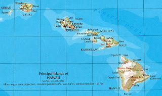
If a place like this existed, it would probably be some place young. It would probably also be someplace isolated, where the exchange of species would not have been easy. Sounds like an island to me; probably a volcanic island(s), something like the big island of Hawaii, in a chain that is only 300,000 years old….. O.K., it is the big island of Hawaii.


Hawaii is the biggest and youngest of the Hawaiian Islands.
Formed from five active volcanoes, it is growing larger
everyday.
Since they are islands, it makes sense that many of Hawaii's species came from somewhere else. A key question is, how did they get there? If seeds were brought by migratory birds or washed up on shore, or if an animal wasn’t quite dead when a predatory bird dropped it on the island, that’s one thing. But if humans brought plants or animals and released them by accident or deliberately, that is something else.

History shows the latter mechanism has been responsible for most of the diversity in Hawaii. Estimates are that the rate of species introduction in Hawaii has outpaced the natural rate of diversification by 2 million times. Over half of the island’s plants species are there because of people, not nature.

Many of the plants and animals that have been brought to Hawaii were introduced deliberately, but that is not the definition of an introduced species. Whether an organism is imported and released to serve some specific purpose, or whether it is a stowaway on a ship or otherwise unknowingly allowed to get loose, it is an introduced species (also called neozoon, alien, exotic, non-indigenous, or non-native species). Introduced species are those plants and animals living outside their native range due to some human intervention, whether intentional or not. For Hawaii, this began in the 19th century.


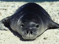
The Hawaiian monk seal is known as
Ilio‐holo‐i‐ka‐uaua in Hawaiian,
meaning, “the dog that runs in
rough waters.” They are critically
endangered, with only about 1100
remaining individuals.
Hawaii has only two native mammals, the Hawaiian monk seal (Monachus schauinslandi) and the Hawaiian hoary bat (Lasiurus cinereus semotus), and no native terrestrial (land) mammals. Many other mammals were introduced in the name of making money, like cattle and goats, while others were brought in specifically to make hunting more enjoyable.

In the last few posts, we have been discussing the activity patterns of different organisms, and we suggested that the organisms that interact most likely have the same or overlapping activity patterns. A tragic story illustrating this concept has played itself out in Hawaii since the late 1800’s.

Ladd & Company (from Maine) had established a lucrative sugar industry on the big island by 1834, and this increased the ship traffic in and out of Hawaii. The ships brought rats, an all too common introduced species.

In Jamaica, the problem of rats in the sugar cane fields was an old one. A successful sugar planter, W.B. Espeut, thought that introducing the Indian mongoose to the sugar cane fields could help with their rat problem. Apparently it did, and Espeut told everyone he could find. Subsequently, 72 mongooses were brought to the big island in 1883. There were some objections raised in the local papers, but as with most good ideas, they were roundly dismissed.


It is sad that mongooses
in Hawaii are causing damage, while
at the same time, mongooses are
endangered in their native India
due to habitat loss.
The problems became apparent not too long after mongoose introduction. In Jamaica there is a predator that eats mongooses, the fer-de-lance snake, but no such predator in Hawaii. What is the number one cause of death for mongooses in Hawaii? Old age!

No predator means unfettered reproduction, and female mongooses can have two litters each year. The result has been lots of mongooses, all looking for a meal and a mate.

Did all these Hawaiian mongooses do their job, did they get rid of the rats? Not really, and this is where a biology class could have helped. Mongooses are diurnal, they hunt during the day, but the rats on Hawaii are nocturnal. Oops. You would have thought someone might have noticed that beforehand.

I’m sure the odd mongoose runs into the odd rat as one goes to bed and the other begins its day, but that isn’t enough to force either species to adapt; everyone is happy keeping to his old schedule. Now Hawaii has too many rats and too many mongooses.

Mongooses (mongeese?) will eat almost anything (fruits, snails, mammals, insects, amphibians, lizards, spiders), but they really like bird eggs. Many species of bird in Hawaii, including the Nene (Hawaiian goose, the state bird) are on the brink of extinction because of the mongoose.

The story of the mongoose and the rat illustrates another point. Not only are there few mammals on Hawaii, there are even fewer native predators. As a result, many animals and plants that happen to be from Hawaii originally (or least for a long time) have adapted to this lack of predators by not developing defense mechanisms. This is an advantage, in that energy normally spent on defense can be saved for other metabolic or reproductive activities. Nature always wants to reroute energy if it is being used unnecessarily.

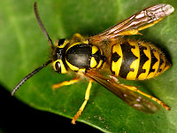
But, if predators are then introduced (and they were by the bucketful after Western Europeans and Americans became involved) the native animals are particularly at risk. The lack of native predators and the introduction of alien predators is illustrated by the case of the western yellow jacket. Though often mistaken for a bee, the yellow jacket is actually a wasp, and a nasty one at that.


Western yellow jackets have a smooth
stinger, so they can sting multiple
times. However, they rarely sting when
away from their nest.
The yellow jacket is an example of an accidentally introduced species, arriving in a shipment of Christmas trees. Law required that a percentage of the trees be shaken to knock off insects before shipment, but the required percentage of shaken trees was apparently too small, or the time shaken was too short, because it didn’t work.

In the continental U.S., the yellow jacket forms an annual nest and starts over building a colony each spring, but in the warmer climate of Hawaii, the species has become perennial, with nests as large as an SUV – more like a Lincoln Navigator, not the small Honda CR-V.

The yellow jacket is a carnivorous wasp only as a larva. The adults eat only nectar, but acquire meat to feed their young. This has decimated native Hawaii insect species, which in turn has reduced the amount of food available to bird species. In addition to insects, the wasps will devour dead birds and other large vertebrates, but will kill lizards and amphibians to feed their young.

In addition to accidentally introduced species, there are many feral (fera in Latin = wild beast) species on the big island. Feral species are animals that were domesticated, but have returned to the wild and propagated there. Their freedom could have come by accident or on purpose, as with cats and dogs in the cities. We call them strays, but they are correctly referred to as feral.


The stories of sewer alligators inspired sculptor
Tom Otterness to create a slightly scary piece in
bronze. It is located in a14th Street subway station
in NYC.
All those alligators in the sewers of NYC aren’t feral, because they were never domesticated. And yes, alligators have been caught in the sewers of NYC; the latest Time and Post articles I could find were from August of 2010. They don’t live for years, grow to be monsters, or reproduce - but small ones, probably recently released, are found just about every year.

In most cases, feral animals cause problems and don’t offer many advantages. They can act as reservoirs of disease, compete with native species for resources, prey on indigenous species and eat native plants.

If there are benefits to feral animals, they will depend on your view of things. Some ranchers can make money by rounding up feral cattle or horses. Feral canines provide an income for the town dogcatcher. Stray cats can help keep the rodent population down; there seem to be many advocates for feral cats. There is even a website that advertises all the wonderful things about sterilized feral cats. Really? They want to catch them, sterilize them, and then release them again?

Literally, thousands of plants and animals of all kinds have been introduced to the big island, and now we get to the crux of the issue – introduced species that become invasive species, ie. those that do damage to the natural ecosystem by becoming dominant by killing or displacing native species. The question is – which is the exception, introduced species that become invasive and do damage, or introduced species that do little damage or even result in a benefit?

On the negative side, we have talked about the Hawaiian mongooses, rats, and yellow jackets. There are others; feral pigs and cattle graze on native grasses and other plants. The Formosan ground termite causes millions of dollars of damage to trees and structures each year. Alien plants, such as Florida prickly blackberry and molasses grass smother native vegetation and prevent their re-establishment. As a result of these and other invasives, Hawaii has more endangered species per square mile than any other place on earth. This is due, at least in part, to invasive species.

Other introductions have been moot. For instance, over 4,600 species of plants have been introduced into the Hawaiian Islands over the last 200 years. However, only 86, less than 2% of the total, have become serious problems for native ecosystems.


Horses were introduced to the Americas
by the Spanish in the 16th century, as were sheep and
cattle. De Soto brought 13 pigs to Tampa Bay in
1539; these were the ancestors of the razorbacks
of the Southeast… and the University of Arkansas.

Some introductions have been wildly successful. Indeed, most of the cultivated crops (except for corn, turkeys, tomatoes, potatoes, and peanuts) and livestock animals in the continental U.S. are introduced species. Your pet cat, dog, bird, fish, or snake is probably an introduced species as well.

Of the approximately 5,000 alien animal and plant species in Hawaii, only about 300 to 500 have gone on to wreak significant damage and some have been beneficial. So the question remains, which are the exceptions- the failures and accidents that have resulted in destruction, or the successes and the accidental introductions that have had negligible effects?

Websites are full of lists of invasive species, the species they are displacing and the lost resources due to their introduction. We don’t see lists of introduced species that have worked out just fine. Perhaps this is as it should be; attention should be paid to those problems that need to be resolved. Attention should also be focused on the failures as a learning opportunity when future introductions are contemplated. But don't assume that a species is a problem just because it was introduced.

Gimme Some Dihydrogen Monoxide

$
0
0

Birds need water just like the rest of us,
but beaks make it harder. They may suck
it up like a straw or scoop it up like a bucket,
or by leaning back and letting the rain fall in.
At some point or another we've all said, “I’m about to die of thirst.” Of course we can only survive for a few short days without water, but do you know why?

Cells are full of salt water (saline), but are also crowded with proteins, carbohydrates and lipids (saline + organic molecules = cytoplasm). This suggests the importance of H2O, but it doesn’t say anything about the reasons behind its importance.

Water is the solvent (the liquid part of a solution), while the proteins and carbohydrates are the solutes (the solids dissolved in the solvent). Lipids (a type of fat) are insoluble in water; therefore, they are good for building cell membranes. They help keep what is in in, and what is out out. With a lipid membrane, our cytoplasm doesn't leak out on to the floor.


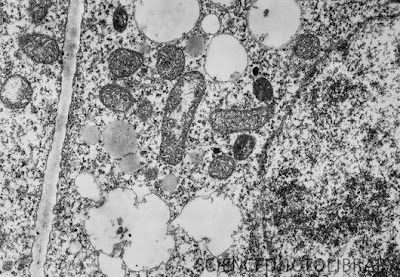
Cytoplasm isn’t water plus some organelles. As shown in
this electron micrograph, it is more like a gel, packed
with organelles, proteins, minerals, sugars, and nucleic
acids. There is water, but just enough to separate the other
constituents. Photomicrograph credit: Dr. Jeremy Burgess/Science
Photo Library.
The intracellular solutes are surrounded by water. It’s like the green jello with pineapple that your Aunt brought every Christmas, except that it's packed to the gills with pineapple. Cytoplasm is more crowded than the public pool on a 104˚F day when the ice cream vendors have gone on strike. In some cases, there may only be a few molecules of water separating different cellular components, but this water layer is crucial.

Water is the solvent in which most cellular reactions take place. Water is made up of an acid (H+) and a base (hydroxyl, OH-). Together, they are two hydrogen atoms and one oxygen, H2O! Having the H+ around keeps the bases in check, while the OH- keeps the acids in check. This helps keep the cytoplasmic pH within a small range (buffers it), about 7.35-7.45. Buffering the cytoplasm ensures that that reactions proceed in the proper direction and at the proper rate.

Water transports materials within the cell, from cell to cell, and through the blood and lymph. The partial negative and positive charges, the high surface tension, and the cohesive properties of water make it good at its jobs.


Water being sucked up in a capillary tube
uses cohesion (water sticking to water) and
adhesion (water sticking to the glass tube).
Water likes to bond to itself (cohesion) via hydrogen bonds formed between the positive H+’s of one water molecule and the negative OH-‘s of two others. Cohesion is what makes water form drops as it rains, and what gives water its strong surface tension. Surface tension is why some insects can land on water and take off again. Water striders (family Gerridae), walk on water and you can actually see the depression in the surface, like when you stand on your bed. They are helped out in this endeavor by hydrophobic (water-fearing) tiny hairs on their legs and feet.

Water also likes to hydrogen bond other surfaces; this is called adhesion. If you pour water into a small diameter glass, you can see it cling to the side (meniscus, Greek for crescent), and even seem to rise up the side of the glass (see the image above). If the glass tube is narrow enough, like in a capillary tube, the water will climb up the tube against gravity. The force that drives this is adhesion.


Water striders spread their weight over a large area to
reduce their pressure on the water. They are also helped
by the hydrophobic proteins on their legs. But mostly, the cohesive
force of the water raises the surface tension so the strider
remains on the surface.
The adhesive force is driven by the bipolar (a negative end and a positive end) nature of water, just as with cohesion. The positive H+ is attracted to any negative molecules, and the negative OH- is attracted to anything positive. Together, they are attracted to most everything, not just other water molecules.

Hydrogen bonding and the adhesion and cohesion they produce are important for plants. How does water absorbed by a redwood’s roots get to its leaves way up high? The mechanism has several features, the most important of which is suction. When water in the leaves evaporates, it creates negative pressure that actually pulls the water up from the roots through the plants vessels.

The negative pressure alone isn’t strong enough to keep the water moving against gravity, but when you add in the cohesion of water molecules to one another, and adhesion of the water molecules to the sides of the vessels, it all works out. The sum total of these actions is called transpiration, and is responsible for moving water against gravity in plants.

Water also participates in many cellular reactions, most famously photosynthesis. During the Calvin cycle of photosynthesis (dark reactions) glucose is produced, water is split into hydrogen atoms that are incorporated into the growing carbohydrate and gaseous oxygen (O2) that is released. It is this transformation of water to gas that drives transpiration.  In cellular respiration, when carbohydrates are used to produce chemical energy (ATP), the exact opposite occurs – water is formed from oxygen and hydrogen.

Other cellular reactions, such as the hydrolysis (hydro = water and lyse = split) of fats or proteins are occurring inside cells all the time. In these types of reactions, a water molecule is split into H and OH while the target molecule is also split in two; one part gains a hydrogen and the other gains a hydroxyl group. This is crucial for the normal degradation of cellular proteins by protease enzymes, amongst other things.

If that wasn’t enough, water acts as temperature buffer, helping organisms hold a more constant temperature. Water does not warm up fast and it does not cool down fast; it tends to keep an even temperature. It has a high specific heat (1 calorie/gram C˚), meaning that you must add a lot of energy in order to change its temperature. Water’s high specific heat evens out temperature fluctuations in the body and allows reactions to proceed in a controlled fashion.

Finally, many organisms use water pressure to hold their form, an example of the turgor pressure we learned about several weeks ago (Plants That Don’t Get A Good Night’s Sleep). For instance, you return home from a trip to find your plants have turned brown and are drooping in their pots. Your goldfish are belly up, and the expensive six-pack in your fridge is now a two pack – the neighbor you asked to look after them did a bang up job. If you’re lucky, the plants stand back up a few hours after a good soaking, especially if you fertilize them with your goldfish carcasses. Your plants need the water for everything we have discussed, but also because the water pressure in the cells keeps them the plant stem and leaves standing rigid.


The tube feet of starfish and other eichinoderms have a
suction cup on the end of the podia. The internal portion
is the ampulla, the tube that holds water to regulate the
tube movement.
In a similar fashion, starfish store and move water through a series of hollow tubes to form a hydrostatic skeleton. In the general sense, this type of skeleton is any fluid filled cavity surrounded by muscle, in which the actions of the muscles work against the fluid pressure in the cavity. Worms, and many other invertebrates have this type of support system.

But starfish take the concept a bit further. Not only is water used to maintain the form and structure of the animal; it makes up the water vascular system for locomotion (tube feet), food transport, and respiration. By moving water in and out of specific tubes in the different arms, the muscles contract and extend the tube feet, pushing them against a surface. The movement of water in and out of the tube feet is also the primary way to move oxygen into the tissues of the starfish, and the water pressure can be used to evert their stomach (it will protrude out their mouth and turn inside out) to surround and engulf food. Ugh!


Many types of animals use hydrostatic skeletons, where the pressure of water substitutes for a rigid skeleton. Muscular movements are generated against the in agonist/antagonist form against the pressure of the water, using muscular fibers positioned in several planes. A recent review by William M. Kierdemonstrates how the hydrostatic skeletons and muscular arrangements of several different animals work to generate stiffness as well as movement.

For instance, in the tube feet of the starfish, Ludia clathrata, muscular fibers are oriented in longitudinal and circular directions, allowing for extrusion and contraction. But he also discusses the connective tissue fibers that are just as important for the limiting of movement and generation of tension.

We always knew water was crucial for life, and now we know why. Its importance is reinforced when you consider how much water there is in different organisms. Humans are about 60% water by mass, but it varies from person to person. Younger children are normally have a slightly higher percentage of water, maybe 70%, while morbidly obese people have much less water, remember that fat is stored in the absence of water (Is it Hot in Here or is it Just My Philodendron?).


The golden barrel cactus has ribs that can expand and
contract, depending on the hydration state of the plant.
It is also called a mother-in-law’s cushion….that’s just mean.
Plants require even more water. Cactuses can be more than 90% water after a good rainfall. The places where cacti grow have variable water availability, so when water is present, they must take advantage. The endangered golden barrel cactus has ribs that can expand to take in more water. In addition, the golden barrel cactus is round to reduce surface area and has a thick waxy surface, both of which reduce water loss.

Despite these dehydration prevention measures, cacti still lose water over time, and it might not be replaced for a long time. Therefore, cacti have evolved mechanisms to withstand the loss of almost 60% of their water without any negative ramifications. In this area, they are the exception. Typical flowers and trees can only withstand a 20% water loss without damage; however, this is still much better than humans can do.

No matter what your personal water percentage might be, you can only afford to lose about 5% of your water without suffering symptoms. At mild levels of dehydration (5%), you may feel groggy or get a headache. Higher levels of water loss will bring tingling in the muscles, nausea, and confusion. If the loss reaches 10-15%, there can be muscle spasms, delirium, and the kidneys may be permanently damaged (if water loss is held for a sufficient period). Held above 15%, dehydration is usually fatal. However, athletes can lose up to 30% of their body water in the short term, but it must be replenished immediately so that performance or normal function will not be compromised.

When we say normal function, we mean those functions of water we have mentioned, but also several others we haven’t. Water, along with surfactant proteins, works to keep our lungs absorbing oxygen. Water lubricates our joints and tissues to avoid friction damage. People with xerostomia (Greek, xero = dry and stoma = mouth) or xerophthalmia (dry eyes) use artificial saliva or tears to prevent damage to mucous membranes. Finally, water acts as a cushion, absorbing pressure and force to protect our organs from traumatic damage, like a punch to the gut.

Damage can come in many forms when water is low, so all living organisms require water intake to function and remain safe, right?……Or is just most organisms? Next time.


Kier, W. (2012). The diversity of hydrostatic skeletons Journal of Experimental Biology, 215 (8), 1247-1257 DOI: 10.1242/jeb.056549


For more information, classroom activities, or laboratories about water in biology, the properties of water, transpiration, or the Calvin cycle, see:

Water in biology –

properties of water –

transpiration –

calvin cycle –
http://www.educationalrap.com/song/photosynthesis.html

Sorry, I Don’t Drink

$
0
0
Biology concepts – water conservation, kidney function, metabolic water, adaptation, water uptake


“Koala” in aborigine means “no drink.” The
moist eucalyptus leaves are poisonous 
to most animals, but koalas have a special 
bacteria that can break down the toxic
eucalyptus oil.
We all know we need water to survive (see Gimme Some Dihydrogen Monoxide), so why is it that koala bears have decided they don’t need to drink?

Koalas eat eucalyptus leaves, as well as mistletoe and a few other leaves. The leaves contain a good amount of water, and the koalas can survive on just this source of moisture. It also helps that they sleep about 18 hours each day, have a very slow metabolism, and feed about 80% of the time they are awake - it is apparent that they have evolved into teenagers. This doesn’t mean that koalas can’t or don’t drink, they just don’t require drinking to get their daily requirement of water unless a drought dries up the leaves.

However, there exist species that never drink. The kangaroo rat and the spinifex hopping mouse take temperance to the extreme. These rodents can live out their entire life (5-7 years) and never use the water fountain. They have chosen their lifestyles wisely, considering that the hopping mouse lives in the Australian outback and the kangaroo rat lives in Death Valley! We will use the kangaroo rat as our exemplar for this exception.

Unlike the koala that gets its water from its diet, the kangaroo rat eats seeds- not a great source of water. Therefore, it must have other strategies for survival. Foremost, it has developed ways to prevent water loss. Its kidneys super-distill its urine so it is up to 17 times more concentrated than its blood; the best we can do is 3-4 times concentration.


Please meet the nephron. The blood vessels form a
glomerulus, which is surrounded by the Bowman’s capsule.
Notice how the blood vessels surround the Loop of
Henle to take the retained water and salts back into
the blood.
The kidney is made up of thousands of filtering units called nephrons (Greek nephros = kidney). Each nephron has a Bowman’s capsule that filters the blood of waste,and removes some of the water and salt. The filtrate then flows through a series of tubules that adjust the concentration of the salts and water according to what the body needs to retain or dispose of at that particular moment. The portion of the kidney that removes water from the urine back to the blood are called the Loop of Henle, and these loops are much longer in the kangaroo rat’s kidney as compared to those in human kidneys. Therefore, more water is returned to the blood and the urine wastes are more concentrated.


The kangaroo rat doesn’t look thirsty, 
even though it doesn’t look like his 
burrow has seen water for years. 
I would imagine that despite the hot 
weather and the fur coat, kangaroo 
rats don’t sweat; they can’t afford the 
water loss.


The kangaroo rat doesn't stop there. He burrows deep and keeps his burrow small. This helps to trap and moisture that escapes via his exhalations. If you breathe on a mirror, it will show condensation; you invest a lot of water in keeping your lungs moist and functional. The rat can reabsorb some of the moisture present in its burrow via its skin, respiratory tract, and his seeds. 

The dry seeds that the kangaroo rat finds are stored in a pouch in its mouth and taken back to the burrow. Here they are stored for several days in a corner, during which time they also absorb moisture from the burrow’s air. This is just another way the rat recycles some of its own moisture. 

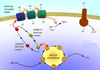
Finally, the kangaroo rat makes the most of the water it produces. Yes, it generates water – but so do you. Think of the production of ATP (aerobic respiration) as the opposite of photosynthesis. In the building of carbohydrates (during photosynthesis). In photosynthesis, water is split and the hydrogen is added to the growing carbohydrate. But in the electron transport chain for oxidativephosphorylation (making ATP) oxygen accepts an electron and then reacts with hydrogen to form water. Water made this way is called metabolic water. In humans, metabolic processes like generation of ATP produce about 2.5 liters of water each day. In the kangaroo rat, this process is more efficient and the water produced is kept in house.


As the electrons from the breakdown of glucose travel down the
electron transport chain in the mitochondrial membrane, they
help to move protons (H+) out. As they leak back in through the
ATPase, they help make ATP. The electron needs some place to go,
and an oxygen atom is a good place to go. This makes 
the oxygen reactive; it picks up hydrogens to form water.
Add all these measures up and the kangaroo rat changes its habitat from Death Valley to Life Valley. Unfortunately,  not many other organisms can join it there.

Just because it doesn't drink or eat watery foods doesn’t necessarily mean that an organism doesn’t take in water. Amphibians absorb environmental (air or surface) water through their skin. Frogs are a group of amphibians that can be used as good examples. Frog skin is smooth, without hair or feathers, and is permeable to water. A ventral patch (sometimes called a seat patch) of skin is located on the underside of the frog between its two hind legs. This skin patch has a higher concentration of blood vessels just beneath the surface, ready to suck available water into the bloodstream.

To get to the blood vessels below the skin, the water passes through a series of aquaporin (aqua = water, pore = opening) protein channels in the skin cells. These proteins also control water entry into bacteria; they are evolutionarily very old and therefore must be important. The frog splays its legs and lays down on a surface that is moist from dew or rain, and the water flows through the ventral patch aquaporins and into the bloodstream. Interestingly, water doesn’t flow the other direction, although some water does evaporate through amphibian skin. That is why frogs must live close to water. Toad skin is much less likely to lose water, so they can live farther from water.

Some plants also garner water in unconventional ways. Non-vascular plants (mosses, lichens, liverworts, hornworts) as well as many epiphytes (bromeliads, orchids, some ferns and mosses, mistletoe) are plants without roots. However, a lack of roots or vessels doesn’t stop these plants, they have evolved marvelous adaptations to procure the water they must have.

Non vascular plants are just that – plants without vascular tissues (xylem and phloem). Plant vascular tissues are tubes inside the stem that transport water (phloem) and sugars (xylem) throughout the plant. Non-vascular plants don’t have roots and vessels to absorb and transport water and minerals, although mosses and ferns may have rhizoids that serve that purpose. In general, non-vascular plants grow close to water so that they can use all their structures to absorb water by capillary action as well as by absorbing water directly from the air.

Epiphytes are even better at pulling water from the air, although they still use pooled rainwater as well. This group of plants may have dense root systems, but some are not anchored in the ground to give support to the plant. Instead, many of them use other plants for support. Orchids are particularly good at storing water in their thick stems and absorbing water through their exposed roots. Velamen (latin for veil or cover) layer root cells of orchids are adapted to prevent water loss while a few cells in this layer and the layer below are hollow and allow water to pass through.

Bromeliad epiphytes are better at absorbing pooled water and humidity through their leaves than in taking water in through their roots. In tropical regions, they have two adaptations to aid this process. One, many bromeliads have near vertical leaves shaped to trap water at their bases (together called a tank) that may hold over a liter of water. Second, they have specialized cells at the base of the leaves to transfer this water (and minerals) to the interior of the plant. The most economically important of this Bromelioideae subfamily is the pineapple, which is a terrestrial bromeliad. It can absorb water through its roots in the ground, but if you are growing one, try to keep the tank from drying out as well.


The top picture is looking down on a bromeliad trichome. 
The middle picture is looking from the side. See how they 
curl up to allow water in. When they fill with water, 
they fold down (lowest picture), to prevent water loss 
from the cells underneath.
Bromeliads living in areas with less rain, such as Spanish moss, have a different adaptation. Their leaves store the water that is absorbed through specialized structures called trichomes on the surface of each leaf. Trichomes have shields made of non-living cells, much like our outer layers of skin. Other cells form a disc and are mostly a void, capable of rapidly taking in water. When these cells swell, their tips curl downward (remember turgor pressure from Plants That Don’t Sleep Well).

Curling forms a small cavity under the disc that draws water in to the protected foot cells under the disc by capillary action. These cells also have aquaporin proteins that draw the water into the interior tissues. When there is less water around, the disc cells flatten out and cover the stalk cells, preventing water loss. The whole structure acts like an anti-umbrella!

So organisms can get water from air, food, or metabolism - but we can go them one better. There is an animal that doesn’t eat or drink during its entire adult life, can you imagine? O.K. – so its life is only five minutes long, but it doesn’t eat or drink during that five minutes.

Adult female sand burrowing mayflies (Dolania Americana) emerge from their water-borne larval form and seek two things, a male for mating, and a place to deposit her eggs. Since all larvae are evolved to mature at once, males are around in large numbers; problem 1 solved. And since they live near water, place to lay eggs are also plentiful; problem 2 solved. Within five minutes, her work is done and she dies – not a glamorous life.


The American sand burrowing mayfly lives a year or more
as a larvae in the water, but when it metamorphoses into
the sexually mature form and leaves the water, 5 minutes
is all she gets. There may be species with shorter sexual
reproductive life span, but it would be hard to spot, and
harder to study.
Different species of mayfly live varying amounts of time – some live as adults for up to 2 days - oldtimers! But even if the mayfly wanted to invest some of their precious time in eating and drinking, they couldn’t do it. Adult mayfly mouthparts are vestigial (having become nonfunctional through evolution) and their digestive systems disappear as they mature. So in this biological case, a lack of form follows a lack of function.

There is another crucial element of life that interacts with water, and ocean going organisms are intimately familiar with it. Salt is just as important for life as is water, but why? We will begin looking into the functions of salts and how they interact with water next time.


Banta MR (2003). Merriam's kangaroo rats (Dipodomys merriami) voluntarily select temperatures that conserve energy rather than water. Physiological and biochemical zoology : PBZ, 76 (4), 522-32 PMID: 13130431


King RF, Cooke C, Carroll S, & O'Hara J (2008). Estimating changes in hydration status from changes in body mass: considerations regarding metabolic water and glycogen storage. Journal of sports sciences, 26 (12), 1361-3 PMID: 18828029


For more information, classroom activities, and laboratories about water uptake, renal function, trichome, or mayflies:

Animals that don’t drink –


Kidneys –

Aquaporins –

Trichomes –

Mayflies -

Keeping Your “Ion” The Ball – Salts and Life

$
0
0
Biology concepts – salts in biology, osmotic potential,action potential, transpiration


Dietary salt – crucial for survival;
Veruca Salt – not so much.
In Latin, verruca means wart, so Roald
Dahl was probably trying to tell us something
when he wrote her character into Charlie
and the Chocolate Factory.
We have learned that one of the crucial functions of water in living organisms is to help regulate the salt concentration in and between the cells (Gimme Some Dihydromonoxide). But why do living things require salts? We all know that we must have a source of salt (sal in Latin) in our diet or we die; the Romans gave it so much importance that part of a soldiers pay was to be used specifically for buying salt – his salary.  But what are its functions?

Water tends to flow from where salts are in low concentration (high water concentration) to where salts are high concentration (low water concentration). Just like other molecules, water diffuses to where its concentration is lower (It’s All In The Numbers-Sizes in Nature). Osmosis (osmo = push in Greek) is the special name given to the diffusion of water, for every other molecule it is just called diffusion.

Too much salt is destructive to cells and organisms, so water helps control the salt held in the body. On the other hand, too much water is also bad for living things (water toxicity), so salts help to control the water concentration. Together, this ratio of salt and water inside and outside of the cell leads to a controlled imbalance called the osmotic potential of the cell. Every living thing has systems to maintain this osmotic potential within a small range (osmoregulation, we will discuss this in more detail soon).


The osmotic potential is measured in units
of pressure (bars). It is equal to the amount
of water that will move in response to a
difference in solute concentration across
a membrane.
When in water, sodium chloride (NaCl, table salt) dissociates into Na+ and Cl- ions, and it is these ions, along with K+ (potassium ion from KCl) that perform many functions in living organisms. Sodium is 10x more concentrated outside the cell, while potassium is 20x more concentrated inside. The slight difference in the charges of the two ions (and the fact that most Cl- is outside cells) sets up a membrane potential in cells.

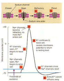
An important function of this membrane potential is in the neuron (nerve cell), as rapid reversal of the potential along the cell membrane (through ion specific channels) produces an electrical current that we know as the action potential (neural impulse). It is the rapid change in concentrations of Na+ and K+cations (positively charged ions) inside and outside of the neurons that sends the messages from our muscles to our brains and back, as well as all the thought processes in our brain.


The action potential of the neuron is not simple.
Sodium is higher outside and potassium is higher inside.
When a signal is received (usually from another neuron),
sodium leaks in and potassium leaks out. The slight
difference in the the charge of each means that the neuron
goes from -70 mV to +40 mV. This depolarization travels
down the neuron’s membrane for the entire cell.
Salt's importance is illustrated when their concentrations get out of whack. Too little salt produces symptoms similar to dehydration, with cramping, nausea and confusion. Too much salt results in hallucinations and insanity. The classic example of too much salt intake is being lost at sea. Not having a supply of freshwater, people may start to drink seawater. The salt concentration is too high; their kidneys can’t get rid of all the excess, and the action potentials in the brain begin to misfire. People will see things that aren’t there, and will make critically bad decisions. Many end up swimming away from relative safety and subsequently drown.

We can get rid of some salt through our skin. Is your dog is happy to see you when licking your face after you arrive home, or does he just want the salt? Athletes will often eat bananas to augment their potassium stores and keep the cramps away after exercising. They should really follow that run with a bowl of lima beans; they have much more potassium.

However, munching on black licorice is alot like running a long distance. Glycyrrhizin is the main glycoside (a sugar bound to a non-carbohydrate) in licorice root and is 20x sweeter than sucrose. Glycyrrhizin prevents potassium reuptake in the kidney, so you end up urinating out most of your potassium stores. You could cramp up due to excessive snacking.

The source of glycyrrhizin’s effect on potassium reuptake has to do with cortisol, a stress hormone. Cortisol is converted to cortisone, but glycyrrhizin inhibits this conversion. The increased cortisol makes it appear like your body has too many salts in the blood, and you adjust. This isn’t just a problem for the people who eat a lot of licorice.

A 2010 study indicates that pregnant women who eat licorice can permanently affect their children’s hormone control in their brains. The hypothalamic-pituitary-adrenocortical axis (HPAA), is a relay that controls the child’s production of cortisol, aldosterone and other hormones. These work to control the osmotic potential of the blood and therefore the blood pressure (as well as other things).

The researchers data shows that maternally ingested licorice inhibits the fetal barrier to maternal cortisol. More cortisol then passes to the fetal blood system, and programs the HPAA to have a higher baseline. From then on, the babies make more cortisol, a stress hormone that puts pressure on the physiology, sodium and potassium levels, and can lead to weight gain. Moms – take care – what you eat does affect your baby.

Na+ and K+ work in muscle function; cramping and paralysis may result from too little or too much salt. Your heart is a muscle, so changes in salt concentration in the cell can cause heart attacks as well. Many a mystery movie has included the injection of potassium chloride to induce a heart attack. Sodium and potassium cations help maintain proper blood pressure, proper acid/base levels, and proper movement of carbon dioxide from the blood to the lungs. There are precious few functions in which these positive ions don’t play a role.


Collagen and elastin help to make your skin and
joints pliable. O.K., maybe not this elastic – this is
the result of Ehlers-Danlos syndrome, which is
often a genetic disease.
When we think of salt, we usually think of table salt (NaCl), but there are more functions for K+ than there are for Na+, and it is present in higher concentrations in the cell. Potassium is important for the formation and crosslinking of collagen and elastin proteins. These connective tissue proteins hold all your tissues together; they keep your skin from tearing when someone pokes you in the arm, and allow your lungs to expand without ripping when you inhale. So K+ is pretty important even when not working with Na+. It is interesting then that potassium is the only major mineral nutrient for which there is not a recommended daily allowance.

Remember that we often take in these salts as NaCl or KCl. Does the Cl- play a role in organism function? – you bet it does. Chloride anion (a negatively charged ion) is used to produce the hydrochloric acid (HCl) that breaks down the food in our stomachs. Chloride also works in the immune system, hypochlorite (the same active molecule as in bleach) in the white blood cells helps to kill infectious agents and activates other immune system molecules. Chloride is required for the uptake of vitamin B12 and iron and helps control your blood pressure; therefore, Cl- isn’t just that other ion that comes in with Na+ or K+ (or Ca2+).

Chloride ion is elemental chlorine that has gained one electron. This doesn’t seem like much of a change, but it is the difference between life and death. Chlorine itself is a yellowish green gas and it can kill you in a matter of seconds. Chlorine really wants that extra electron, and it doesn’t care if it has to rip it from your lung proteins to get it. When you breathe in chlorine, it reacts with the water in your lungs to produce hydrochloric acid that eats away the cells. It will also react with almost any carbon-containing molecule and further destroy the lung tissue. It was suggested during the American Civil War that chlorine gas could be useful, but it wasn’t until World War I that it was used as a weapon.

Chlorine is poisonous, but we use it to disinfect drinking water and pools. When diluted greatly in water, chlorine does not have the strongly deleterious effect on our cells as it does as a gas, but can still react with and kill microorganisms. Chlorination of water began in the Chicago stockyards around 1908, when the decaying meat and gut bacteria were getting into the drinking water and making the residents sick. The bleach used to disinfect surfaces is much the same as the chlorine used to disinfect 75% of the drinking water in the U.S.; it’s just there in lower concentration. Now chlorine is used in pools as well, and you know it is working because your eyes get red and sting.


Did you know that plants had openings in their leaves called
stomata? Turgor pressure caused by the flow ions in and
out of the guard cells makes the stomata open or close. Their
shape changes based on the amount of water in the guard cell.
There are no exceptions to the rules of salt requirements (weird, isn’t it). All living things need to take in Na+, K+, Ca2+, and even Cl-. Plants use potassium and sodium for water balance, especially to bring morphologic changes like the blooming of flowers. These cations, along with chloride, work in the opening and closing of pores in the leaves (stomata) for the uptake of carbon dioxide and the release of oxygen and water during transpiration (Gimme Some Dihydromonoxide), and in the chemical splitting of water during photosynthesis. It seems that other organisms rely on these ions even more than animals.

All bacteria require potassium and sodium for osmotic regulation and cellular activities.
As the concentration of Na+ in a bacteria’s environment goes up, its dependence on Cl- becomes greater. Fungi, protists, and even viruses depend on salts to remain alive, even though viruses are technically not a form of life. Viruses carry nucleic acid, and salts are needed to balance the charges of the DNA or RNA so it can be stuffed into the viral package, a function within the area of molecular biology.
 

Giardia lamblia and other protozoa use salt ions
to control their osmotic potentials and for other
biochemical functions. Giardia can also change
your potassium levels by causing intense diarrhea
after drinking contaminated stream water.
Molecular biology involves replication of DNA, the transcription of DNA to RNA, and the activities of RNA translation to proteins. K+, Cl-, and Na+ are involved in all these areas. In a feedback mechanism, salt ions control the switches that turn on genes that then control the levels of the ions. If one ion is too high, it will turn on the genes that code for proteins which remove that ion from the cell. Isn’t evolution nifty?

Tightly regulating salt concentration in the cell is important for life, and we have to drink water (kangaroo rats excepted) in order to stay alive. These are the peanut butter and jelly of biology and we will start to see how they work together next time.


Räikkönen, K., Seckl, J., Heinonen, K., Pyhälä, R., Feldt, K., Jones, A., Pesonen, A., Phillips, D., Lahti, J., Järvenpää, A., Eriksson, J., Matthews, K., Strandberg, T., & Kajantie, E. (2010). Maternal prenatal licorice consumption alters hypothalamic–pituitary–adrenocortical axis function in children Psychoneuroendocrinology, 35 (10), 1587-1593 DOI: 10.1016/j.psyneuen.2010.04.010

For more information and classroom activities on salts in biology, osmotic potential, action potentials, or chloride ion in biology, see:

Salts in biology –

Osmotic potential –

Action potential –

Chloride in biology -

stomata –
http://www.apsnet.org/edcenter/intropp/topics/Pages/OverviewOfPlantDiseases.aspx

Don’t Eat The Yellow Snow

$
0
0
Biology concepts – osmoregulation, tonicity, phytohormones, avian kidney, pinnieds, cetaceans


African elephants are larger than asian elephants, but their
urine production is similar. A 2007 study found that
African elephants can differentiate family members
based on their urine. It is similar to marking territory,
but they use urine to keep track of family members who
may be out of sight.
The asian elephant can urinate as much as 55 liters/day. That's about 3/4 of the volume of the average size bathtub! By comparison, the vaunted racehorse can only manage about 6 liters/day. Maybe we should rethink that old saying.

We know from the posts of the last few weeks that both salts and water are necessary for life, and that they work together to keep their concentrations within safe limits; a process called osmoregulation. You suspect correctly that kidneys and urination is involved, but what about plants – they don’t use the restroom.

For many animals, the kidney is the major organ of osmoregulation. The average adult human voids 1-2 liters of urine each day, but an uncontrolled diabetic with polyuria (poly=much and uria=urine) might expel 5-6 liters. Maybe we should bet on diabetics at the racetrack.

We get rid of water and soluble wastes via our kidneys. The kidneys filter the blood; nearly 800 liters of the red stuff each day. The basic filtering unit of the kidney is the nephron, who we met previously (Sorry, I Don’t Drink), made up of the Bowman’s capsule and sets of tubules.


Solutions of different tonicity have similar effects on plant
and animal cells, but plant cells can handle it better because
they have a rigid cell wall.
If the body is low on water, more water is reabsorbed in the tubules. Likewise, if the body has too much salt, few of the salt ions are reabsorbed in the tubules. In this way, our kidneys are basically concentrating our wastes in a small amount of water for excretion from the body. The amount of water depends on many factors, including the need to keep the cells at the right level of tonicity (concentration of salt relative to outside the cells).

Solutions can be hypertonic, meaning they have more salt than the cytoplasm, and water will flow out of cells by osmosis. Solutions can also be hypotonic, with less salt than in the cells (water will flow in to the cells) or isotonic, with the same osmotic pressure inside the cells as outside.

We all know that we don’t urinate the same amount all the time – drink more, go more. However, you don’t urinate the same amount you drink; your urine is concentrated by your kidneys in order to conserve water. Therefore, there must be some control mechanism. The answer is hormones. A hormone (“to set in motion” in Greek) is a small protein that is released from one cell and then acts as a chemical signal on other cells, either through the bloodstream (endocrine hormones) or through a duct (exocrine hormones) to the bloodstream or directly to other cells.


The angiotensin system. 1.The body senses that water
is low. 2. The kidney releases renin. 3. Renin  and
angiotensin converting enzyme produce angiotensin II
from angiotensin I in the lung. 4. Angiotensin II stimulates
aldosterone in the adrenal glands. 5. Aldosterone causes
more water and salt to be reabsorbed in the Loop of Henle;
this increases the blood volume and solves the problem.
Aldosterone is produced by the adrenal glands and acts on the distal collecting tubules of the kidneys. This endocrine hormone acts to conserve sodium and water and secrete postassium, thereby reducing urine volume but increasing the loss of potassium.  Aldosterone is released in response to angiotensin levels in the plasma, which in turn are controlled by sodium and water levels in the blood.

Arginine vasopressin (AVP, also called antidiuretic hormone) is another endocrine hormone that reduces the amount of water to be lost in the urine. This hormone is produced in the pituitary gland of the brain and also works to conserve water. By reducing the amount of water lost, the blood volume (which is mostly water) is increased, so blood pressure increases. This is why people are given intravenous fluids when they have lost a lot of blood.

The exceptions to this mechanism of kidney function are the mammals that live in hypertonic (saltwater) environments, like whales and dolphins (cetaceans) and seals or walruses (pinnipeds, latin for feather- or fin-footed). It is hard to study the urination in these animals in their native environment; they urinate in the ocean. Are you going to measure their individual contribution to the ocean – I think not.

Water wants to flow out of the cells and into the sea (hypertonic as compared to the cells), trying to balance the salt concentrations in both places. Therefore, the marine mammals must conserve freshwater or they become dehydrated. Both pinnipeds and cetaceans have large kidneys with enough renal tubule length to produce very concentrated urine, thereby conserving water. However, it appears cetaceans don’t really take advantage of this. Instead, they make a lot of metabolic water (Gimme Some Dihydromonoxide) and can keep from dehydrating by using the water they produce through cellular respiration.


Here is an inside view of a seal kidney. It’s huge! The many
lobules provides much tubular area to take up freshwater
and concentrate the urine.
Pinnipeds don’t drink water saltwater to any degree at all, they get their freshwater from their diet and their metabolic water.  Scientists use to think this was also true for cetaceans, but recent studies show that they do drink a small bit of seawater – not enough meet their water needs, but also not more than their kidney’s can handle.

Don’t think that marine (saltwater) mammals have it so bad. If they were to abandon the seas for freshwater sources, they would just trade one problem for another. Freshwater mammals have too much of a good thing, they run the risk of losing too much salt by being in so much salt poor (hypotonic) water all the time. This is why the kidney is so amazing, it can adapt functionally and anatomically to get rid of too much water or too much salt, depending on where you are. That is not to say the kidney is the only anatomic mechanism needed to maintain osmolarity within a tight range. Many organisms need more than kidneys, and have developed completely different mechanisms of osmoregulation.


Bird kidneys may be small, but they represent an evolutionary
intermediate, Some parts have short loops, like most mammals,
and some have long loops, like pinnipeds and cetaceans. However,
most of the kidney has reptile-like nephrons with long loops.
Birds share some water conserving and salt regulating apparatus with mammals. Avian (bird) kidneys have about 75% of their nephrons with reptilian structure, and 25% mammalian nephrons, containing a Loop of Henle. Therefore, avian kidneys are not as good at removing water and regulating salts as mammals are. Mammal urine can be concentrated 20-50x as compared to blood (the Kangaroo rat can produce a 9000x concentration), but birds can only manifest about a 2-3 fold concentration.

Therefore, birds have another mechanism to get rid of salt and maintain an osmotic potential within its limits. The salt gland is found in birds and reptiles. In many birds it is located near the eyes or nostrils (in crocodiles, salt is excreted through their tongues – everything tastes salty to them).  The salt gland removes Na+ and K+ from the blood, allowing birds and reptiles to consume saltwater or animals that live in saltwater.

Some organisms have it easier, like amphibians. With semi-permeable skin, they just leak salt out through their entire skin surface. Other organisms aren’t so lucky, like plants.

Plants must also regulate salt concentration, but they don’t have a familiar excretory system; in fact, they don’t have a specific osmoregulatory system. Water is lost via transpiration (Sorry, I Don’t Drink), and adjustments can be made to alter the amount of evaporation that occurs. Unfortunately, transpiration of water is linked to moving nutrients such as salts up the plant from the roots to the leaves. Therefore, shutting down transpiration will also shut down movement of nutrients. 

Plants in high temperature, low humidity, high wind environments have the highest rates of transpiration and are in danger of losing too much water. Once again, hormones are the answer. Plants do have hormones (phytohormones), so they probably have to deal with teenager issues just like human parents. Abscisic acid is an important hormone which shuts off transpiration. This phytohormone closes the stomata (stoma = mouth in Greek) on the upper sides of leaves, from which water evaporates and gases are exchanged. Abscisic acid also promotes water absorption from roots and root growth.


Some plants are cryptophytes by surviving unfavorable
seasons either underground (geophytes), hide their
seeds in the marshy mud (helophytes) or underwater
(hydrophytes). Hydrophytes in general are plants that
have their roots in water or water-logged soil.
Many xerophytes (plants that live in hot, dry places) have adapted to resolve these issues. They have leaf modifications to reduce water loss; needle-shaped leaves, sunken stomata, and waxy cuticles to cover the leaves. On the other hand, in hydrophytes (plants that live completely or almost completely in water), salts and water can be absorbed in the entire plant, not just the roots.

In terms of cations (Na+, K+), plants have a problem. They use potassium as their primary intracellular cation, but dirt is usually potassium-poor. Therefore, plants have K+ transporters to actively take up this ion. Unfortunately, the transporters don’t discriminate very well between K+ and Na+, so often times too much Na+ is taken up into plants.


Red mangroves have impermeable roots that help keep
out salt, and can also secrete some salt from there leaves,
but their most visible mechanism is the yellow salt leaves.


Excess Na+ can be toxic to cells, so measures must be taken to deal with these ions. Glycophytes are plants that are salt-sensitive, and include many of the plants that we cultivate. Therefore, soil salinity is an important factor in agriculture and gardening. Much research and breeding continues to an effort to produce crops that are better at differentiating uptake of K+ and Na+. Halophytes (halo=salt, phyte=loving), on the other hand, will allow the uptake of the excess ions, and then sequester them in vacuoles to prevent cellular damage.

Some plants live in extremely high salt environments. One example, the red mangrove tree, is a facultative halophyte. Facultative is a fancy way of saying “optionally.” These trees live in estuaries, where the river meets the sea. The water is quite salty there, and the mangroves are rooted in the water, so excess salt could be a problem. To deal with the toxicity of the excess Na+, the mangrove will store the salts in selected leaves, called the “kidney leaves.” When a toxic level is reached, the leaves turn yellow and just drop off. The tree must constantly invest energy in producing new leaves, so there is a cost to this way of life, but it seems to work for them.

If plants that live in or near seawater have adaptive mechanisms to maintain proper salt concentrations, then how about fish? We'll look at the osmoregulatory tricks by these organisms next week.


Ben Hamed-Laouti I, Arbelet-Bonnin D, De Bont L, Biligui B, Gakière B, Abdelly C, Ben Hamed K, & Bouteau F (2016). Comparison of NaCl-induced programmed cell death in the obligate halophyte Cakile maritima and the glycophyte Arabidospis thaliana. Plant science : an international journal of experimental plant biology, 247, 49-59 PMID: 27095399

Peña-Villalobos I, Valdés-Ferranty F, & Sabat P (2013). Osmoregulatory and metabolic costs of salt excretion in the Rufous-collared sparrow Zonotrichia capensis. Comparative biochemistry and physiology. Part A, Molecular & integrative physiology, 164 (2), 314-8 PMID: 23103672

Takei Y (2015). From aquatic to terrestrial life: evolution of the mechanisms for water acquisition. Zoological science, 32 (1), 1-7 PMID: 25660690


For more information, classroom activities or laboratories about osmoregulation, tonicity, abscisic acid, avian kidney, pinnipeds, cetaceans, see:

Osmoregulation –

tonicity and osmotic pressure –

abscisic acid –

avian kidney –

pinnipeds –

cetaceans –
http://what-when-how.com/marine-mammals/osmoregulation-marine-mammals/

Do You Drink Like A Fish?

$
0
0
Biology concepts – fish osmoregulation, shark osmoregulation, semelparity, iteroparity


The irony of fish drinking is not lost on this café in
the Hotel Portofino at Universal Orlando. What I
really like is the eye patch.
You’d think that fish would never be thirsty; if he needs a drink, he just opens his mouth. But some fish don’t drink a drop! Wouldn’t that be similar to some birds never breathing? Ridiculous.

Fish are good examples of the problems of maintaining proper water and salt concentrations. Some fish live in freshwater, and some in saltwater. These are opposite sides of the same coin when dealing with osmoregulation.

Freshwater fish live in a hypotonic (low salt) environment. The flesh of the fish contains more salt than does the water. Diffusion and osmosis work to equalize salt concentrations in different compartments. Therefore, water will move from the lake or river into the fish’s tissues in order to balance the salt concentrations by osmosis. Salt will not move out of the tissues, since there are molecular mechanisms that work to keep the inside.

Like the kangaroo rat, freshwater fish don’t drink. They do take in water when they eat and move water across their gills, but they don’t take in water just for the water. Even without drinking specifically, freshwater fish take in way more water than they need. Anywhere freshwater contacts a fish cell, water will move inward; this includes the gills, the mouth and gut, and the skin.

In a situation like this, kidney-mediated concentration of urine would be counterproductive; why retain water when water is exactly what you have too much of? Therefore, freshwater fish excrete large amounts of urine. Their kidneys have large glomeruli, which move lots of water into the collecting tubules for excretion.


Saltwater and freshwater fish have different ways of
dealing with salt and water loss and conservation.
Freshwater fish must conserve salt, while saltwater
fish must conserve water. The kidneys play a role,
but so do the chloride cells in the gills.
But if the freshwater fish aren’t drinking, how do they get their salt, which is present in low concentrations in the water? You’d think they would have to be drinking all the time just to collect enough salt.  To get around this, they conserve the salt they ingest through the food they eat. They also take in salts through their gill chloride cells, actively pumping sodium and chloride out of the freshwater and into cells that have a lot of mitochondria (to provide energy to pump the salts). The relatively short collecting tubules of the freshwater fish kidney allow for reuptake of a lot of salt, while excluding almost all the water.

Marine (saltwater) fish have the opposite problem. Their tissues are of much lower salt than they surrounding hypertonic ocean, so osmosis wants to dry them out, sending water out of their bodies. The amount of available drinking water is extremely low - can you imagine dying of dehydration while surrounded by water. Just ask anyone who has survived a shipwreck and prolonged float in the ocean; drinking seawater can be lethal.

However, marine fish must drink all the time in order to keep enough water in their body. Retaining water would be an essential function of marine fish kidneys. They are all fish, but their kidneys work in exactly opposite ways.  Marine kidneys have small or absent glomeruli, so little water is taken out of the blood, but long collecting tubules in order to excrete as much salt as possible.

Drinking a lot of saltwater leaves marine fish with way too much salt; more than their kidneys can get rid of. To aid in salt excretion, they also have chloride cells in their gills. In the opposite fashion of the specialized gill cells of freshwater fish, the chloride cells of saltwater fish actively sequester salts from the blood, and then pump the sodium and chloride out into the seawater.


Sharks have unique ways of maintaining
salt and water. I have no idea of their
mechanisms for pepper regulation.
But sharks are an exception among marine fish. They have a different way to combat high salt concentrations. Remember that osmosis means that water moves from areas of low solute (high water concentration) to areas of high solute (lower water concentration). For many marine fish, this would mean a constant loss of body water to the ocean and quick death by dehydration; much like pouring salt on a slug.

To overcome this movement, sharks produce and retain a huge amount of a chemical called urea; it is one of the soluble wastes that animals normally get rid of. This molecule doesn’t affect the electrical potential that salts create, but increases the solute concentration in the shark’s tissues at levels higher than in the seawater, so water (without the salt) will diffuse into the shark’s body. This is its source of fresh water.

Therefore, sharks are osmoconformers; they maintain an osmotic balance with their environment. If the shark becomes too salty and salt needs to be excreted, it has a salt gland, much like that of birds and reptiles, but the shark’s gland is located in it anus, not near its eyes or nose – that’s a big difference! Taken together, there is no force for movement of water in or out of the shark’s tissues, and the shark remains shark-shaped instead of shriveling or swelling up.


Here is a bullshark caught in the Potomac River.
And you thought that sharks in Washington D.C.
were just in the federal buildings.
An exception to this rule for sharks is the bull shark; it can live in both saltwater and freshwater. Most sharks put into in freshwater would absorb too much water and die of water toxicity. However, the bull shark’s kidneys can adjust to the salinity of the water within a short period of time. Their kidneys will remove less salt and more urea from their blood and tissues and into their urine. They move from being osmoconformers to osmoregulators.

A shark that can live in freshwater; this can present a real problem. There have been many bull shark attacks in rivers and estuaries (video), where people don’t expect to encounter sharks. It is suggested that this behavior and physiology is an adaptation that gives the bull shark a protected nursery for their young, away from predators.

Most fish are stenohaline (Greek, steno = narrow and haline = salt), which means they are restricted to either salt or fresh water and cannot survive in water with a different salt concentration than to that which they are adapted. However, there are exceptions- like the bull shark mentioned just a second ago.

Some salmon species are born in freshwater, then move to saltwater for several years, and then return to freshwater to spawn. Other fish, like some eels, are born in a marine environment, move to freshwater, and then go back out to sea to reproduce. If freshwater and saltwater fish kidneys work opposite of one another, how can there be fish that can do both?


Salmon returning upstream to spawn have many obstacles
to overcome. Their spawning grounds are usually a thousand
feet or more above sea level so they must leap up many
waterfalls. Oh, there are hungry bears too.
Salmon are famous for migrating to and from the sea. Almost all the species are semelparous (in Latin, semel = once and parous = breeding); this means that they return to their freshwater streams to spawn only once, and the trip and the reproduction kills them. The one exception is the Atlantic Salmon (Salmo salar). This species is spawned in, and returns to, the calm streams along the Atlantic coast several times in its life to spawn. This reproductive strategy is call iteroparity (itero = repeated). Iteroparous species lay fewer eggs at a time, the advantage is that survival chance is increased by repeated spawning – one bad year doesn’t destroy a big proportion of the population.

The migratory species of salmon are osmoregulators, as are most freshwater fish; their physiology demands a certain salinity level, and use energy to produce that level in their tissues. However, they can also adapt to various salinity levels. As such, these salmon as well as bull sharks are known as euryhaline (eu = good, haline = salt). Their physiology changes with the salt concentration.

While in freshwater the salmon will not drink, and will produce copious amounts of urine to get rid of the excess water it absorbs through osmosis.  But when it migrates to the ocean, it drinks all the time, and its kidneys work hard to remove the excess salts.


Chloride cells in euryhaline fish can sequester or
excrete salt, based on the hormone signals they receive.
This helps some fish move from aquatic to marine
environments and back again.
But the gills are the key to survival in the both the freshwater and saltwater environments. Energy consuming reactions will transport both Na+ and Cl- against their gradients, so they pump Na+ and Cl- into the fish’s tissues in freshwater and out of the fish’s tissues in saltwater. It is an adaptation of the marine fish’s chloride cells to work in both directions. This switch, as well as the kidney’s change in urine concentration, takes time. Therefore, salmon will spend days or weeks in intermediate zones, or estuaries, before going out to the ocean, and before returning to the rivers.

These are difficult lifestyle choices for salmon, the trips and the spawning kills them. So what is the advantage? The movement to oceans provides the growing salmon with readily available sources of food, so competition is reduced. The return to where they were spawned is just a good bet; if the stream was good enough to spawn them, then it is still probably a good place to lay eggs. Finally, working so hard to get to the spawning ground just a single time allows for selection of strong individuals, allows for huge numbers of eggs to be laid (the chance that some survive goes up), and the death and decomposition of the adults provides nutrients for the hatched fry (baby salmon). But these are human interpretations, I bet there are other advantages and disadvantages. However,  one thing is for sure, the balance sheet for these species comes out in favor of these adaptations – if it did not, nature would adapt further.


The eggs that don’t hatch and the carcasses of the mated
Adults create nutrient rich waters for the fry to develop in
before they head out to sea.
How about one more exception for today? Some individuals in semelparous species of salmon (Chinook, Coho, Pink, Steelhead, etc.) will not die after spawning, and will return again to the ocean. These individuals are often females, and are often smaller than average. These gals reverse their salt and water conservation strategies several times in their lives, making them prize winners for osmoregulatory exceptionality.

Next week, let’s tackle how the properties of hard water affect all life on Earth.


Sakamoto T, Ogawa S, Nishiyama Y, Akada C, Takahashi H, Watanabe T, Minakata H, & Sakamoto H (2015). Osmotic/ionic status of body fluids in the euryhaline cephalopod suggest possible parallel evolution of osmoregulation. Scientific reports, 5 PMID: 26403952

Cozzi RR, Robertson GN, Spieker M, Claus LN, Zaparilla GM, Garrow KL, & Marshall WS (2015). Paracellular pathway remodeling enhances sodium secretion by teleost fish in hypersaline environments. The Journal of experimental biology, 218 (Pt 8), 1259-69 PMID: 25750413


For more information and classroom activities on osmoregulation in fish and sharks, chloride cells, and reproduction strategies, see:

Osmoregulation in fish –

Chloride cells –

Osmoregulation in sharks –

semelparity and iteroparity –
http://web2.uwindsor.ca/courses/biology/weis/55-324/lecture9.htm

I Am Your Density -- Life On Ice

$
0
0
Biology concepts – density of water, latent heat, stratification


Ernest Rutherford showed that atoms were
mostly space by shooting alpha particles at
a sheet of gold foil. Only a few particles struck
something solid, most just passed straight
through – because the atom is mostly the
absence of matter.
It is amazing to know that atoms are mostly empty space. Atoms make up everything around us, including the stuff that hurts when it hits me in the head, but even those things are mostly empty space... or maybe its my head that's empty.

When atoms fit together to form molecules and molecules fit together to form solids and liquids there is also space. How massive the molecules are and how much space is between them determines a substance’s density.

Density (mass per unit volume) has a big impact on biology, and we have been talking about water for a few weeks, so let’s talk about the density of water. Simply put, without water’s unique density properties, life as we know it on Earth would not be possible.

Pure liquid water has a density of 1 g/cm3 (or 1 g/ml). This is 800x times the density of air, so moving around in water is much harder and requires more energy than moving around on a land. Try running in the pool – we just aren’t built for moving in water.


Gram for gram, fish have more muscle than
any other vertebrate animal. Notice how the
muscle fibers are arranged in different
directions to provide forward movement as
the skeleton changes orientation.
But fish have adapted streamlined shapes and big muscles in order to move through water a little easier. The skeleton of a fish is the most complex of all vertebrates. The skull anchors the waving of the vertebral column and the attached muscles. The muscle fibers (myomeres) are arranged so that the muscles can contract in several different directions as the swaying motion passes down the fish body. In all, a fish is about 80% muscle. If you are a marine fish, you’d better be even stronger, since ocean water is slightly more dense (between 1.02 and 1.03 g/cm3, depending on the salinity).

But here is the amazing part - when water freezes, its density goes down. Most substances are denser as solids than as liquids, but water is the exception. As ice crystals form, the water molecules arrange themselves in a very particular order, and this order places slightly more space between them as compared to when they are in liquid form. More space means less mass per unit volume, ie. lower density (0.92 g/ml)….. and this is a key to life on Earth.


Water will form ice crystals in a definite structure,
with more space between the molecules than when
in liquid form. Snow crystals form from water vapor,
not liquid water, and retain a more hexagonal lattice
shape that may stack on one another.
Imagine for a moment that ice was denser than water. Then as the winter came, the winds would blow, the surface water in the pond behind your house would start to cool down, but the deeper water would be a little warmer (remember that water has a high specific heat, it likes to retain its heat. As the surface water arranged itself into a crystal form, ie. turned to ice, it would sink. The warmer water would then be pushed up higher and exposed to the colder temperatures, freeze, and fall to the bottom. Eventually the pond would fill with ice, and be completely frozen.

Few animals or plants could survive in a solid block of ice, so life would cease to exist in the pond. What is more, when spring came, the sun’s energy and warmer temperatures would have to penetrate to bottom of the pond in order to melt all the ice, and this would take longer than a spring summer and fall to occur. Most bodies of water would stay somewhat frozen all year long.

Our food webs (who eats who) depend so much on the growth in water, and half of the Earth’s oxygen’s production oxygen depends so much on phytoplankton, the one celled plants that float on the water’s surface and release oxygen as a by product of photosynthesis. So we couldn't survive for long with completely frozen bodies of water. What is more, frozen lakes and bays would eliminate huge heat sinks that normally keep the surface of the earth warm, so we would plunge into another ice age.

Can you imagine if the massive number of aquatic organisms died as a result of their environment being frozen year round? The animals that feed on them would then die, and the animals that feed on them would die, etc. Eventually the animals on the land that feed on the amphibians and fish would die, and so on.  What’s more, we humans would be looking for more warm clothing while we gasped for enough oxygen to survive! Relax, we are all just fine, and it is because ice floats. Surface water freezes, trapping heat below and keeping the aquatic organisms comfy and cozy until spring.


The North American wood frog can freeze
solid in a long Arctic winter, but once it thaws,
it has work to do. It must find a find a mate and
then fertilize the eggs. The fertilized eggs have
to develop from to tadpoles and then to adults
during the short warm period. Then they can
freeze next winter.
You might have noticed that above I mentioned that MOST organisms can’t survive being frozen, but there is an exception. The wood frog (Rana sylvatica) winters in shallow burrows that are not protected from the cold. To survive, the frog actually freezes solid!

Nucleating proteins in the frog’s blood act as point for ice to form as soon as the frost touches the amphibian’s porous skin. Since the frog is still above 0˚C at this point, the freezing is slower, and the frog can control it. As the liquids freeze, the water is pulled out of the frog’s cells.

It replaces the water with huge amounts of glucose and sugar alcohols, that keep the cells from forming ice crystals (they are sharp and would puncture the cells causing permanent damage and death). Eventually, the frog is 65% frozen and the internal organs are surrounded by a pool of ice until spring, when it takes about 10 hours for the frog to thaw and hop away. Scientists are now using this process to freeze and thaw rat hearts and livers without damage, in hopes to use to the process in human organs for transplant.

But freezing and thawing a whole organism is harder than using a glucose bath to freeze individual organs. Research from early 2013 shows the energy that R. sylvatica must spend to accomplish this feat. In response to cooling near the freezing point, the wood frog increases its metabolism to prepare for freezing. But this increase in metabolism is nothing compared to the increase the frog undergoes when freezing is first detected in its tissues. Carbon dioxide (a sign of metabolism) is increased by 5.8 fold during freezing, as to the period just before freezing. This increase is needed to mobilize glucose into the tissues as the cryoprotectant.

The same thing happens when R. sylvatica thaws, metabolism increases to exactly the same degree as during freezing. But in this instance, the increased cellular activity is necessary for re-establishment of homeostasis and for tissue repair (no anti-freezing strategy is perfect). We have a long way to go to mimic the wood frog's entire preservation strategy, especially since the frog may go through these increases as many as twenty times each winter!

The wood frog takes advantage of freezing in order to survive. Humans can also take advantage of freezing water (other than keeping your drink cold); in fact, your orange juice may depend on it. Freezing of oranges or grapes ruins them for the same reason it kills animals, it causes frostbite. Ice crystals stab through the cell membrane and cell contents spill out. This isn’t conducive to continued function.

To prevent oranges and grapes from freezing, farmers will spray them with water when their frost warning systems sound the alarm. Does that make sense, spraying with water to keep something from freezing? It has to do with a property of freezing called latent heat. This is an amount of energy taken up or given off when a substance changes phase (solid to liquid to gas). The energy goes to changing the arrangement of molecules with no change in temperature.


Oranges can be protected from freezing by
spraying them with water which then freezes!
In a controversial use of genetic modification,
bacteria that do not permit ice crystal formation
can be sprayed on the oranges to compete with
the normal bacteria there. These "ice-minus"
Pseudomonas syringae can reduce frost damage
on oranges, but have not been used commercially.
As water surrounding the orange or grape changes from liquid to solid, the formation of crystals gives off heat (539.4 gram-calories per gram of water frozen). The latent heat of the freezing mist is enough to keep the fruit above 0˚C. This technique doesn’t work if the temperature falls much below 0˚C or stays at 0˚C for an extended time, but it does work well enough to save millions of dollars per year in freezing damage.

Thermal changes have more to do with differences in water density than salt concentration does, so seasonal changes can alter density in both freshwater and salt water. Even if the changes are not enough to form ice or boil the water, differences in temperature can result in different layers of water within a freshwater body or an ocean.

Both salt water and freshwater are affected by the sunlight that strikes their surfaces. As water warms, it’s density decreases, and the nutrients in the water stay close to the surface. This supplies phytoplankton and algae with lots of food, and blooms can occur.

As winter approaches, the surface water cools and becomes more dense (down to 4˚C). The dense water drops to the bottom and taking nutrients down to the benthic organisms. When all the water reaches 4˚C, the surface can begin to freeze.

In the spring, the process is reversed, and the temperature layers (stratification) can churn again. In salt water, the differences in salinity are added to the differences in density to bring complex stratifications, both in salt content and temperature.


Stratification shows how temperature can set up
layers of water of different density (least dense is
the epilimnion). In the winter, the water is churned,
and then churned again in spring. These churnings
based on changing density move the nutrients around
so everyone gets fed.
Different organisms thrive in different temperature and salinity layers. In order to stay put, some floating organisms (planktonic) and swimming organisms (nektonic) can adjust their buoyancies. Fish can use swim bladders, which are air filled cavities to help them stay buoyant. The size of the bladder is regulated by the CO2 and O2 in the blood that can remain dissolved or leave the blood as a gas.

Bladderwort plants also use air filled cavities to keep part of themselves afloat. Sharks, on the other hand, produce large amounts of oil in their livers to reduce their density; oil is less dense than water, just look at your salad dressing layers.

Plankton can also slightly adjust their densities, but floating is easier for very small things. To them, water is thick, the polar charges have a larger effect on their small bodies. It would be like us trying to swim in molasses. They still have to adapt to seasonal changes in density, but they do it in more subtle (and harder to explain) ways.

Just because there is water around, it doesn’t mean that life will be easy. Next week we will look at a continent-sized exception to idea of water availability.


Sinclair, B., Stinziano, J., Williams, C., MacMillan, H., Marshall, K., & Storey, K. (2012). Real-time measurement of metabolic rate during freezing and thawing of the wood frog, Rana sylvatica: implications for overwinter energy use Journal of Experimental Biology, 216 (2), 292-302 DOI: 10.1242/jeb.076331


For more information, classroom activities and laboratories on the density of water, latent heat, North American wood frog, or stratification, see:

Density of water –

latent heat –

North American wood frog –

stratification –
http://www.lmvp.org/Waterline/spring2002/stratification.htm

Water, Water Everywhere, But….

$
0
0
Biology concepts – symbiosis, mutualism, water storage


“Gobi” means desert in Ural-Altaic,
so when you say, “Gobi Desert,” you
are really being redundant.
Sometimes the places with the most water are the most lifeless areas. Everyone thinks of sand and heat, but Lawrence of Arabia wouldn’t even recognize most biological deserts.

The term biological desert is misleading, since places like the Gobi Desert in Asia support over 600 species of plants and hundreds of animal species, vertebrate and invertebrate. Death Valley in the USA has over 100 plants species; it could hardly be called dead! A biological desert has less to do with the climate and more to do with the adaptability of organisms to adverse conditions of oxygen, salt, water, light, or too often - pollution.

Take for instance the South Pacific Gyre. This area of about 34 million square kilometers (10 million sq. miles) has very little life in the pelagic zone (the below the surface waters to just above the sea floor). In the last posts we learned why water and salts are crucial for life, and the extreme evolutionary adaptations that have occurred in many organisms in order to conserve body water and maintain safe salt levels. But here we are in the ocean – water everywhere, salt everywhere, but almost nothing lives in the gyre.


The north and south Pacific gyres represent
a huge portion of the Earth’s surface, and
these are relatively life free areas, the largest
deserts on Earth.
The reason for this paucity of life has more to do with nutrients than with water or salt. Because the current moves counter-clockwise, the center of the gyre is isolated from the upwelling of nutrients from the ocean floor, and the winds can’t help to churn the waters. Even if they could, it would help little. The waters of the gyre are rigidly layered due to salt and temperature differences (stratification, I Am Your Density), so nutrients find it difficult to travel to the surface from below. Adding to the problem, there is little landmass in the South Pacific, so windblown organic material and terrestrial runoff are limited. Nutrients are coming from neither above nor from below.

With limited nutrients, there is a ceiling to the amount of primary productivity of phytoplankton (phyto = plant, and planktos = wandering in Greek) that can take place. Fewer producers means that few primary consumers can be supported, and so forth up the food chain. Little life on the surface means few nutrients drop to the ocean floor (waste and dead organisms), and so on.


The ocean gyres have little upwelling if nutrients
and therefore little plankton production. The bad
news - with global climate change, the gyre-related
low productivity zones are growing in size.
Strangely enough, the lack of producers in the gyre has benefited humans in at least one aspect. The chlorophyll of the producers changes the color of the ocean, and this affects the trapping of heat and the wind currents. With a loss of living things in the North pacific gyre, a 2010 study states that typhoon formation has decreased in this region by more than 70%............Don’t get too excited, global surveying also says that the biological deserts of the gyres are growing much faster than global warming models would predict. As they grow, global productivity will be reduced, and that can’t be good for any of us.

We don’t make things any easier by letting chemicals run into the oceans either. Man made dead zones from increased nitrogen and phosphorous. These nutrients are needed for growing phytoplankton, but you can have too much of a good thing. The overgrowth of phytoplankton and algae in these areas, along with the decomposers they support, deplete O2. The result is that there is no oxygen left for succession organisms, so larger animals cannot live there (neither can the plankton or algae after a while).


Man made dead zones correspond to areas of
runoff from sprayed fields. For instance, the estuary
of the Mississippi River in the Gulf of Mexico forms
the second largest man made dead zone in the world
each summer. Not to be outdone, the Baltic Sea dead
zone in Northern Europe is the largest, and it is
present all year round.
So the gyres are “almost dead” zones, and some polluted estuaries are considered dead zones. What about a body of water with dead in its name, the Dead Sea? At 423 meters (1388 feet) below sea level, the Dead Sea is officially the lowest body of water on Earth. Water flows into it, but not out of it, so all the salts and minerals just accumulate.

The temperature of the desert surrounding the Dead Sea is warm enough that evaporation plays a factor in increasing the salinity and mineral content of the remaining water. Only certain types of bacteria and algae can survive in the 33.7% saline waters (~8.6 x the salinity of the Mediterranean Sea).

 Dunaliella salina algae are particularly abundant in the Dead Sea after the rainy season. These green algae produce antioxidant carotenoids to protect themselves from the intense sun exposure of the Jordan Rift Valley as well as huge amounts of glycerol (a three carbon carbohydrate) to counteract the osmotic pressure which would otherwise move all the freshwater out of the algal cells.

The algae is a good food source for halophilic (salt-loving) bacteria. However, during dry years, both the alga and bacteria are present in much lower numbers. But isn’t just the high salt that prevents larger plants and animals from living in the Dead Sea. The minerals that accumulate, such as magnesium chloride, calcium chloride, magnesium bromide, and calcium sulfate, are toxic to animals that drink the water. Fish from the freshwater feeders of the Dead Sea sometimes swim into the mineral-laden waters and are killed almost instantly.


The Dead Sea has receded a mile in the past twenty
years, and environmentalists warn it could be
completely gone by 2050. As it recedes, it leaves
salt on the rocks after the water evaporates.
The exception to this is the recently discovered freshwater springs that also feed the Dead Sea. Along the sea bottom near these vents lives a multitude of Archaea (often called extremophiles) that used to be classified as bacteria, but are now known to be a different kingdom of life. Spreading along the seafloor, mats of Archaea form biofilms, previously unknown in the Dead Sea.

The Great Salt Lake in Utah is similar to the Dead Sea biologically, but the lower salinity (some places are 5% salt, while others are 25 %; a railroad causeway has separated it into a more saline north arm and less saline south arm) allows more types of organisms to thrive in the water. Still no fish, but more types of algae, as well as some brine shrimp and brine flies.

Surprisingly, there is abundant flora and fauna around both the Great Salt Lake and the Dead Sea. The Jordan Rift Valley boasts camels, leopards, and ibexes, as well as fig trees and the rose of Jericho. In the western hemisphere, the Great Salt Lake has millions of shore birds, mostly fed by the 100 billion brine flies that hatch each summer. It is just the exception that here you have to move away from the water to find the life.

The above two examples indicate areas that have a lot of water, but too much salt for it to be useful. There is another place on Earth that has plenty of H2O, but not enough liquid water to support much life – does that make sense?

Antarctica. It is hard to believe that with all that ice, miles thick in some places, there is not enough free water to keep plants and animals alive, but in many parts of the continent, that is the case.


McMurdo Station is the largest community on
Antarctica, if you don’t count the penguins. It is
located near the McMurdo Dry  Valleys, the driest
places on Earth. This is due to the katabatic winds.
Cold air is more dense, and is pulled downhill. The
wind can reach speeds of 200 mph, and as it warms,
it evaporates all the moisture on the ground and in the air.
Some areas of Antarctica do support a little life; two vascular plants exist on the frozen continent, hair grass (Deschampsia antarctica) and the pearlwort (Colobanthus quitensis). These plants only grow on the west coast peninsula.

In the McMurdo Dry Valleys, east of McMurdo Station and the Ross ice sheet, almost nothing grows. There are hypersaline lakes here that put the Dead Sea to shame, including the Don Juan Pond that is 18x the salinity of the ocean.  

There are no vertebrate animals in the valleys; microbes make up all the biology there. In all of Antarctica, only 67 species of insect are found, and most of these live as parasites on penguins.

The exception is the wingless midge (Belgica antarctica). At an average of 6 mm long, this fly is the largest purely terrestrial and year-round animal on the entire continent (penguins only live on the continent for part of the year).  This flightless fly relative lives in algae mats, on rocks, and in the mud… just about anywhere it wants to. There are no competitors on Antarctica; this walking fly reigns supreme!


Belagica is well adapted to life in Antarctica. It is
black to absorb heat, and it is wingless so it won’t
be blown out to sea by the strong winds. It has a
short egg laying time and adult life span so that it
can complete its life cycle in the highly variable
summer season.
Other adaptations allow B. antarctica to thrive in this harsh environment. While the vast majority of plants and animals die with a relatively low level of dehydration (5-25%), these midges can survive a 70% water loss event - I suspect they can’t expectorate! In the winter…… WINTER? Isn’t it always winter there? Well, no; there is a colder season....the midge can react to winter by dehydrating and then coming back to life in the spring.  Something like having a piece of beef jerky moo after you start salivating on it. Amazing.

Recent evidence shows just how adapted B. antarctica is for the dehydration. The midge has one genetic response to thermal stress, whether it be hot or cold. They turn off some pathways and increase glucose metabolism pathways.

But in dehydration, it has different responses to different patterns of dessication. If it is a rapid dehydration, glucose metabolism pathways are up regulated, but if it is slow and steady, a whole different set of pathways are upregulated, including those for different osmoprotectant molecules (trehelose and proline).

The dry valley temperatures (-10˚C to -51˚C) could easily cause havoc with the midge’s protein function, including the pathways that protect it from dehydration stress. Heat shock proteins help to stabilize protein function in temperature extremes, usually they are expressed (transcribed from DNA and translated from mRNA) for short periods of time, only when there is an abnormal event. But Belgica’s heat shock proteins are expressed all the time. This is a huge energy investment, and an investment that few animals are willing to make. But in areas with too much salt or too little water, sacrifices must be made.

Next time we will talk about one of the greatest exceptions in biology, an organism that can live in the Atacama Desert, the Jordan Rift Valley, the Great Salt Lake, and even at Antarctica. It's not a bacteria, not a fungus, not a plant, not an animal – this is one heck of an exception.


Teets NM, Kawarasaki Y, Lee RE Jr, Denlinger DL. (2012). Expression of genes involved in energy mobilization and osmoprotectant synthesis during thermal and dehydration stress in the Antarctic midge, Belgica antarctica. J Comp Physiol B DOI: 10.1007/s00360-012-0707-2 


For more information or classroom activities on biological deserts, life in the Dead Sea, and life on Antarctica, see:

Biological deserts and gyres –

Life around the Dead Sea –

Life in Antarctica -

I’m Likin’ The Lichen

$
0
0

Biology Concepts – symbiosis, mutualism, lichens


The lycan is a subject better relegated a cryptozoology
blog. Along with the Loch Ness Monster, vampires, and
the Easter Bunny, cryptids are those animals for
whom there is little or no solid evidence, yet the search
for them by some devotees continues.
A current movie craze has been to replace werewolves with lycans, animals that can control there physical changes to wolf, and can survive under difficult conditions. I know of another organism that has even greater powers, but wouldn’t make a great movie monster – they don’t move and are very slowgrowing.

Lichens (not lycans) are some of the most intriguing species on Earth, and may very well be the most amazing organisms off Earth as well. Lichens don’t necessarily break a lot of biological rules; they just refuse to acknowledge that our rules apply to them. They write their own rulebook, and humans can’t come close to playing by their rules. They make us look like such wimps. In lichen gym class, we wouldn’t be picked last - we wouldn’t picked at all.

Lichens are symbiots of two completely unrelated organisms; one is the mycobiont, which is always a fungus. The other component is the photobiont, and can be either a green algae or a cyanobacteria. The fungal partner of the lichen makes up about 80% of the mass, but the algae or bacterial component is photosynthetic. Therefore, when they become a mutualistic symbiot, the mycobiont provides a structure and a foothold to a surface, while the photobiont supplies energy through photosynthesis.


Lichens provide food for many animals. For instance, the
Cladina Stellaris grows in the desolate Arctic. It provides
food for the resident reindeer, who we know from past posts
can disconnect its biological clock and feed all through the
day. The reindeer must be particular though, because it will
take the reindeer lichen decades to recover from grazing,
since it grows only 3-5 mm each year.
This is the first exception when dealing with lichens – what are they? They certainly aren’t plants, since they contain a fungal element and not plant element. But they aren’t fungi, since they also contain a bacterial (the cyanobacteria) or protist element (the algae). They are kings in search of a kingdom. Just like Lady Gaga, they defy classification as normal life!

Fungi are decomposers; they break down organic materials to produce nutrients and carbohydrates. But in the lichen, the photobiont produces glucose by photosynthesis, so there is no need for the fungi to decompose for energy. The lichen stores most of its soluble carbohydrate as sugar alcohols, which are made by the fungal component from the algae/cyanobacteria-produced glucose. Therefore, the fungus provides a carbohydrate storage mechanism as well as a structure. These aspects give lichens the ability to live where neither the fungus nor the algae could live on its own.

The second amazing aspect of the lichen symbiosis is that the lichen doesn’t look like either the fungus or the algae that makes it up. It also doesn’t look like a mix of the two. The lichen creates a whole new morphology, with the photobiont housed below a layer of the modified fungus. In the case of lichens, you add 2 + 2 and get a Chevy.

The thallus is the body of the lichen (latin for “green shoot”). In most cases, the thallus is a layer of the fungus, called a cortex, with the photobiont house just below the cortical layer. Enough light still reaches the algae or cyanobacteria in order to make photosynthesis possible.  Below the photobiont layer is the medulla, and can include a stringy (hyphal) fungus layer or maybe just the gelatinous photobiont. Finally, some lichens will have a lower cortex layer of fungus as well. The take home message is that neither the fungus nor the algae or cyanobacteria take on any of these forms UNLESS they are part of a lichen – it is a completely different structure.

Not every lichen has a lower cortex layer, but almost all
have the top cortical layer of tough fungal material. This
layer protects the lichen from predation and dessication
(it does nether spectacularly well). The photobiont lives
primarily in the subcortical symbiont layer, while the
medulla is spongy and has many fungal filaments. The
rhizine connects the lichen to its substrate, but many
lichens are erhizinate, they do not have rhizines.
The mycobiont is the more flexible of the two components; literally thousands of different fungi can act as the mycobiont. On the other hand, only 100 or so different photobionts exist. Most common of these are of the species Trebouxia. They are green algae which rarely live on their own, they have become specialized for symbiotic life as a lichen.

The combination of these two components yields the over 17,000 different lichens that have been identified. The combinations are also flexible, a lichen may use different photobionts during its life, and identical lichen types may use different photobionts even within the same general area.

The combinations of decomposer and autotroph that make up lichens are hearty and diverse. Fully 8% of the Earth’s surface is covered with lichens, not bad for something so small. More amazing is that lichens can survive in places that support almost no other life. Lichens and endolithic bacteria are only living things in the McMurdo Dry Valleys of Antarctica, as well as the Atacama desert of Chile, often called the two driest places on Earth (I think they forgot about Lynchburg, TN).

The McMurdo Valleys (4,800 sq. km) are a cold desert environment (Water, Water, Everywhere). They are almost ice and snow free, even though they are on the frozen continent of Antarctica. Less than 200 mm (8 in) of precipitation is available each year, and most of this is from summer glacier melt.


The Atacama Desert in Chile is a desolate wasteland,
no offense to any inhabitants. It probably has its
nice parts too.  Parts of the desert have had no
recorded rainfall..... ever. This leads to some
interesting formations, like these geometric salt
patterns, very appropriate for this series of posts.
The average rainfall in the entire Atacama Desert is even less, only about 1mm (0.04 in) per year, and many weather stations have never recorded any precipitation at all. The lichens survive on the water vapor that reaches them from the coastal fog,which comes from 150 km (80 miles) and a mountain range away. Interestingly, an extreme Antarctic cold front brought 80 cm (31.5 in) of snow to the plateau in July of 2011! This was enough to bring wildflowers to the Atacama, in places they had never been seen before.

Despite (or perhaps because of) these arid environments, lichens are the major form of life in the Atacama Desert and McMurdo Valleys. Most organisms cannot survive a loss of 20% moisture, but lichens can do just fine when 90% dehydrated. While their growth may be retarded, they quickly make up for it by absorbing up to 35x their mass in water when it is available. Lichens dry out slowly because of the dense cortex of fungus on the outside, so they can still photosynthesize despite long arid periods.

Even more exceptional, the lichen symbiot is less than 50% water, even on a good day. Mushrooms are 92% water, and algae or bacteria are typically 96% water, but when you put them together as a lichen, their normal water content is some 40-45% lower. This is how the lichen can live in places that would not support either of its components on their own – amazing.

The deserts, both cold and hot, allow the lichens to show off another of their skills. Lichens can withstand extreme temperatures and wild swings in temperature. Scientists keep thinking up new ways to torture them. Lichens survived a bath in liquid nitrogen at -195 ˚C. Not satisfied that they had been treated harshly enough, European Space Agency scientists strapped some lichens to a rocket and exposed them to the cold and radiation of outer space for 14.6 days. Cold, hot (shielded re-entry), vacuum, UV, cosmic rays – the lichens survived just fine. Because of this will to live, exobiologists (scientists who study what life on other planets might be like) study lichens as a model alien life form or as an organism with which we might seed other planets.

Lichens (or something similar to them) are likely to be found on other planets, but they also may affect other forms of life off Earth. A recent study by performed in Italy and the UK has shown that the few animal types (rotifers, nematodes) that are able to survive dessication as lichens can are influenced greatly by their environment. They may have different ways to survive drought, but statistical modeling shows that the type of lichen they are found in has more to do with their survival in drought or even in space than their own tolerance mechanisms.


Lichenometry is the art and science of investigating
how long a surface has been exposed. For example,
moraines are gatherings of stones at the edges of
glaciers. How long has it been since the glacier receded
from that spot? Lichens grow at a predictable rate given
a known environment, so measuring the size of a lichen
will give good estimate of how long the surface has been
available to be lichenized (just made up that word).
As a result of the poor environments where lichens can be found (although they also grow just fine in temperate areas- just look outside your front door), lichens are the slowest growing life forms on Earth. Usnea sphacelata, which looks like a small forest of bonsai, grows about 0.01-1 mm per year. Usnea can only grow on about 120 days per year, but they live a very long time. An age of 200 years is not unusual, the record is about 4500 years.

In a defined area with a defined weather pattern, lichens may grow at a very slow rate, but it is a very consistent rate. This predictability makes them good for dating other structures, a process called lichenometry. For instance, lichens can be use to estimate how long a rock face has been exposed by a retreating glacier. Once the rock is uncovered, lichens will soon colonize it and grow at a consistent rate. Once you know the size of the lichen, identify the type of lichen, and know its growth rate for that area, an age for the exposure can be calculated.

Next week we will talk more about the amazing properties and abilities of lichens, but one last tidbit for today. For anyone who has read Peter Rabbit or Benjamin Bunny to their child, Beatrix Potter is a familiar name. Before becoming a famous author, Beatrix made a living by illustrating other author’s books and doing some scientific illustrations. She was an outdoorsy girl, and her pictures of lichens led her to study them on her own.


Beatrix Potter wrote more than 20 childrens classics; the
illustrations were her own and are perhaps more iconic than
her prose. But she started out working on lichens, and was a
devout “Schwendenerist,” a follower of Simon Schwendener’s
idea of lichen symbiosis. I got the chance to collaborate with
one of Simon’s distant relatives a few years ago. Hi Reto!
While the dual hypothesis of lichens had already been put forth by Simon Schwendener, it was not well received in England. Potter used microscopy and her drawings to generate evidence for Schwendener’s hypothesis. However, she was not a scientist, and worse, she was a woman – so she couldn't present her evidence to the botanists of her time. Her uncle was Sir Henry Roscoe, the eminent scientist who developed the first flashbulbs for photography (along with another scientist named Bunsen – name sound familiar?). He supported her and read her papers into the scientific record, but she could never make name for herself as a scientist in that environment, so she turned to writing. It was a lucky thing for us all – a world without Flopsy and Mopsy is too horrible to imagine.


Fontaneto, D., Bunnefeld, N., & Westberg, M. (2012). Long-Term Survival of Microscopic Animals Under Desiccation Is Not So Long Astrobiology, 12 (9), 863-869 DOI: 10.1089/ast.2012.0828
For more information or classroom activities on lichens, exobiology, or lichenometry, see:

Lichens -

Exobiology –

Lichenometry –
www.geog.uvic.ca/dept2/faculty/smithd/.../06%20Geog%20477.pdf

More Than The Sum Of Its Parts

$
0
0
Biology concepts – symbiosis, lichen products, weathering, pedogenesis


During the Depression, the Civilian Conservation
Corps got the idea to have unemployed people earn
some money by planting kudzu vine in the South to
reduce erosion. It seemed like a good idea at the
time, but in biology, not all good ideas stay good
ideas. Yes, there is a cabin under all that vine.
Will today’s good idea be tomorrow’s bust? In nature, an adaptation may provide an advantage today, yet be the cause of extinction tomorrow. Conditions rarely remain the same and never duplicate themselves. Very few organisms could develop identically at different times and places – but lichens are the exception.

Genetic studies of lichens from different places and of different ages show us that these amazing organisms have developed numerous times. This doesn’t mean that different lichens have appeared and gone extinct, only to make a comeback. It means that at least seven times in the history of life on Earth, a fungus and a photobiont (algae or cyanobacteria) have developed the exact same symbiotic relationship that we see in today's lichens.

Each of these original ideas has used different fungus types and sometimes different photobionts, but their relationship is identical in each case. Think of that, lichens are such a good idea that over 4 billion years, in deserts, forests, and coastlines lichens have invented themselves again and again. That must be one good idea!

One reason lichens have been so exceptional is that they can survive in places that can’t support much life. This may be the link in the separate development of lichens time and time again. One reason for their success in desolate environments is that they are veritable chemical factories. They make many products, some of which have uses in their stark homelands. Many of these products are unique to lichens.

Lichen acids (also called lichen substances or lichen products) are chemicals made by the lichen by further processing of regular cellular products, making them secondary metabolites. Lichens make 600-800 of these products, and all but 60-80 of them are unique to lichens.


The lichens themselves can be different colors, based on their
constituents. However, their colors may be hidden under the
different lichen products excreted from the cortex onto the thallus.
Here the lichen products are white and crystalline, and probably
mean that the conditions aren’t great for lichen growth.
Even more amazing, neither the fungus nor the algae (or cyanobacteria) that make up the lichen produce lichen acids when they are on their own. Many are made by the fungal component of the lichen, but the type of photobiont included in the symbiosis will control which lichen acids can be made. Most are produced as crystalline powders that are deposited extracellularly, on the stalks or the thallus bodies.

Making lichen acids would be wasted energy if they did not confer some advantage to the lichen, and evidence suggests that they do have specific functions. Lichens that are growing rapidly (for them, still might be only 1 mm/year) make very little of these lichen products. When exerting energy to make biomass, the lichen doesn’t need the lichen acids. This suggests that the acids are most needed when the going is tough, when lichens are trying to survive in poor conditions, ie. on difficult substrates, in drought, in extreme temperature or radiation, etc.

What is more, there must be very specific functions for the different acids based on what the lichen needs to do to survive. Lichens of identical morphology and made up of the same component fungus and algae can make very different acids, depending on their location or environment. Lichens can be grouped into complexes of similar organisms, but they may make different lichen acids.

For example, the Ramalina siliquosa complex of lichens is found on the Atlantic coastline of Europe. Low on the cliffs, most exposed to the sun and saltwater, R. cuspidate produces a lichen acid called stictic acid. However, high on the cliffs, away from the wind and facing toward the continent, R. crassa produces lots of hypoprotocetraric acids, but no stictic acid at all. Finally, R. stenoclada lives in the region between the other lichens, and produces a different lichen acid, norstictic acid. These different positions must present different growth challenges, and the lichens respond by making different acids.

So what do these lichen products do for the symbiots? They can dissolve rock to help anchor the lichen, and they can increase membrane permeability to permit flow of carbohydrates from the photobiont to the mycobiont. Many functions have been proposed and demonstrated, and one lichen acid, usnic acid, is a particularly good example of many of these functions.


Usnic acid is a dye that provides many advantages to the lichen,
but has also been a traditional dye for yarn for hundreds of years.
Usnic acid was first described in 1844, and is a yellow-green dye. Its color provides protection for the lichen from damage by visible and UV light, but this is just the beginning. Usnic acid is also important for protection of the lichen from predation. It has anti-herbivore properties, meaning that tastes bad or is toxic to the snails that like to make a meal of lichens. It has the same effect on insects and many fungi and microbes.

Antibacterial properties are particular strong for usnic acid. Many lichen acids are effective against a group of bacteria called Gram+. These include Mycobacterium tuberculae, several streptococci and staphylococci and some pnemuococcus. But a 2011 study indicates that usnic acid can go even further, and is toxic to Helicobacter pylori, the organism responsible for causing many stomach ulcers. Importantly, the usnic acid is not toxic to the photobiont component, whether it be cyanobacteria or algae. In addition, usnic acid has demonstrated anti-inflammatory properties and is a painkiller (analgesic).

But the most promising and surprising activity of lichen acids is as a degrader of prion proteins. Misfolded prion proteins are lethal to humans and other organisms (see -An Infectious Genetic Disease) and are resistant to being broken down by all known human protease enzymes. But a few lichens can produce a protease that destroys prions, some down to the level of undetectability.  Fungi themselves are susceptible to prion diseases, so this may be why the lichens produce anti-prion enzymes, but no one has checked lichens for prions. Not enough is known yet to predict if lichen products could be used as treatments for Creutzfeld-Jakob or fatal familial insomnia; here’s your chance to win a Nobel Prize!

This may be an important human use for lichens, but humans have been using lichens for thousands of years. Many of the dyes we use are lichen acid based, as is the litmus dye used in pH paper. Other uses have been more inventive, like as stuffing in Egyptian mummies and in Native American Indian diapers! If these don’t appeal to you, perhaps the Iceland-made lichen schnapps will be more your style. It supposedly tastes a lot like mouthwash.

However, we are amateurs compared to lichens in using the lichen acids. Lichens also use these products to grown on rocks. No soil needed. Crustose lichens are firmly attached to rock surfaces; they can’t be separated without damage…. to the rock.


Some lichens can protect themselves from poor environments by
Living within the rocks. Euendolithic lichens, like the one shown
above, bore into the rock using lichen acids, and then grow under
the surface of the rock. “You make a better door than a window,”
apparently doesn’t apply to rocks, because the endolithic lichens still
get enough light to perform photosynthesis.
Lichen acids can chemically weather rock by literally dissolving it. This provides crevices for the lichen to attach itself. Lichens can live on top of the rock (epilithic, epi = on top, and lith = stone), or they can be endolithic (within the stone). Within endolithic types, they can be chasmolithic, meaning they limit themselves to the fissures in the rocks and between the mineral grains, or they may be cryptoendolithic, meaning that the lichens grow within natural cavities in the rock. Finally, there is euendolithic lichens, and these are the toughest guys. They can dissolve the rock to the point of boring directly into the rock and creating its own cavities. Interestingly, the lichen will absorb much of the dissolved minerals, up to concentrations that would kill other organisms. This may prevent predation by making the lichen toxic to things that might eat it.

This ability to grow below the surface of a rock is exceptional. The photobiont must still be close enough to the surface to receive sunlight, but growing beneath the surface can protect the lichen from destructive forces of nature. Together with lichen acid protection from UV radiation and the lichens ability to survive extreme temperatures, the ability to grown inside rocks has implications for space travel. 

We know that lichens can survive in space (I’m Likin’ The Lichen) and growing inside rocks would protect them from re-entry temperatures, so could lichens have arrived on Earth from outer space? Pangenesis is the theory that life on Earth arriving from other planets, and lichens seem like a natural for this process. Unfortunately, pangenesis is most often considered with bacteria alone, and as a theory it has not got much to support it. But it is still an interesting proposition.


This rock is getting a good does of biological weathering. Tree roots,
lichens, and probably burrowing animals are all working to reduce
this noble boulder to gravel.
Growing inside rocks and dissolving the rock as needed promotes weathering breaking down of rock). Two type of weathering are brought about by lichen grown. Physical weathering comes primarily from turgor pressure. When the lichen takes up water (when it can get some), it swells and puts pressure on the fissures of the rock. Over time, this will lead to cracks and parts of the rock falling off.

There is also chemical weathering. This comes from the lichen acids dissolving the rock. Some of the minerals are mixed with the bits of rock that break off due to physical weathering and the organic material left over from dead lichens. All together, this makes soil. The process of pedogenesis (soil formation) is an important aspect of lichen growth. Making soil promotes the succession of bigger and more complex life forms, which then continue the weathering and soil formation. Look around you, all that dirt outside your window, deeper than you could dig, is there because of lichens started it all off – amazing. Lichens could be the most important player when it comes to human terraforming (terra = Earth and form = make like) another planet for future colonization.


Lichen growth on Easter Island. This ancient
statues can’t survive much Moai!
Not everything about weathering rock by lichens is good; consider their effects on stone statues. Some people say that the covering is protective, keeping the wind and sun from damaging the statue, but other say the chemical weathering promotes their breakdown. At Mount Rushmore, workers actively scrub the mountain to remove lichens and prevent the aging of Presidents Lincoln, Roosevelt, Jefferson, and Truman. How would you like to have that job, hanging off a cliff scrubbing out Theodore Roosevelt’s huge nostrils?

There is a strange dichotomy with lichens. They have arisen many times. They live thousands of years. They live in space, surviving radiation, extreme temperatures, and dryness. But lichens are very susceptible to pollution, it kills them or stops their growth.  Lichens have billions of years of success behind them, but it took humans to find a way to kill them off. As such, we now use lichens as indicator species, to determine if pollution concentrations are affecting nature. They built our world, now maybe they can help us to save it.

Heng Luo, Yoshikazu Yamamoto, Hae-Sook Jeon, Yan Peng Liu, Jae Sung Jung, Young Jin Koh and Jae-Seoun Hur (2011). Production of Anti-Helicobacter pylori metabolite by the lichen-Forming fungus Nephromopsis pallescens Journal of Microiology DOI: 10.1007/s12275-011-0289-9

For more information on lichen products, biological weathering, or pedogenesis, see:

Lichen acids –

Biological weathering –

Pedogenesis –
files.dnr.state.mn.us/education.../activities/.../lichens_studyguide.pdf

Cells Are Great Multitaskers

$
0
0
Biology concepts – compartmentalization, organelle function, cellular biochemistry


This chart represents a portion of the cellular reactions
that are taking place every second in a mammalian cell.
It looks more like a multicolored plate of pasta, but
shows you how complex a single cell is, and remember
that this doesn’t even count all the reactions for one cell
to be able to talk to another cell.
It is hard to estimate the number of reactions that must take place in a cell every second in order to keep a cell alive and performing its jobs(s)...... but I bet it is more than seven or eight.

The typical mammalian cell contains 2000 or more different proteins as well as many thousands of non-proteins (lipids, carbohydrates, and nucleic acids). Each molecule is crucial for carrying out chemical reactions, and each individual molecule is itself produced, modified, and destroyed by chemical reactions.

When I started to think about all this chemical activity, I looked to see if someone had counted, or at least estimated, the number of reactions taking place in the cell at any one time. I got no answer, not even a reliable guess from a credible source.

Think of it in this light, a plant cell has to perform more than twenty reactions to convert one photon of light into the chemical energy that will later be used for the synthesis of glucose. Each of these twenty reactions is occurring simultaneously at least hundreds of times in every chloroplast of the plant cell, and a single plant cell might have more than one hundred chloroplasts. The numbers add up fast, but remember that production of carbohydrate from light energy is just one of thousands of functions of a plant cell. There are chemical reactions occurring every second for all of these functions.

All this chemistry results in perhaps hundreds of thousands or millions of reactions each second, and all taking place within the confines of a cell that is too small to be seen with the naked eye. Wow!

When talking to students, I often use the analogy that a cell is like a factory, producing many different products at the same time. Not unlike a factory, a cell has to perform many functions, such as energy production, product manufacture, oversight and management, transportation of products, quality checks, and cleanup. What complicates matters is that all these different jobs have to be able to occur simultaneously.


I often use the analogy that a cell is like a factory, with
different departments. Other like the cell as a city
analogy. I even had one student make the analogy
that the cell is like a movie set, where the nucleus is
the director, and the plasma membrane is the fence
around the movie lot, etc.   She got an A.
How can a factory, or a cell for that matter, keep all the parts for all the different products, all the different workers, and all the different processes and jobs from messing each other up? A factory does this by setting up departments, where individual jobs take place, and then creating management teams that coordinate the work of the different departments—although to often there is too much management and too little production, but that is another matter.

The business and manufacturing industries stole this strategy from the cell, just as most our good ideas have been copied from nature. The cell uses compartments to increase the efficiency of all its needed chemical reactions. In eukaryotic (eu = true, and karyo= nucleus)cells, the compartments are called organelles (organ = instrument and elle = small), most of which are membrane bound containers.

Remember in our discussion of why cells must be small (It’s All In The Numbers) we said that mixing rate (time needed for a molecule to become evenly dispersed in a cell) and traffic time (time needed for two molecules needed for a certain reaction to find one another) are important for determining the maximum size of a cell.

Membrane bound organelles sequester needed components and create different local environments so that their mixing rates and traffic times are reduced. The result is a cell that is more efficient and can be bigger. This is evidenced by the fact that prokaryotic (pro = before)cells, such as bacteria, don’t have organelles and are about 50 times smaller than eukaryotic cells which have evolved organelles.


The nucleus has two membranes that form an envelope.
The outer membrane is continued as the endoplasmic
reticulum (ER), another vital cell organelle. The ribosome
attached to the ER, so it is easy to see how the organelles
work together to make a functioning cell.
The membranes of organelles look a lot like the membrane that surrounds the cell itself, but organelle membranes are often modified for their particular job. Take the nucleus (Welsh for "kernel of a nut," meaning the central part of a thing) for instance. It has two membranes and nuclear pores that run through both membranes are very specific for what they will let into and out of the nucleus.

The plasma membrane of the cell also limits the passage of molecules, but the nuclear pores are a complex of many unique proteins and this structure that is nowhere to be seen on the cell’s plasma membrane. Just like the membrane of the cell separates what is in the cell from what is outside the cell, the membrane of the organelle separates the needed components of their reactions from all the unneeded components of the cell.

In addition, many chemical reactions in organelles require the membrane as a workbench. Thousands of reactions take place in or across the membrane. This is an important function of many types of organelles, they increase the membrane surface area of a cell without making it bigger.

Some cellular reactions produce or use an intermediate molecule that must be separated across a membrane in order for the rest of the reaction to take place. This is the case for the mitochondrion– the energy producer in eukaryotic cells. To produce ATP (adenosine triphosphate, the chemical currency unit of energy in the cell), the mitochondrion sets up a gradient of protons between two membranes (remember that the nucleus has two membranes also) of the mitochondrion. The energy from the leaking of protons back into the inner space is used to produce ATP. We will talk more about these organelles with two membranes in an upcoming post.


Second messenger systems allow for messages from outside
the cell to be transmitted throughout the cell. There are three
general types, including one for gases like nitric oxide. In all,
there are more than two dozen different signal transduction
cascades, each with its own set of reactions.
Likewise, the outer membrane of the cell has many jobs that require messages to be transferred from one side of the membrane to the other. Called second messenger systems, these reactions are mechanisms to bring messages from outside the cell to the inside of the cell without the need for anything to cross the cell’s boundary.

In some cases, the membrane is not enough compartmentalization. The lysosome is an interesting organelle whose job is to break down many complexes that are brought into a cell and to recycle old organelles so the cell can reuse the parts. To do this, the lysosome contains proteins that can eat up other proteins, lipids, and carbohydrates. Unfortunately, these are the exact same molecules that make up the cell and the lysosomal membrane themselves. So why doesn’t the lysosome digest itself, and the entire cell for that matter?

The protein enzymes in the lysosome work efficiently within a narrow range of acid pH. Therefore, this organelle is acidified when produced. If the lysosome ruptures, the 7.2 pH of the cytoplasm will inactivate the lysosomal acid hydrolases, so the cell is protected. In addition, the lysosome membrane has many sugars stuck to it that act as a buffer between the lipids and proteins of the lysosomal membrane and lysosomal enzymes. There probably is some damage to the lysosome membrane, but repair reactions also help to keep the membrane intact. The cell often has redundant systems for safety.

So, we have seen that many of the organelles function to keep things sequestered in the cell, either for protection, organization, efficiency, or function. However, there are other reasons why organelles are a good idea.


Osteoclasts and osteoblasts are hard workers, so much so
that they needed more than one set of instructions for their
work. The osteoclast above shows multiple nuclei for many
DNA copies. Sometimes separate osteoblasts will join
together to form a multinucleated giant cell.
Organelles increase specificity, both for individual reactions and for cellular activity as a whole. Many cells in multicellular organisms are specialized for a certain function, and their organelles help them carry out this function. For instance, muscle cells are specialized for contraction, and this requires lots of energy. Therefore, they need many mitochondria, but few other types of organelles. These cells might contain 10-100 times more mitochondria than other cell types.

Likewise, osteoclasts (osteo = bone, clast = break) cells break down bone – and yes, you are breaking down and rebuilding your bones every second of every day. This activity requires many proteins to be produced, and one set of DNA instruction housed in one nucleus is often insufficient for the job. Therefore, these cells often have two or more nuclei in order to get the job done.  In these ways, specialization of organelle compartments and combinations allows for specialization of cellular function.


Centrioles are organelles important for the cellular
division. They are also a target for cancer therapy,
since many cancer cells have more than the regular
set of two centrioles.
As we have seen in every topic we have investigated, there are exceptions in the world of organelles. Some organelles are not membrane bound bags that carry things around or house certain reactions. Ribosomes are cellular organelles that make proteins, but they have no membrane. The cytoskeleton elements help the cell hold its structure, help the cell move, and help move other organelles move around within the cell, but they are not membrane bound either. Other cellular components, like the mitotic spindles of the centrioles that pull chromosomes apart when the cell undergoes mitosis are proteins that are present at only certain times in the animal cell. Even more confusing, plant cells divide similar to animal cells, but don’t have centriole organelles.

The take home message is that these organelles, whether membrane bound or not, perform vital services for the cell and make the many cellular reactions possible. The general modus operandi for organelles is that they carry out their functions with in the cell, but one type of organelle is the exception, it’s the traveling organelle.


Song RL, Liu XZ, Zhu JQ, Zhang JM, Gao Q, Zhao HY, Sheng AZ, Yuan Y, Gu JH, Zou H, Wang QC, & Liu ZP (2014). New roles of filopodia and podosomes in the differentiation and fusion process of osteoclasts. Genetics and molecular research : GMR, 13 (3), 4776-87 PMID: 25062413

Saltman LH, Javed A, Ribadeneyra J, Hussain S, Young DW, Osdoby P, Amcheslavsky A, van Wijnen AJ, Stein JL, Stein GS, Lian JB, & Bar-Shavit Z (2005). Organization of transcriptional regulatory machinery in osteoclast nuclei: compartmentalization of Runx1. Journal of cellular physiology, 204 (3), 871-80 PMID: 15828028


sciseekclaimtoken-4f69bbdd1d073

For more information on organelles, see:

Organelles –
http://www.teach-nology.com/teachers/lesson_plans/science/biology/cell/

Cell To Cell Tanning

$
0
0
Biology Concepts – vacuoles, phagocytosis, melanin, cephalopod camouflage

Sometimes, the name of an item becomes the same
as its function. Have you ever asked for a Puff when you
needed a facial tissue? But we all ask for a Kleenex.
Vacuoles are the same, but in reverse, they are all
very similar, but are named based on their function.
Last week we discussed the specific characteristics of organelles that allow them and the cells in which they work to specialize their activities. But being specific is not always more accurate. We ask for a Kleenex no matter what brand of facial tissue is handy. Many people ask for a Coke, when they really mean any soda at all.

There is an organelle that gets the same treatment. The vacuole is a generic membrane bound compartment; its function and name brand is usually defined by what it is carrying. Lysosomes are vacuoles, named for the lysosomal enzymes they carry. Peroxisomes are vacuoles, named for the peroxidase enzymes they carry. Branding can be important, even in biology.


The vacuole is a membrane bound compartment inside
the cell. The central vacuole of the plant cell is shown above,
and can occupy a huge percentage of the cell volume. It can
store water or carbohydrates, and can change size very rapidly.
Most cell types have many vacuoles or vesicles (small vacuoles), but there are exceptions. Plant cells generally just have one vacuole - not one type - but a total number of 1. This is called the central vacuole, and can occupy up to 90% of the cell volume. We have discussed its functions several times; the central vacuole is the part of the cell that fills and empties in order to induce movement of plant components, like flowers blooming and leaves folding (Plants That Don’t Sleep). Vacuole functions seem to be quite varied, from digestion to structure.

The melanosome is another type of vacuole. Take a guess how it got its name…… O.K., it’s because it carries melanin. Melanin (melas in Greek means black) is the pigment that gives color to your skin, hair, iris (the colored part of your eye), and even parts of your brain.  One crucial function of melanin is to protect your skin cells from ultraviolet (UV) damage. This is why natives in sunnier areas have darker skin, while people from latitudes farther north and south have lighter skin tones.


Inuit (translated as “the people”) are a group of different
peoples of the north latitudes. In Alaska, the term Eskimo is
often used because its meaning includes the two main groups
that live there. All Inuit seem to have darker skin tones than one
would think was called for.
However, even in this there is an exception. Inuit natives in the Arctic have darker skin on average as compared to white Americans or Europeans. This may be because the amount of sunlight that reflects off the snow increases their UV exposure, or because they have been living in this environment for only 5000 years and haven’t had time to adopt a lighter skin color. Either way, you don’t see many red-headed, freckled, Inuits.

In your skin, melanin is produced by a specialized skin cell type called the melanocyte. We discussed in last week’s post that the different combinations of organelles allows cells to take on specialized function, and this is the case with melanocytes. Melanin is synthesized from the amino acid tyrosine within the melanosome, and remains stored in this organelle when it is stored, moved, and when it performs it jobs.

Only 5-10% of skin cells are melanocytes, and they are located only in the deepest  layer of skin, called the stratum basale. (stratum = layer and basale = base or lowest) But if you look at dark skin or at a well-tanned person, it seems that there is pigment all over. You would think that freckles would be more natural, the melanocytes that produce melanin are spotted over the skin, and so are freckles. So how does a person get tanned skin evenly, or how is it that dark complexions are homogeneous over a large area?

This is where the melanosomes as organelles are so exceptional; they can move from melanocytes to keratinocytes (skin cells). This is unique amongst organelles and is still not fully understood. However, much evidence has been uncovered in just the past few years.


Melanocytes are rare in the skin, but can project up into the
upper layers of the skin in order to spread out their melanin.
The melanocyte is stimulated to make melanin due to UV exposure of keratinocytes. When the DNA of the skin cell is hit with UV radiation, it triggers production of a hormone called alpha-melanocyte stimulating hormone, alpha-MSH. This hormone acts on the melanocytes through receptors on the cell membrane (2nd messenger system, see Multitaskers). The message is transferred to the melanocyte nucleus and melanin is produced in melanosomes.

Next, the melanosome grows dendrites (from Greek for tree), sometimes called filipodia (like a foot). These extensions snake their way between keratinocytes and reach up into the higher layers of skin cells. The dendrites are rich in melanosomes, but research has yet to show if the melanosomes are responsible for the formation of the dendrites. This may be so, because it is not until the keratinocytes acquire melanosomes that they also start to form these filopodia.

The melanosomes end up inside the skin cells, and this is where current research is focused. Recent hypotheses for their movement include ideas that they are released from melanocytes and then taken up by keratinocytes, or that there is fusion of the keratinocyte and the melanocyte.

However, evidence from a 2010 study indicates that the keratinocyte actually swallows (phagocytoses, phag = eat and cyto = cell) the ends of the dendrites, and the included melanosomes become skin cell organelles. The keratinocyte membrane expands around the end of the dendrite, then pinches together until the two sides meet each other. Part of the melanocyte, its cytoplasm, its membrane, and its melanosomes ends up as part of the keratinocyte. Your trip to the beach causes your cells to eat each other, cool!

The phagocytosed melanosomes can have two fates, but neither is what you would expect. Usually phagocytosed vacuoles are merged with lysosomes and the contents are degraded. But not so for the melanosome.

Some are pushed out into new keratinocyte filopodia. These dendrites can then be phagocytosed by other keratinocytes. In this way, the melanin produced by the few melanocytes can be spread through out the layers and surface area of the skin cells and result in a continuous skin tone. However, if you have fewer melanocytes, or they have a mutated alpha-MSH receptor that forces them to produce more local melanin, you end up with freckles.


These micrographs show that melanosomes aggregate around
the nucleus of keratinocytes that have been exposed to UV
radiation in order to protect the DNA in the nucleus.
The second fate for the melanosomes occurs when they get the right signal, and is just as amazing. The UV rays that can stimulate the DNA to make alpha-MSH can also induce DNA damage; this is the main reason for melanin production. In response to increased sun exposure, the keratinocytes that take up the melanosomes will move them into position between the UV source and their DNA, like a hat worn by the nucleus. There the melanin absorbs the radiation like nature’s own sunscreen.

In truth, melanin is really three different pigments. Eumelanin (eu = true) is dark brown and is the most common type of melanin. But the same cells and melanosomes also produce pheomelanin (pheo = dusky) which is more reddish in color. Pheomelanin is responsible for red hair and for the freckle color in fair-skinned individuals.

Finally, there is neuromelanin in the brain, which gives a dark color to the portions of the brain like the substantia nigra (Latin for black substance). This brain structure coordinates muscle movement, and when these cells die or malfunction, the result is Parkinson’s disease. The melanin in these cells is actually just a byproduct of dopamine production. Parkinson’s disease can be treated, at least in its early stages, with synthetic dopamine.

Although it serves no known function in the midbrain, melanin does help in ways other than UV absorption. Different stressors, like chemicals, oxidative damage, and high temperature are also suppressed by melanin.

Melanin is particularly important for cephalopods, like squid, cuttlefish, and octopuses (yes, the plural of octopus is octopuses or octopodes, not octopi). These animals can disguise themselves within their environments and this task requires melanin. Under their skin, cephalopods have three or four layers of cells that allow them to create many colors and surface characteristics.

Chromatophores are the top level. The have saccules of melanin that can change shape. When stretched out, they show much color, but when relaxed, only pinpoints of color show. Each saccule is attached to many muscles and each muscle is innervated by several neurons; the octopus has fine control of each and every saccule, and each saccule can rapidly assume a different size and shape. This makes many shades and patterns possible


Cephalopods use several specialized cell types to hide
themselves from predators or to display for a mate or
rival. These special functions are possible, in part, because
of the organelles they possess. In the case of the blue ring
octopus, the blue is not from melanin, and is used to warn
of its toxicity, not to hide.
Below the chromatophores are the iridophores. These are like little mirrors that can also change angle position through many muscle and nerve controls. This allows the cephalopod to change the reflectivity and shine of its surface. Under this layer are the leucophores. They specialize in reflecting the dominant light wavelengths that they receive. These are the cells that help the octopus to match its surrounding colors so well (amazing video). Lastly, only some cephalopods have a layer of photophores that produce light (bioluminescence). We will talk about this amazing feat in a future series of posts.

Working together, and coordinated by their fantastic eyes, cephalopod skin cell layers can perform some amazing tricks to help the organism to survive. All this muscle control and sensory input requires a big brain, and cephalopods have the biggest brains of all the invertebrates (no backbone) and bigger than many vertebrates.  I think they should be worthy of a post or two in the future, especially since almost all cephalopods are color blind!

But back to melanin. Squid and octopus ink is also made of melanin, and is most often used to confuse predators, but I especially like its effect when used with eggs, water, and flour – try squid ink pasta sometime, you’ll like it.

Finally, there is a new way that melanin is helping science. Melanosomes are big, and they tend to fossilize well, so scientists are starting to learn what the coloration of dinosaurs might have been based on the preserved melanosomes and their included melanins.

We have looked at several organelle types, and have seen how they allow for specialized cell functions. But what if you had to get along without organelles, could a cell cope? We'll see about this next week.


Suman K. Singh, Robin Kurfurst, Carine Nizard, Sylvianne Schnebert, Eric Perrier and Desmond J. Tobin (2010). Melanin transfer in human skin cells is mediated by filopodia—a model for homotypic and heterotypic lysosome-related organelle transfer FASEB Journal DOI: 10.1096/fj.10-159046


For more information and classroom activities on vacuoles, melanin, melanosome movement, or cephalopod camouflage, see:


Vacuoles –

Melanin –

Melanosome movement –

Cephalopod camouflage –
http://tolweb.org/treehouses/?treehouse_id=4225

Simple Ain’t So Simple Anymore

$
0
0
Biology Concepts – prokaryotes, simplicity, complexity, organelles, microcompartments


Not everything new is better; new doesn’t
necessarily mean improved. Remember the
“New” Coke debacle?
Newer is better, right? Everything old is simple and plain. Back in the good old days, you had to read a book, but today you can browse the internet and pick from 8000 songs while you drive to the superstore to pick up a Kindle. Today is faster. Better. More complex?

How about living in 1800. Could you catch and kill your dinner with a trap of your own making, followed by gutting and dressing it on the back porch of a house you built with your own hands, while you try to keep your entire family from being eaten or dying from an infected scratch?  Now whose world seems complex!

This same belief has been applied to forms of life. Bacteria are old and simple; we are new and complex. Plants and animals can do millions of things that bacteria can’t, because they are so simple and primitive and we are so high tech, biologically speaking.

But it is a mistake to call bacteria simple or primitive. They may not have all the bells and whistles that eukaryotic (eu - true and karyo = nucleus) cells have, but they have survived much longer than other life forms, and they outnumber us by billions. There are more bacteria in a handful of rich soil than people who have ever lived on Earth. So don’t confuse complexity with success.

A cursory look at bacteria would suggest that they are indeed simple. They are bags of chemicals, without the complex organelles that mark eukaryotic cells. Plus, they're small; the whole organism is just one cell. They have just one chromosome and fewer genes than eukaryotic cells. It would be easy to see them as simple.

Even at the biochemical level prokaryotes (pro = primitive) appear simple compared to eukaryotic cells. Our more modern cells aren’t satisfied with just making more proteins, they also modify many of these proteins, adding carbohydrates, acetyl groups, phosphate groups, sulfur groups, etc. These post-translational modifications (after peptides are translated from mRNA messages) are crucial for different functions and for interactions with messengers and DNA.


Histones are protein complexes that help DNA to coil up into tight 
configurations. But DNA it is tightly packaged, it is hard for 
individual genes to be transcribed and made into protein. Histone 
acetyltransferases are enzymes that add acetyl groups to the histones 
and open DNA to be read. Histone deacteylases do the 
opposite, they add acetyl groups and cause the DNA 
to tightly coil.
The exception here is that less than a decade ago scientists found that many prokaryotes also do some kinds of post-translational modifications, includingphosphorylation and acetylation. Acetylation, the addition of a -COCH3 group to a molecule, is important in eukaryotic cells for several reasons, not the least of which is in determining which DNA is open to be replicated or transcribed (copied to mRNA).

Data from 2004 was the first to show that prokaryotes can carry out phosphorylation (addition of PO3 groups) to proteins. What is more, acetylation and phosphorylation are reversible modifications, so an additional layer of complexity is added. Prokaryotic proteins have one function when modified and another when not modified, just like modification of eukaryotic proteins. Sounds like prokaryotes have more going on than we thought.

Prokaryotes are the real success stories of life on Earth. They can do things some things eukaryotes can’t do (more on this next time). Even more amazing, every deficit we have said they have - they can’t do this, they don’t have those – can be seen as a reason they are more amazing.

Prokaryotes are single celled organisms, so they have less specialization. But this means that the cell has to carry out every function that the organism needs. Could your fat cells produce antibodies and kill off protozoan invaders? I think not. We also poke fun at prokaryotes because they don’t have organelles; but this means they have to find a way to do all their chemistry in one big open environment, much more difficult .……….or maybe not.

That classic rule of biology, "eukaryotic cells have organelles and prokaryotic cells don’t," may not be completely true. This would be a big exception.  Evidence shows that many kinds of prokaryotes do have local environments, called microcompartments. We have all been living a lie!

The most studied of the microcompartments is the carboxysome. This hollow shell, first described as far back as 1956, holds enzymes (RuBisCo, see When Amazing Isn’t Enough) that many prokaryotes use for carbon fixation. Photosynthesis is the most obvious type of carbon fixation, where carbon in a gas form (CO2) is converted to carbon in an organic, solid form (carbohydrates).


Carboxysomes as seen by electron microscopy. They really
do look geometric. The faces and corners are specific groups
of proteins, and hold the enzymes inside the microcompartment.
There are minute pores where the proteins come together
to let reagents and products move in and out of the carboxysome.
RuBisCo is a fairly inefficient enzyme, so sequestering it with its substrate inside a microcompartment works to increase the production of energy. Doesn’t this sound a lot like one of the key reasons for the development of organelles – the bringing together of reagents for increased efficiency of reactions?

But it is not just photosynthetic bacteria (cyanobacteria) that use carboxysomes. Many other autotrophic bacteria (auto = own and troph = food) use carboxysomes to fix carbon during chemosynthesis. Chemoautotrophs, for instance, are organisms that use chemical energy rather than sunlight energy to fix carbon.

In many prokaryotes, the oxidation of hydrogen sulfide or ammonia (a nitrogen containing compound) provides the energy for producing organic carbon; Thiomargarita namibinesis from our posts on giant bacteria uses sulfur for chemosynthesis. But there are also organisms that use the energy from the production of methane to drive carbon fixation. You have undoubtedly had experience with intestinal prokaryotes that produce methane gas (methanogens) – don’t try to say you haven’t.

The carboxysome (as a model of many microcompartments) is not a membrane bound bag as organelles are in eukaryotes. Carboxysomes are more like soccer balls made of protein, but in this case they hold a rigid polyhedral form and don’t get bicycle kicked into a prokaryotic net by Pele.

Each face of the shell is made up of a two dimensional polymer of protein hexagons. However, as architects will tell you, this is a difficult shape to close using only hexagons, even with 10,000 of them, like the typical carboxysome has. Soccer balls and the dome at the Epcot Center use strategically placed pentagonal faces that allow for the turning of the hexagonal faces and a closing of the compartment (see cartoon above).


These are cartoons showing the structure of a
carbon fullerene (right) and a carbon nanotube
(left). Each green sphere represents a carbon atom.
These structures are very strong, like for making
bicycle helmets. They may also become useful for
things like space elevators, nanoelectrical circuits,
and solid lubricants.
We have used this hexagonal and pentagonal combination for decades, but it was identified in bacteria less than five years ago. This arrangement is also seen in viral protein coats, as well as in carbon fullerenes, which are superstable carbon nanostructures described in 1985 and named for the inventor of the geodesic dome, Buckminster Fuller.

Could this be the exception – nature stealing an idea from humans? Probably not, I’m guessing Dr. Fuller independently happened upon the same solution that nature had worked out millions of years ago – but it took a heck of an intellect to recognize a good thing.

It might be lucky for us that Fuller’s domes had us looking for this combination in other areas. Carboxysomes are present in up to 25% prokaryotic pathogens (disease causing organisms), and current research is aiming to disrupt the formation of the hexagonal/pentagonal compartments as a way to kill, or at least slow down, the microbes. So many prokaryotic pathogens are developing resistance to traditional antibiotics that a new approach will be heartily welcomed.

There are other microcompartments besides the carboxysome. The bacterium Clostridium kluyveri is proposed to have a metabolosome compartment for the conversion of ethanol into carbohydrates. Furthermore, Salmonella enterica, is capable of producing two different metabolosomes; one for propane-1,2-diol and one for ethanolamine, for conversion of these substrates into energy-containing carbon sources.

The evidence of these additional microcompartments makes one wonder just how many different species of protein shelled microcompartments there may be. To investigate this question, a group from UCLA recently published a study using comparative genomics (comparing genes of similar and dissimilar organisms to find groups of genes of similar function) to point out possible enzyme pathways that may be sequestered in microcompartments.

Their late 2012 study suggests that new types of microcompartments for different types of propanediol metabolism, and the identification of microcompartments in organisms for which they were previously unknown, like mycobacteria. The genomic evidence also suggests new types of protein shells, differing compartments being used for differing variants of enzyme function.

It is in these final examples that we see a more concrete purpose for the microcompartment. During the metabolism of alcohol, propane-1,2-diol, or ethanolamine, a compound called acetaldehyde is formed. This is a toxic product that needs to be converted to acetic acid in rapid order to avoid toxicity to the cell. By isolating the acetaldehyde in the metabolosome, S. enterica improves its own living conditions. This is also important to us humans.


This is not a before and after picture for an embarrassing
karaoke incident. This is a demonstration of the facial
flushing reaction when a person has an ALDH2 mutation, and 
can’t metabolize alcohol efficiently.
Many Asians and Ashkenazi Jews have a mutation of the acetaldehyde dehydrogenase (ALDH2) gene that produces the enzyme that rids the body of acetaldehyde after the consumption of alcohol. The mutation produces a poorly functioning enzyme, so acetaldehyde builds up in their systems and causes a facial flushing reaction. If both ALDH2 genes (one from mom, one from dad) are mutated, the person gets violently ill from consuming ethanol. As you might imagine, populations in which this mutation is prevalent have very low rates of alcoholism.

So we have the exception that prokaryotes are not really without organelles; theirs just look different. Could you guess that the exception goes the other way too? Well, it does. The nucleus of eukaryotic cells works with microcompartments that allow certain things in and out, but keep your DNA inside the nucleus.

The pores of the nucleus (Cells Are Great Multitaskers) are complex openings made up of many proteins. Why? Nuclei could just use receptors to allows certain things in or out, similar to the system used by the cell plasma membrane. But evolution went with a more complex solution.


The vault complex is made of 78 identical protein chains.
One chain is shown in white. Together, they form a
microcompartment that is crucial for our nucleus function.
There is a protein microcompartment called a vault complex that works with the pore complex. This is a highly regulated way of moving RNAs and ribosomes (made in the nucleolus which is inside the nucleus) out of the nucleus, while keeping your DNA inside. I don’t think it is a hard concept to grasp that you cells are happier when your DNA stays inside the nucleus; do you keep your valuables on your front lawn?

Next time we will see how the nucleus, its pore complex, and its microcompartment carriers helped us make the jump from prokaryote to eukaryote. The nucleus is a later evolutionary development, but it still uses a prokaryotic system. This is clue that helps us investigate our cellular family tree. 


Jorda, J., Lopez, D., Wheatley, N., & Yeates, T. (2012). Using comparative genomics to uncover new kinds of protein-based metabolic organelles in bacteria Protein Science DOI: 10.1002/pro.2196


For more information or classroom activities on bacterial microcomponents, post-translational modification of proteins, alcohol metabolism, or the vault complex, see:

Bacterial microcomponents –

Protein post-translational modification –

Alcohol metabolism –

Nuclear vault complex –
http://en.wikipedia.org/wiki/Vault_%28organelle%29

Extremophiles Are Key, Or Archaea

$
0
0
Biology concepts – archaea, bacteria, domains of life, hydrothermal vent ecosystem, chemosynthesis

What is a bigger mistake – to overestimate or to underestimate? If you overestimate someone, you may be disappointed with the result. If you underestimate, you may never realize what they are capable of accomplishing. What is more, your underestimation may cause you to miss incredible things already taking place.


Underestimate the power and importance of
wee small things at your peril. The atom holds
extreme amounts of energy, and we depend on
the tiniest of prokaryotes for our survival on Earth.
Itwould be a mistake to underestimate the grit and power of some of nature’s smallest organisms. We could talk about this for months, but why don’t we stick to the discussion of prokaryotes and their ability to get along without conventional organelles that we began last week.

We can go farther in praise of the prokaryote by looking at how some of them manage to live in the most inhospitable environments; places that would kill us in seconds, or at least we hope they would. These are the “extremophiles;” the name makes them sound like Saturday morning cartoon superheroes.  For example, Thermococcus gammatolerans is the most radiation tolerant organism on Earth. It can laugh at gamma radiation levels 100x higher than other resistant organisms, even though it lives at the bottom of the sea.

As a result of the molecular biology revolution, many of the extremophiles are now called Archaea (Greek for “ancient”) or archaeabacteria, a completely group of organisms. Archaea are older than bacteria, and but they have some similarities to bacteria. Archaea are generally smaller than bacteria, but the cell wall of most archaea looks just like that of Gram+ bacteria. This is a thicker cell wall than that of Gram- bacteria, and takes up the Gram stain, hence the name Gram+.


The archaea cell wall is thick, and is contiguous with the cell membrane, 
like that of Gram+ bacteria. Gram- bacteria have thinner walls and 
they have a periplasmic space between the wall and the membrane.
Just looking at archaea and bacteria through a microscope makes it hard to tell the difference between these two distant relatives. It is at the molecular level that most of their differences become apparent. The way that archaea make RNAs is more eukaryotic than bacterial and while they both have cell walls, the lipids that make up archaeal membranes are quite different. Archaea lipids are hydrocarbon based, not fatty acid based like those of eukaryotes and bacteria. Also, archaeal cell walls lack the peptidoglycan that is characteristic of bacterial cell walls. Peptidoglycan synthesis is a common target of antibiotics, like penicillins, cephalosporines, and vancomycin.

This last difference might work out O.K. for us as humans. Not a single disease can be attributed to an archaea – yet. This is a big exception. Every other group of organisms on Earth has at least some members that can do humans harm, even if only inadvertently. Fungi, protozoa, bacteria, even plants can all cause us harm. One study says it is unlikely that we have just missed disease-causing archaea. About 0.38% of bacterial species cause disease, so if diversity in archaea is similar to that in bacteria, we should have found about 20 disease causing archaea by now.

Gum disease (periodontitis) has an outside chance of having an archaeal cause, but the evidence is sketchy. In a couple of studies, the presence of archaea in the mouth has correlated with gum disease; if archaea were present, then there was disease. Also, higher archaea number correlated to more severe disease. But archaea were only present in 1/3 of all cases of periodontitis – this is not good evidence to say archaea are the cause of periodontitis. This is the closest we have come to finding an archaeon with an anti-human bent.

Some archaea are thermophiles (heat loving); they don’t just like it hot, some require it really hot. Many thermophiles live in near undersea hydrothermal vents, where heat from the Earth’s mantle and core escapes into the ocean; basically ocean volcanoes.


The hydrothermal vent is an ecosystem that one
would be hard pressed to call home. Varies from 700˚C
to 4˚C, it is acidic, toxic, and radioactive. Yet many unique
prokaryotic and eukaryotic organisms live nowhere else.
To each his own.
Near a thermal vent, the temperature can reach 400-410˚C (700-720˚F) . The water doesn’t boil because of the great pressure exerted on it by all the water above it. No eukaryotic organism can survive at these temperatures, but thermophiles like T. gammatolerans do just fine. The hydrothermal vents pour out high levels of gamma type ionizing radiation from deep in the Earth, so it is handy that this archaeon is a multi-extremophile.

Only a few feet away from the vent the temperature of the ocean bottom will remain near freezing, about 4.5˚C. Other archaea (and some true bacteria) thrive in this cold environment. Called psychrophiles, cold tolerant archaea have cell walls that resist stiffening in water that is even below freezing temperature, and can fill there cytoplasm with anti-freeze proteins (AFPs; they create a difference between a solution melting point and its freezing point, called thermal hysteresis).

Between these two extreme environments, you can find quasi-conventional animals. As the hydrothermal vent water gives up its heat to the surrounding ocean, it creates an area that holds a temperature of about 10-15˚C. Many interesting animals have been found in this area, including the yeti crab and tube worms. Data from January 2012 describes a pure white octopus found at a depth of 2,394 meters. At this depth there is no light, so the octopus has no need for the elaborate camouflage mechanisms of color and texture. This octopod may represent a new species, but other white, vent-dwelling octopuses have been described previously, just not this far south.


This is the yeti crab (Kiwa hirsute). It is white because it lives
in the dark. It is furry because……..well, it makes the name
appropriate. Actually, the setae (hairs) contain bacteria that
may act to detoxify the water from the hydrothermal vents
where it lives. And it isn’t really a crab either, but I’m not
going to tell it so.
Ultimately, even these animals depend on the archaea for survival. No photosynthetic producers can survive at these depths, so the food chain starts with the chemosynthesizing prokaryotes, particularly those that use hydrogen sulfide to produce energy. Hydrogen sulfide is a major constituent in the hydrothermal vent output…. and would kill us quickly by binding to the enzymes in our mitochondria that perform ATP synthesis.

Some animals, like snails, eat the chemosynthesizing prokaryotes directly, while others predate the snails, etc. On the other hand, tube worms (Riftia pachyptila) get their energy directly from thermophilic proteobacterium that live inside the worm in a symbiotic relationship.

Other archaea live in high salt environments, like in the Dead Sea or the Great Salt Lake. They must be lonely, because given the high salinity, they are the only things living there (Water, Water Everywhere, But….). On the grosser end of the scale, some archaea thrive in human sewage plants, working well in environments without oxygen and high nitrogen contents.

Archaea have also been found in natural asphalt lakes, like near the La Brea region of Trinidad and Tobago. With toxic gases, high temperature, and practically no water at all, it was surprising that scientists found so many different kinds of prokaryotes, including several types of archaea. These 2010 findings suggest that life on other planets might not necessarily depend on water – that would be one heck of an exception!

But not all archaea are extremophiles, and they turn out to be much more common than we had thought. This isn’t just a numbers game, it turns out that we have been underestimating their effects on our lives all along. For instance, nitrogen fixation is crucial for crop production. A 2006 study by Schleper et al. in Norway suggests that there are many more ammonia oxidizing archaea in the soil than there are nitrogen fixing bacteria.


Archaea are responsible for much of the primary production that 
occurs in the soil and in the water. Just the methanogenic 
archaea alone are responsible for nearly 2% of all the carbohydrates
produced on Earth. Archaea contribute to the primary production 
of every ecosytem.
Further, current evidence suggests that archaea may represent 25-84% of all primary production (creation of carbohydrates and other organic compounds from inorganic carbon sources, whether by photosynthesis or chemosynthesis) in the upper layers of seawater. Primary production is the beginning of every food chain, so ultimately all of our food depends on archaea as well. To bad that we have been underestimating our dependence on these oldest of life forms. Who knows what our effects our life choices have been having on them all these years.

On the other hand, not all extremophiles are archaea either. Thermus aquaticus is a bacterium that lives in hot sulfur springs and geysers. It is a chemosynthesizing bacterium that has become important in molecular biology. Since its enzymes can tolerate high temperatures, it is useful for replicating DNA sequences in the lab using the polymerase chain reaction. One step in this reaction requires high temperature and would kill most other enzymes. 

Amazingly, this PCR technology and T. aquaticus polymerase has been crucial for helping us see how important the archaea have been in our evolution. In 1977, scientists Carl Woese and George Fox began DNA sequencing of some the extremophiles. They recognized that archaea were very different from eubacteria. The two groups must have diverged long long ago.


The three domains of life are shown here. The length of line shows 
the evolutionary distance between domains. You can see that 
Archaea are more like us than are the bacteria. You can’t tell from 
this chart, but Archaea are older too. They are the roots
of our family tree.
It turns out that Archaea are as closely related to eukaryotes as they are to eubacteria. This stood science on its ear. Up to this point, scientists had been arguing as to whether there were four, or five, or six kingdoms. Now they had to impose a higher classification which superseded all the kingdoms.

Woese’s evidence has led us define to the three domains of life. One domain is the eukaroytes, all the cells with a nucleus (with exceptions, but we can talk about those later), with linear chromosomes instead of one circular piece of DNA (again with exceptions), and with organelles. The second domain is the archaea and the third domain is the bacteria. Six kingdoms follow from these domains; archaea, bacteria, protista, fungi, plantae, and animalia.

Archaea, bacteria, and eukaryotes; we have shown that they are all different, and yet they all developed from some single precursor cell. Next time we will see if our discussion to this point gives us a roadmap to get from that ancient first cell to us.


Rogers, A., Tyler, P., Connelly, D., Copley, J., James, R., Larter, R., Linse, K., Mills, R., Garabato, A., Pancost, R., Pearce, D., Polunin, N., German, C., Shank, T., Boersch-Supan, P., Alker, B., Aquilina, A., Bennett, S., Clarke, A., Dinley, R., Graham, A., Green, D., Hawkes, J., Hepburn, L., Hilario, A., Huvenne, V., Marsh, L., Ramirez-Llodra, E., Reid, W., Roterman, C., Sweeting, C., Thatje, S., & Zwirglmaier, K. (2012). The Discovery of New Deep-Sea Hydrothermal Vent Communities in the Southern Ocean and Implications for Biogeography PLoS Biology, 10 (1) DOI: 10.1371/journal.pbio.1001234

For more information and classroom activities on archaea, hydrothermal vents, chemosynthesis, and domain/kingdoms, see:

Archaea and extremophile bacteria –

Hydrothermal vents –

Chemosynthesis –

Domains/kingdoms -

The Evolution Of Cooperation

$
0
0
Biology concepts – biological timeline, serial endosymbiosis, endocystosis, evolution


Taxonomy, the placing of species in different
groups based on their characteristics, changes
everyday – literally everyday – organisms are
placed in different groups and groups are created
and eliminated. That better be a temporary tattoo!
If we look at the 3.5 billion year history of life on Earth, we see that out planet was lifeless for almost a quarter of its span, and animals have been around just a short blip of time, a mere 760 million years. Often, it seems that the big numbers to get in the way of understanding the time line as a whole.

If we treat the entire history of earth as one year, we might get a clearer picture. Earth coalesces from space dust on January 1st, but it isn’t until March 22nd that we find the first evidence of life. These most primitive fossils are of the prokaryotes called Archaea (Greek for “ancient”). Not long after this, maybe a week or so, the eubacteria and Archaea separate from one another.

Then we have to wait until August 7th to find a big change; the first eukaryotic organisms are seen. These represent a fundamental change in the organisms, having nuclei and membrane bound organelles. It's amazing that we must travel 3/4 through our one year time line before we see a cell that looks somewhat like ours!


Here is one of the Namibia sponge fossils recently
discovered in Africa. It represents the oldest animal
in the fossil record. Just how that was recognized as a
fossil is beyond me – I think I have six of those in my
garden!
Later in the year, around October 30th at noon, we see the first animals. Fossils of Namibia sponges in Africa were first reported in February of 2012. This fossils are 100 million years older than the previously oldest animal remains, so our new data means that animals have been around for an additional week in our time line of a year.

Insects appear about Nov. 26th, while mammals first show up around Dec. 8th. The dinosaurs became extinct sometime in the afternoon of Dec. 26th, so they had very little time to play with their Christmas presents. Homo sapiens (us) didn’t appear on the doorstep looking for holiday cheer until 11:40 pm on New Years Eve, Dec. 31st!

Our time line analogy shows us that prokaryotes are the wise old ancestors; we aren’t even old enough to be rebellious teenagers, although we still think we know everything. The key question is: how did we progress to analogy-makers from single celled Archaea? If we put together several of the topics we have been discussing in the past three weeks, we may come up with an interesting step in the process. Our clues include:

1) Microcompartments exist in bacteria, like organelles, and they also exist in eukaryotic cells, especially in nucleus' function. This links eukaryotes to prokaryotes.

2) Sometimes cells will engulf objects, parts of other cells, or other cells. Depending on the size of the particle or cell, we may call this endocytosis or phagocytosis, and is similar to how we saw keratinocytes take up melanosomes.

3) Three eukaryotic organelles, the nucleus, the mitochondria, and the chloroplast have double membranes, and they each have their own DNA.

4) There are two different types of prokaryotes, archaea and bacteria.

Bacterial microcompartments give prokaryotes some compartmentalization in order to carry out necessary chemical reactions. Eukaryotes also have some prokaryotic microcompartment remnants, like the nuclear vault complex. This shows crossover between prokaryotes and eukaryotes, and gives us clues about eukaryotic origins. In fact, the currently accepted theory about the evolution of organelles - the very thing that makes cells eukaryotic - has to do with both types of prokaryotes - archaea and bacteria.


There are three types of endocytosis (with exceptions).
Endocystosis of large objects and cells is called phagocytosis.
Internalization of very small molecules and fluid is called
pinocytosis. Other molecules of various sizes have specific
receptors that recognize them on the cell surface. They are
brought in by receptor-mediated endocytosis. Notice that no
matter what method is used, the internalized particle ends up
surrounded by part of the cell membrane.
The key to their interrelationship has to do with endocytosis (endo = into, cyto = cell). Most prokaryotic and eukaryotic cells eat other cells; they do it all the time – it is how heterotrophicorganisms (those that can't make their own carbohydrates, ie. non-plants) gain their nutrients. We do it too, just on a larger scale; we eat millions of cells at a time; often these millions of cells can take the shape of a steak or a carrot.

When a cell, protein, other molecule is engulfed by another cell, it is wrapped in a portion of the aggressor cell’s membrane. The naked molecule is now contained in a vesicle, a membrane bound sac, like the melanosome. If the endocytosed material is an entire cell, something that has its own membrane, then it ends up with two membranes, just like the mitochondrion, chloroplast, and nucleus.

Most often, when one prokaryote phagocytoses another, the story is over….gulp, yum, digest. But scientists believe that long ago (sometime in the first week of August in our time line) an endocytosed cell did not go gentle into that good night. Instead, it took up residence in the cell that ate it. In this rare case, it turned out that both cells gained from the situation.

The endocytosed cell was protected from other predators and had a ready supply of nutrients from the parent cell. The captured cell made lots of ATP, but it didn’t need much because it was being supplied with everything it needed; it didn't need to make energy to move or hunt or escape. Most of its ATP production went unused. Perhaps it moved this excess ATP out into the parent cell. So the parent cell gained a source of ATP production. This was mutualism, a type of symbiosis in which both parties benefit.


Clownfish clean the sea anemone and keep it
parasite free. The poisonous anemone provides
a safe environment for the clown fish; no
unwanted house guests! This is a good example of
mutualistic symbiosis. Bet you didn’t know you
learned things from Finding Nemo.
Imagine if the same thing happened with a cyanobacterium, a cell that could perform photosynthesis. The same sort of symbiosis might be set up, with the endocystosed cell providing carbohydrates and the parent cell providing protection.

Now imagine that these captured cells, the photosynthesizer and the ATP maker, replicated themselves inside their parent cells just as they would if they were outside, living on their own. They could easily do this since they still retained their own DNA and cell division mechanisms.

This is in fact what scientists believe happened. The endocytosed cells that produced extra ATP evolved into our mitochondria. Endocytosed cells that could do photosynthesis became the chloroplasts of plants. Not all cells are plants because not all cells with an ancestral mitochondria also ate a cyanobacterium. The fact that plants cells have mitochondria as well as chloroplasts tells us that plant cells developed AFTER cells with mitochondrial ancestors.

But the nucleus may be a tougher nut to crack. It may be that an endocytosed cell good at keeping DNA safe and producing ribosomes became the nucleus, by endocytosis. The data suggests that our DNA is closer to archaeal DNA than bacterial DNA, so it would have been a eubacteria endocytosing an archaea. Or perhaps the archaea invaded the bacterium rather than being endocytosed. The nucleus does have a double membrane and uses some prokaryotic microcompartments to this day, so this could make sense.

But other theories also exist, including one that says an intermediate eukaryotic cell, theoretically called a chronocyte, had developed some organelles on its own or by endocytosis, including a cytoskeleton. This internal structure allowed the cell become bigger, and engulf a cell large enough to evolve into the nucleus.

Another theory uses an evolutionary exception as its basis. Some aquatic bacteria, called planctomycetes (planktos = drifting and mycete = fungus-like), have an organized interior, with something that looks like a nucleus with pores, called a nucleoid. In fact, when they were first discovered, planctomycetes were mistaken for small fungal cells. However, we know they are prokaryotes by DNA sequencing. I thought prokaryotes didn’t have nuclei! Remember that in biology, there is almost always an exception. The planctomycete nucleoid structure suggests that the nucleus may have evolved on its own, without endocytosis.


The planctomycete species, Pirellula (latin for small pear),
is an exceptional bacterium. It has a primitive nucleus
and a stalk that makes it look like a eukaryotic
fungal cell. It was misidentified for a long time, and is
a prime example of why the tattoo above was a bad
idea!
Finally, another theory posits that the nucleus originated from a virus infecting a primitive prokaryote, and this internalized virus forming a nucleus or causing the cell to be predated by another cell. Even though there are different theories for the nucleus, we can see that the three organelles that have double membranes look like they could have been endocytosed cells, that then evolved into the organelles we see today. Endocytosis resulted in symbiosis, so the theory of organelle development is called endosymbiosis.

Endosymbiosis is a cool idea and has lots of support. Besides the double membrane evidence, lets look at how dividing cells get more mitochondria and chloroplasts. These organelles replicate on their own by binary fission, just like bacteria. They can replicate on their own because they have their own DNA. Mitochondrial DNA (mtDNA) and chloroplast DNA (chDNA) are smaller pieces of DNA than nuclear chromosomes, mtDNA and chDNA look much like the small genomes of bacteria. They are also circular pieces of DNA, not linear like our nuclear chromosomes.

By replicating through binary fission, they can be portioned in the dividing cell so that each daughter gets some of these crucial organelles. But it isn’t as if mitochondria and chloroplasts of today look just like the engulfed ancestors. Mitochondrial and chloroplast genomes are greatly reduced from what they used to be.


Serial endocytosis is also called secondary (2˚) endocytosis.
This refers to the movement of DNA from internalized
cells to the nucleus of the endocytosing cell by lateral
gene transfer. This strengthens the symbiotic relationship
between the two organisms until they can be considered
one total organism.
The mitochondria only codes for about thirteen proteins, just enough for it to replicate on its own. The DNA that codes for the rest of the 1500 or so proteins needed for mitochondrial function have been transferred to the nucleus over time. For a discussion of the chloroplast and its horizontal gene transfer to the nucleus, see the posts on C. litorea, the photosynthetic sea slug.

We know that these gene transfers were actual events based on the structure and nucleotide ordering of the mitochondrial and photosynthetic sequences in the eukaryotic chromosomes; they are structured and coded in ways that are typically bacterial. Because of this slow transfer of DNA to the nucleus, endosymbiosis has evolved over time, changing again and again until we got today’s organelles. Therefore, our idea of organelle development is sometimes called serial endosymbiosis theory (SET), because it must have had several different changes through evolution.

Now that we have laid out the evidence and sense for the serial endosymbiosis theory, next week we can talk about some exceptions that show us that that some organisms just can't stick with something that seems to work. Some life just has to take the road less traveled.


Okie JG, Smith VH, & Martin-Cereceda M (2016). Major evolutionary transitions of life, metabolic scaling and the number and size of mitochondria and chloroplasts. Proceedings. Biological sciences / The Royal Society, 283 (1831) PMID: 27194700

Kostygov AY, Dobáková E, Grybchuk-Ieremenko A, Váhala D, Maslov DA, Votýpka J, Lukeš J, & Yurchenko V (2016). Novel Trypanosomatid-Bacterium Association: Evolution of Endosymbiosis in Action. mBio, 7 (2) PMID: 26980834

Erbilgin O, McDonald KL, & Kerfeld CA (2014). Characterization of a planctomycetal organelle: a novel bacterial microcompartment for the aerobic degradation of plant saccharides. Applied and environmental microbiology, 80 (7), 2193-205 PMID: 24487526


For more information or classroom activities on history of life time lines, endocytosis,  serial endosymbiosis theory, evolution of eukaryotes, or planctomycetes, see:

History of life on Earth timelines -

Endocytosis –

Serial endosymbiosis theory –

Evolution of eukaryotes –

Planctomycetes –

On Geometry And Genomes

$
0
0
Biology concepts – linear chromosomes, circular chromosomes, taxonomy, replication, telomere


Organization is helpful in learning and work,
and apparently in crafts. But there is a fine
line between organization and obsessive
compulsive disorder.
Everyone (teenagers excepted) knows that getting organized helps you to learn and work. When you group tasks, items, or facts, it helps in remembering or working with them. In biology, grouping organisms has a history as old as language.

In the older grouping systems, the name of an organism was a phrase that described some characteristic of the organism. When a new relative was identified, the name phrase had to be lengthened to separate this new organism from those similar to it. As you can imagine, the names got very long very fast.

In the 1750’s, Carolus Linnaeus developed a much easier system of naming. In his “trivial system,” each organism had two descriptors in its name; a binary naming system. Linnaeus’ system (and others) of taxonomy (taxis is Greek for “arrangement”) is based on shared characteristics.


Carolus Linnaeus (he let me call him Carl) had many
names. His knighthood name was Carl von Linne, his
born name was Carl Nilsson Linnaeus. In his naming
system Linne came up with the name mammal, so I guess
he named himself again.
At first, it was the characteristics people could see that were used to group organisms. Then it was the characteristics on the macroscopic and the microscopic levels. Now it is based on molecular characteristics, forming both a taxonomic classification and an evolutionary tree; this is now called the science of phylogenetics.

Molecular characteristics usually mean DNA. Differences in DNA sequence and in the number of mutations that have occurred provide a relationship between organisms. Using these factors, a time line for their divergence can be estimated. We changed the ways we determine similarity, and that changed the rules. With new rules come new exceptions.

Many of the DNA rules start with chromosomes (chromo = color and soma = body, this comes from the dark and light banding pattern of stained DNA). Cellular DNA is very long and very thin, perhaps only 12-22 nanometers wide (about 1/5000 the width of a human hair). In this form, it can only be seen with an electron microscope.

In eukaryotes, this DNA becomes complexed with many proteins during cell division so that all the DNA can be packed up and moved more easily to the daughter cells.  Called chromosomal packaging, the DNA is wound around proteins called histones, then folded many times over, so that the finished chromosome is packed 10,000 times more compact than the original DNA helix. This is the packed DNA that we see as dark and light bands and gives it its name.


DNA packaging with proteins is a eukaryotic characteristic, unless 
I find an exception! The DNA wraps around the histones, then the 
histones line up into a coil, then the coils fold up into the
chromatid. Total packing – about 10,000 fold; it takes a piece of 
DNA 1.5 cm long and makes it 0.0000002 cm long!
By definition, a chromosome is a piece of DNA that contains genes that are essential for the survival and function of the organism. This implies that there may be other pieces of DNA that contain genes that are not necessary for survival.

The molecular rules of biology state that prokaryotes have one chromosome, a single piece of double stranded DNA that contains all the genes that the prokaryote (archaea or bacteria) needs. This is efficient for the organism; it is one stop shopping for replication of all its instructions and only two chromosomes (after replication) need to be segregated to the two daughter cells that are being made.

And here begins our exceptions. There are several prokaryotic organisms that have more than one chromosome. That is to say, their essential genes are located on more than one piece of DNA.

The first identified example of multiple chromosomes in a prokaryote was Rhodobacter sphaeroides, a photosynthetic species of true bacteria that can also break down carbohydrates it takes up. This bacterium was found to have two chromosomes, although one was more than three times the size of the other.

Genes encoding essential products for making proteins and carrying out day-to-day functions are located on each of the two R. sphaeroides circular chromosomes. There are other genes that exist on both of the chromosomes, but appear to be turned on and off via different signals. This implies that the same gene may serve its function at different times in the organism's life, or under different environmental conditions.

R. sphaeroides is by no means the only prokaryote that possesses multiple chromosomes. More than a dozen different groups of bacteria have at least some members with more than one chromosome. This includes Vibrio cholerae, the causative organism of the disease cholera. V. cholerae is responsible for a diarrheal infection that affects more than 3-5 million people per year and causes 130,000 deaths each year.


This is a crown gall in a birch tree caused by R. radiobacter.
Like in cancer tumor in animal tissues, a gallis unregulated 
growth. In grape vines, it has been responsible for the ruin 
of entire Kentucky vineyards. Kentucky makes wine?
In addition to these organisms there is Agrobacterium tumefaciens, whose name was recently changed to Rhizobium radiobacter. This is a very interesting two chromosome bacterium. It usually is a pathogen of plants, forming galls (tumors) on several cash crops, such as nut trees and grape vines. This is an important tool in the molecular biologist’s toolbox, since it has been found that R. radiobacter easily transfers DNA between itself and the plants it infects, via lateral gene transfer (a subject we have discussed in depth, When Amazing Isn’t Enough and Evolution of Cooperation). But R. radiobacter goes further, it can also cause disease in humans who have poorly functioning immune systems. For folks battling cancers, HIV, or other diseases that wreak havoc with their ability to fight off infections, R. radiobacter can cause bacteremia (bacteria colonizing the blood) or endopthalmitis (infection of the two hollow cavities of the eye).


The second molecular rule of biology is that prokaryotic chromosomes take the shape of a circle; the DNA forms a single loop. This shape is helpful in terms of replicating the prokaryotic chromosome prior to cell division. Start anywhere, and you can keep going to replicate the entire thing.  In point of fact, they don’t start just anywhere, but one start point (called an origin of replication) leads to complete replication.

There are advantages to having a circular chromosome. Prokaryotic chromosomes do not complex with proteins to become more densely packed, so it remains as a thin, long molecule. This means that fewer proteins are needed to maintain a circular, prokaryotic chromosome. In addition, since replication requires the doubling of just one piece of DNA from one origin of replication, this takes less time and fewer proteins to accomplish. Together, these features of a circular chromosome result in a more efficient and simpler process, with fewer chances for mistakes to be made.


Borrelia burgdorferi, a spirochete (spiral) bacterium was
Named for the researcher who discovered, it in 1982, Willy
Burgdorfer. It is one of the few pathogens that can function
without iron; it uses manganese instead. The ways this bug
gets around the rules is astounding.
However, there are exceptions in which prokaryotes have linear chromosomes. The Borrelia burgdorferi bacterium has a single chromosome, but it has the geometry of eukaryotic chromosomes, a line segment with two ends. This was the first prokaryote found to have a linear genome, way back in 1989. This lyme disease pathogen has one major linear chromosome and other pieces of smaller DNA that are circular or linear (which we will discuss in the next post); you just can’t trust a pathogen to follow the rules. Other prokaryotes that have linear chromosomes include our friend R. radiobacter. Even more interesting, while this pathogen has two chromosomes; one is circular and one is linear. How does that happen?

The previous discussions do not mean that all prokaryotes with multiple chromosomes or linear chromosomes are disease-causing agents, just the interesting ones. Since they cause pathology in animals or crops, they hit us in the wallet. It makes sense that we have studied them in more detail and have discovered their hidden exceptions. There are probably thousands of innocuous prokaryotes that have more than one chromosome or have linear chromosomes, we just don’t have a reason to look at them in that much detail.

There may be more than one way that prokaryotes end up with linear chromosomes. In some cases, the linear chromosomes still have bacterial origins of replication, indicating that they may have evolved from circular chromosomes. There is also evidence that some linear chromosomes might have developed from other linear DNAs in the cell, something we will talk about next time.

The rules of defining prokaryotes and eukaryotes also state that eukaryotes have linear chromosomes. The essential genes are stored on more than one piece of DNA, and these pieces have two ends apiece, like a line segment in geometry.

Linear chromosomes are a disadvantage because it is hard to replicate the ends. Because of the way that DNA replicates, the ends of the chromosomes, called telomeres, end up being shortened every time the DNA is replicated. Over time, this leads to shorter chromosomes that might lose DNA sequences that the cell needs in order to function.

Some lines of evidence suggest that telomere shortening is a direct cause of ageing. The loss of important sequences at the ends of chromosomes cases cells to perform at less than optimal levels, and mistakes and toxic products then build up and lead to larger dysfunctions of cells, organs, and systems, ie. getting old.


This is a very simple cartoon depicting recombination. When
sequences are exchanged, it isn’t necessarily a 1:1 exchange.
Sometimes parts of genes are sent one way but not the other,
So new genetic sequences can result. Some help, some hurt, and
some have no effect until the environmental conditions show
them for what they are. Most exchanges do not increase diversity
to any great degree, but the fact that some do has helped move
evolution along.
On the other hand, linear chromosomes may promote genetic diversity. In eukaryotes, the division of the cell requires each chromosome to be replicated, then the matching chromosomes of a pair (one from mom and one from dad) line up together. This is a prime opportunity for the chromosome to exchange some sequences in a process called homologous recombination; a mixing of genes beyond just getting one from each parent.

However, a study published in 2010 indicates that the geometry of the chromosome doesn’t matter when it comes to recombination rates. Scientists took a circular chromosome organism and linearized its genome (they cut it so it had ends). They also did the reverse experiment, taking a linear chromosome organism and circularizing its DNA.

In both cases, there was no change in the rate that its DNA recombined and produced slightly different offspring (the two circular chromosomes after replication can swap some pieces). So geometry does not appear to affect genetic diversity – so why did each type evolve? Good question – that can be your Nobel Prize project.

Next week we will continue the discussion of exceptions in DNA structures, including DNA that isn’t part of a chromosome, and mitochondrial and chloroplast genomes that don’t look like they should.


Casjens SR, Mongodin EF, Qiu WG, Luft BJ, Schutzer SE, Gilcrease EB, Huang WM, Vujadinovic M, Aron JK, Vargas LC, Freeman S, Radune D, Weidman JF, Dimitrov GI, Khouri HM, Sosa JE, Halpin RA, Dunn JJ, & Fraser CM (2012). Genome stability of Lyme disease spirochetes: comparative genomics of Borrelia burgdorferi plasmids. PloS one, 7 (3) PMID: 22432010

Ramírez-Bahena MH, Vial L, Lassalle F, Diel B, Chapulliot D, Daubin V, Nesme X, & Muller D (2014). Single acquisition of protelomerase gave rise to speciation of a large and diverse clade within the Agrobacterium/Rhizobium supercluster characterized by the presence of a linear chromid. Molecular phylogenetics and evolution, 73, 202-7 PMID: 24440816

Suwanto A, & Kaplan S (1989). Physical and genetic mapping of the Rhodobacter sphaeroides 2.4.1 genome: presence of two unique circular chromosomes. Journal of bacteriology, 171 (11), 5850-9 PMID: 2808300


For more information or classroom activities on prokaryotic chromosomes or eukaryotic chromosomes, see:

Prokaryotic chromosomes –

Eukaryotic chromosomes –
http://www.windows2universe.org/earth/Life/genetics_intro.html

Life Outside The Chromosome

$
0
0
Biology concepts – plasmid, linear organelle genomes, extrachromosomal circular DNAs, conjugation,


Planet of the Apes (1968) – a good movie, but not a great movie.
Every ape was a ventriloquist; you never saw their lips move.
But it did have the first reciprocal interspecies kiss. The pan and
scan version loses the, see no evil, hear no evil, speak no evil joke;
you only see what is in the red box.
I love older movies, but only if shown in full aspect (wide screen or letterbox format). So much of old cinema had interesting things going on outside the field of focus.  Take Charlton Heston testifying before the panel of apes in Planet of the Apes. In the pan and scan version, you see one ape covering his ears when he doesn’t like what Heston is saying, but you miss the other two apes – one is covering his eyes and one is covering his mouth! You only get the joke in wide screen.

Biology can be the same. So much emphasis is placed on chromosomal DNA that we sometimes miss interesting things going on elsewhere, or we start to investigate years later than we might have if we would just look at the whole picture.

Last week we focused on the big DNA in prokaryotes, the chromosome(s). But this doesn’t mean prokaryotes don’t have other DNA. Most prokaryotes have extrachromosomal DNA in the form of plasmids (plasma = shape, and id = belonging to). These are smaller loops of DNA that have fewer genes than a chromosome, and the genes are not essential for survival.

However, "smaller than chromosomes" doesn't mean they have to be small. The "megaplasmids" are over 100,000 nucleotides, and can be more than 2 million nucleotides in length, but even these are smaller than the chromosome. The exception might be in bacteria that have multiple chromosomes. Often one chromosome is much smaller; a megaplasmid could be larger than the secondary chromosome.

Plasmids replicate on their own, so sometimes they are called autonomously replicating elements. As such, they do not depend on the chromosome for their existence. Plasmids have internal control features that keep the number of a certain plasmid within limits in any one bacterium. Some plasmids have other controls that keep certain plasmid types from surviving in cells that have other types of plasmids. But this doesn’t mean that a cell may have only one type of plasmid. Our lyme disease-causing example of last week, B. burgdorferi, has 21 different plasmids. What is more, some are linear and some are circular. It just can’t help but be an exception in all things molecular.


The plasmid is different from the chromosome. It is
smaller and is not tethered to the cell membrane.
New data is showing that eukaryotes also possess
plasmids, especially yeast. They are being used to
produce complicated proteins in a system more
like our own cell
Even though plasmids do not carry genes essential for survival, they can still have an influence on the life of the cell. For instance, most antibacterial resistance genes are carried on plasmids. These extrachromosomal elements can be transferred from bacterium to bacterium, and can be passed on to the daughter cells, producing populations of bacteria that can laugh at our puny efforts to kill them.


Plasmids may also transfer metabolic genes, allowing the recipient cell to degrade other sources of food, or virulence genes, allowing them to colonize different portions of the body. This is sometimes what happens with E. coli.  Species that live in the large bowel pick up a plasmid that codes for a system that lets them cling to the wall of the small intestine, higher in the gastrointestinal tract. Having them live here can cause diarrhea in several different ways, but it all depends on the presence or absence of  that plasmid.


One type of plasmid, called the F plasmid, has a role in bacterial sex determination. O.K., it isn’t like the sexes we think usually think of; bacteria with the F plasmid are considered F+ or “male” and those without are considered F- or “female.” The F plasmid codes for proteins that will create a tube (pilus) that can link one bacterium to another and permit the replicated F plasmid to be transferred to the F- cell, thereby turning a female in to a male. Tada – sex change the easy way.


The F plasmid contains tra genes that build the pilus
and control the integration of the DNA into the
chromosome. Helicase, the enzyme that unwinds
DNA for replication or insertion, was first identified
in the F plasmid.
Most of the time this is not such a big deal, but sometimes the F plasmid sequences can integrate into the chromosome of the bacterium, and when it cuts itself back out and becomes circular again, it may bring piece of the chromosome as well. This is now a F’ plasmid. When the F’ gets transferred to a F- cell, it takes those chromosomal sequences with it. This is one important source of genetic diversity in bacteria, called conjugation.

Plasmids are an integral part of the prokaryotic genome, so I have never considered them exceptions. What is more, you and I both know that there are circular DNAs in eukaryotic cells. Remember that the mitochondrion and chloroplast have their own chromosomes, although significantly reduced from what they had when captured by our ancestor cells underwent endosymbiosis.

Since the organelles were derived from prokaryotes, it would follow that their DNA is kept in a single, circular chromosome. In most cases this is true, but there are those organisms that demonstrate linear organelle DNA or multiple chromosomes in their organelles.

For example, the human blood sucking louse Pediculus humanus doesn’t have a single mitochondrial chromosome. Its 34 remaining mitochondrial genes are housed on 18 separate minichromosomes. Why ? – IDK (with a nod to my texting children). Even stranger, the fungus Candida parapsilosis has a linear mitochondrial genome, while its very close relative, the human pathogen C. albicans, has a conventional mitochondrial genome geometry.


The moon jellyfish is a cnidarian. Cnidarins are named
for cnidocytes, the stingers that allow them to defend
themselves or catch food. However, the sea turtle is
immune to the toxin of the moon jelly, so they are
happy with jellyfish sandwiches, like on SpongeBob.
Many other examples of linear organelle chromosomes exist, especially in the cnidarians (animals like corals and jellyfish). The relationships between these groups, phylogenetically speaking, have been hard to work out. The evidence that the hydrozoans (like the fire coral and the Portugese man-o-war) and scyphozoans (like moon jellyfish) have linear mitochondrial genomes indicate that they are probably closely related to each other and are younger than the other groups of cnidarians, like anthozoans (most corals and sea anemones).

Finally, corn (maize, species name Zea mays) cells have been show to have linear, complex, and circular forms of the chloroplast genome. In seedlings, the areas of high cellular division seem to be more active in the linear copies of the chloroplast chromosome. This may indicate that while the circular form is still present, it is the linear form that is functional in the Z. mays cells. Maybe we are catching a peak at evolution in action.

Most prokaryotes have circular chromosomes, and most eukaryotic species have organelles with circular chromosomes. It would follow that the instances of linearization of mitochondrial or chloroplasts sequences occurred after endosymbiosis was established, but why? What is their advantage? What would the text abbreviation be for “nobody knows?”

The above examples indicate that extrachromosomal DNA in eukaryotes can be more dynamic than previously surmised. But we haven’t touched on the interesting part. Eukaryotic linear chromosomes can sometimes give rise to circular pieces of DNA that then replicate on their own and stick around for varying lengths of time, just like plasmids.

Probably for reasons of "species prejudice" we don’t use the term plasmid for circular DNA in higher organisms; it makes us sound too similar to our prokaryotic ancestors. Circular DNA in plants and animals is called extrachromosomal circular DNA (eccDNA) or small poly-dispersed circular DNA (spcDNA) – and the scientists are right, these sound much more advanced: a plasmid that a eukaryote can be proud of.

The sources of these eccDNA sequences are several. They can be formed from non-coding DNA (sequences that don’t lead to the production of a particular RNA or protein), or they can be derived from tandem repeat (two copies of the same gene) DNA that are plentiful in the eukaryotic genome. A June, 2012 study identified a new type of eccDNA in mice and humans that actually has coding sequences that are non-repetitive.

eccDNA has been found in every species in which it has been looked for, so its presence is not unusual. What is unusual is that eccDNA can come and go, and can be formed from normal intrachromosomal recombination (the crossing over of sequences within one chromosome) or by the looping out of sequences from a chromosome and then being cut out. As of now, we don’t know what controls their occurrence or why they form.

Importantly, they do seem to have a function. Small numbers are seen in normal cells, but the number is increased in cancer cells or normal cells that have been exposed to cancer-causing or DNA-damaging agents. This was first demonstrated using a cancer cell line called HeLa, named for the mother from whom they were isolated, Henrietta Lacks. I highly recommend the biography of her tumor cells called, The Immortal Life of Henrietta Lacks, authored by Rebecca Skloot.


Xenopus laevis is a good model organism for
Studying development. Notice how the tadpole
Only takes 3 days to develop into a tadpole, and
every stage can be visualized. Plus, they can lay
up to 2500 eggs at a time.
The function of eccDNA in normal tissues is suggested by a study in Xenopus laevis, the African clawed frog. This animal is a much used model for studies of development because the eggs and embryos are big, the frogs can be induced to mate year round, and the embryos develop outside the body.

During development of the embryo, different levels of eccDNA are seen. Some sequences are seen early, while different sequences are seen later, and most of the eccDNA is gone by the time the embryos mature to tadpoles. This suggests specific functions for eccDNA in normal development. We wish we knew what the specific functions are – again, your opportunity for a Nobel Prize. 

The type of eccDNA in X. laevis is called a t-loop circle. The “t” stands for telomeres, like we mentioned last week. Telomeres have many units of a repeated sequence and are used to help replicate the ends of linear chromosomes. We have talked about how each replication of the chromosome leads to a slightly shorter telomere and how some scientists hypothesize that telomere shortening has something to do with aging defects.

Early in development, embryonic cells are dividing rapidly; in the 4-week human embryo, new cells are produced at a rate of 1 million/second! All this cell division requires replication, and replication shortens the telomeres. Could it be that the t-loop circle eccDNA has a function in preserving telomere length?


The telomere has many copies of a repeat sequence. Each repeat 
is recognized by an enzyme that helps to replicate that end of 
the chromosome. The enzyme called telomerase contains 
an RNA primer that can’t be converted to DNA, so the last
repeat is always lost. The telomere gets shorter with every 
replication. Sooner or later, this is going to cause a problem.

A study in 2002 suggested just that, these eccDNA telomere sequences might serve as a reserve of long telomeric sequences. These repeats could later be added back on to the telomeres through recombination events, thus preserving telomere length despite high levels of chromosome replication.

One the other hand, eccDNA is more plentiful in ageing cells and damaged cells. This might be an attempt to save the cell from the defects induced by telomere shortening or by damaging agents, or it may have a completely different function, perhaps even to induce cell suicide (apoptosis), so as to prevent damage to other cells. Once again, the small DNAs that are so easy to ignore may very well be the ones that allow us to live.

We have talked directly and indirectly about the mitochondria for the past few weeks; a crucial structure for energy production. Next time lets talk about the organisms that think they can do without this organelle.


Shibata, Y., Kumar, P., Layer, R., Willcox, S., Gagan, J., Griffith, J., & Dutta, A. (2012). Extrachromosomal MicroDNAs and Chromosomal Microdeletions in Normal Tissues Science, 336 (6077), 82-86 DOI: 10.1126/science.1213307

For additional information or classroom activities about plasmids, extrachromosomal DNA, or telomeres, see:

Plasmids –

Extrachromosomal DNA –

Telomeres -

A Biological Energy Crisis

$
0
0
Biology concepts –  mitochondria, aerobic respiration, anaerobic respiration, glycolysis, fermentation, mitosome


The bee hummingbird is the smallest bird in the world. 
Living on the 2 largest islands of Cuba, this little 
guy is only 5 cm (1.9 in) long and weighs just a bit more 
than a paperclip. The males and females live in separate
nests and never see each other again after mating.
Birds in flight use an astounding amount of energy, and the smallest birds use the most energy. Hovering hummingbirds must flap their wings 50-80 times a second, which requires a lot of energy. To meet this demand, they use 10x the amount of oxygen that a person uses (per gram of body weight)! To move this much oxygen in their blood when flying, their hearts must beat over 1200 times per minute. At that rate, a red blood cell can traverse the bird’s entire circulatory system in less than one second!

It is a vicious circle; the hummingbird must eat constantly in order to have the energy to hover, and it must hover in order to eat constantly. Hummingbirds convert their carbohydrate intake into cellular energy (ATP) on the fly, using the sugars ingested only a few minutes earlier to support up to 90% of their need. Contrast that to humans; elite athletes can draw only about 15% of their needed energy from the sugars they ate recently. 

So how is all this energy made? Since we have been talking about the mitochondria on and off for several weeks, you would be right to guess that this organelle is involved, but it doesn’t start there.

br />
This is an extremely simple cartoon of glycolysis.
If you want more detail, like which step calls for
glyceraldehyde phosphate dehydrogenase, then
look here.
Dietary glucose ends up in the cytoplasm after it is eaten and transported through the blood to each and every cell in the body. In the cytoplasm, the sugar is broken down in a process called glycolysis (gly = glucose and lysis = splitting). This process takes the carbohydrate from a six-carbon sugar down to two three-carbon sugars (pyruvate). In the process, there is a net gain of two ATP molecules (four are actually made per glucose but you have to invest 2 ATPs to get the process rolling). That isn’t much of a payoff. There must be something more, and this is where the mitochondria figure in the process.

The pyruvates are taken into the mitochondria and a second process begins to consume them. First there is a carbon and two oxygens removed from each pyruvate to form acetyl-CoA in what is called a linking reaction, since it links glycolysis to the next step - the citric acid cycle (Kreb’s cycle).   In this cycle, a series of reactions takes place to sequentially remove carbons from the sugar, leaving a four-carbon molecule (oxaloacetate) that then joins to the acetyl-CoA produced from another pyruvate. The series of reactions results in 2 ATPs and 6 NADH’s formed. This latter molecule (long name = nicotinomide adenine dinucleotide + hydrogen) will become important in the final step.

Remember that the mitochondrion has two membranes, and the inner membrane is folded into many cristae, in order to increase its surface area. The NADH’s produced during the Krebs cycle work with a series of proteins embedded in the inner mitochondrial membrane (called the electron transport chain) to create a proton gradient.


This is a Goldilocks version of the electron transport chain; the level 
of detail is juust right. Keep an eye out for the NADH, the water, and 
the protons moving in and out. They are important, as is the flow of 
the electron, hence the name; the electron transport chain.
When the NADH is broken down, a hydrogen ion (the same thing as a proton) is pushed into the inner membrane space. This is against its gradient and creates a high-energy situation, since it wants to move back into the matrix (the space inside the inner membrane). The ATP synthase allows the proton to move back in, but uses the energy of the gradient to convert ADP into ATP. One ATP is made for every proton that is pushed out and then allowed back into the matrix by oxidative phosphorylation.

The driving force behind NADH’s release of an electron and a proton (hydrogen atom) is that some atom must be waiting to scoop the extra electron, and this something is oxygen (this is why it is called oxidative phophorylation). This is why we have to breathe, the oxygen is a big magnet (metaphorically speaking) for the electron. The oxygen plus the electron plus two hydrogens bind together to form water. This is the metabolic water that is so important to many animals that don’t drink water

All told, the electron transport chain produces 36 ATP molecules per glucose, much more than the paltry 2 resulting from glycolysis (called substrate level phosphorylation as opposed to oxidative phosphorylation). It is a good thing that hummingbirds have mitochondria to wring so much energy out of their food (not so bad for us either).

And herein lies the exception, some eukaryotes have decided to try to live without mitochondria. It isn’t as though they just never underwent endosymbiosis; recent evidence is showing us that all eukaryotes had mitochondria at some point in their evolution. These exceptional organisms just worked out another way to produce energy, and allowed their mitochondria to disappear or change over time.

The human gut pathogen Giardiaintestinalis (or lamblia) is a good example. Look as long as you like, but you won’t find a mitochondrion in this protozoan. Until 2003, scientists hypothesized that the lack of mitochondria in G. lamblia meant that it was a very early eukaryote, diverging from other eukaryotes before the endosymbiotic event that created mitochondria. But, then we discovered it was an even bigger exception.


Meet Giardia intestinalis; he looks happy to see you.
The blue probe binds to DNA, those are the two nuclei.
The green probe binds to the mitosomes. Just like the
duck in A Christmas Story – “it’s smiling at us!”
Instead of mitochondria, Giardia has 2-50 cryptons, also called mitosomes. These are mitochondrial remnant organelles (crypton = cryptic mitochondrion), with no genome of their own. They are completely reduced; all of their DNA has been transferred to the nucleus or lost, so mitosomes do not replicate on their own.

In Giardia, the mitosomes line up and down the sides of the organism’s two nuclei, with some between the nuclei. Yes, you're right -  Giardia doesn’t have any mitcohondria, but it has two nuclei – go figure. This specific and repeated arrangement suggests a specific function for these organellar remants. We aren’t sure what the functions might be, but it is not energy production. G. intestinalis produces its energy by glycolysis and by fermentation– the same process that yeast use to produce alcohol.

In alcohol fermentation of yeast, the 3-carbon pyruvates from glycolysis are converted to 2-carbon ethanol and some NADH is converted back to NAD+. This prevents a critical shortage of NAD+ in the cell. The amount of NAD+ in the cell is limited, so if glycolysis is to continue there must be NAD+ must be recycled from NADH.  The conversion of NADH back to NAD+ is the main purpose behind fermentation; it doesn’t produce any more energy than glycolysis alone.


Notice how fermentation doesn’t make more
ATP than glycolyis alone. In both lactic acid
fermentation and alcohol fermentation change
NADH to NAD+. This is the purpose behind
fermentation. Lots of energy is left on the
table -you can power a car engine on ethanol.
By the way - you ferment too. Yes, you. When oxygen is scarce, mammals will resort to fermentation, we just don’t produce alcohol. Instead, our waste product is lactic acid. In 1929, Nobel laureate Archibald Hill stated that it was the buildup of lactic acid in the muscles that caused muscle soreness after exercise, but his experiment was flawed. It wasn’t until just a few years ago that we discovered that lactic acid is crucial in keeping the muscles working (and brain) working when they are taxed. Lactic acid isn’t the problem, it is part of the solution.

But back to Giardia. Unlike yeast, G. lamblia doesn’t have a choice, it undergoes alcohol fermentation all the time. Make that almost all the time. Without oxygen (even though it doesn’t use it to make ATP) most of the pyruvate is converted to alanine, an amino acid, during fermentation. With even a little bit of oxygen, this switches over to alcohol production.  But there is another way Giardia can make some energy.

A mechanism called the arginine dihydrolase pathway has been seen only in prokaryotes and two eukaryotic anaerobes (Giardia and Trichomonas vaginalis). This speaks to the primitive nature of Giardia; no wonder scientists thought that it didn’t ever have mitochondria, like prokaryotes. In the arginine dihydrolase pathway, a whole bunch of steps lead to a little bit of ATP formation. It must make a difference for the organism’s survival, otherwise they wouldn't invest the energy in maintaining the pathway.

Giardia isn’t the only eukaryote to choose mitosomes over mitochondria. Entamoeba histolytica also causes diarrhea when it takes up residence in your gastrointestinal tract. I think this suggests that we are providing them with all the carbohydrates they need so that glycolysis and fermentation pay off. Was there less diarrhea before twinkies and french fires? Could be – there is probably grant money available for that study.


Entamoeba histolytica and Giardia intestinalis
are not closely related, they are very different
types of protozoa. For instance, Giardia is a
flagellete (moves by flagella), but E. histolytica
is an amoeboid (moves by body movement).
But they both cause diarrhea, and Giardia has
two nuclei and E. histolytica has four!
E. histolytica was also thought to be an ancient eukaryote that never had a mitochondrion, but mitosomes were discovered in this pathogen way back in 1999; the good old days. Another pathogen, Cryptosporidium parvum is also a mitosome-containing amitochondriate. Again, this is an intestinal parasite that causes diarrhea. I think that living in the gut must have turned these organisms into mutants, like the 1950’s animals exposed to radiation in great old movies like Them! and Godzilla.

C. parvum is closely related to the organism that causes malaria (Plasmodium falciparum), but they make ATP in different ways. P. falciparum  has mitochondria and can carry out oxidative phosphorylation via the electron transport chain. So how can they be related?

Here’s how: P. falciparum might have mitochondria, but they look like they are on their way out. They only have a few genes, and at least one principal enzyme is completely missing. In one stage of the infection, Plasmodium survives only by fermentation (although it goes to lactate, not alcohol), so maybe these two parasites are not so different after all. They have another similarity, but we will talk about that in a couple of weeks when we discuss plants without chloroplasts.

Fermentation is one way eukaryotic organisms get along without mitochondria, but there are many paths to the top of the mountain. Next time we will look at organisms that found another path.


Makiuchi T, & Nozaki T (2014). Highly divergent mitochondrion-related organelles in anaerobic parasitic protozoa. Biochimie, 100, 3-17 PMID: 24316280

Raj D, Ghosh E, Mukherjee AK, Nozaki T, & Ganguly S (2014). Differential gene expression in Giardia lamblia under oxidative stress: significance in eukaryotic evolution. Gene, 535 (2), 131-9 PMID: 24321693


For more information or classroom activities on glycolysis, oxidative phosphorylation, or fermentation, see:

Glycolysis –

oxidative phosphorylation –


fermentation –
Viewing all 288 articles
Browse latest View live