Quantcast
Channel: As Many Exceptions As Rules
Viewing all 288 articles
Browse latest View live

One Thing Is Just Like The Other – Sort Of

$
0
0
Biology concepts – undulipodia, convergent evolution, parallel evolution, homologous structures, re-emergent evolution, atavism, flagella, eukaryote, prokaryote


This represents the evolution of cell phones over the last
couple of decades. The latest models aren’t there since
things are changing so fast. Evolution in biology doesn’t
always work this way, one thing leading directly to another,
sometimes you have to go back to a rotary phone go forward
to an iPhone, and sometimes two phones (species) will look
exactly alike although they were designed in secret by
different companies.
Two things look similar and perform the same function. Chances are, they have the same origin; one got copied from, or spawned, the other – see the phones to the right. Evolution in technology makes sense, but it’s not always so simple in biology.

Let’s use our flagellar example from the last few posts. Prokaryotes have flagella and use them for motility, amongst other things. Well, some eukaryotic cells have flagella too. Eukaryotic cells evolved from archaea that swallowed bacteria (see this post), so it makes sense that eukaryotic flagella evolved from prokaryotic flagella. But all evidence says no.

We’ll get into this more in the coming weeks, but flagella and similar structures in eukaryotes have very little in common with flagella from prokaryotes. Eukaryotic flagella are made up of a ring of microtubules surrounding a core of microtubules. Microtubules have nothing to do with bacterial flagella, since they are made up of polymers of flagellin protein with a hollow core, as we have discussed.


These cartoons show the differences between the
prokaryotic flagellum (left) and the eukaryotic flagellum
(right). They look pretty similar from afar, but their
structures are completely different. What you can’t see
are the genes that code for these. There are NO similarities
between the genes for the prokaryotic
and eukaryotic flagella.
Likewise, the base of the prokaryotic flagella is a series of rings that anchor it into the membranes and wall, and the motor spins in one direction and then the other based on movement of ions across a gradient. But eukaryotic flagella have a base that is not involved in the movement, yet is important enough to be derived from another organelle.

If eukaryotic flagella had evolved directly from prokaryotic flagella, then they would be termed homologous structures, having similar function and similar structure because one is directly descended from the other. There might be small or large adaptations that change the genes, structure, or function a bit, but there is still a direct link from one to the other.

Examples of homologous structures are the forelimbs of larger animals. The arm of a human, the forelimb and paw of a cat, the pectoral fin of a dolphin and the wing of a bat. They all have similar structure and function and they come from a common ancestor; they’re all mammals.

Direct descent with adaptation is how evolution works most often. But flagella in eukaryotes and prokaryotes aren’t connected by common genes or structures and you have to go sofar back for a common ancestor. This is one of the exceptions – an exception called convergent evolution.

Convergent evolutionis defined as independent evolution of similar features in species of different lineages. It produces analogous structures– they may look the same and function the same, but were derived separately. An example would be flight. Flying insects, birds, and bats all developed flight and they all use wings, but wings developed for each after their ancestors split away from one another.


You don’t have to be a specialist in dermatoglyphics (fingerprint
analysis) to see that koala prints are very similar to human.
They have to grasp tree limbs and we have to grasp beer
bottles –it’s about the same thing. However, koalas only have
ridges on their fingertips, toetips, and selected parts of their
palms and soles, not like humans that have them all over the
working sides of our hands and feet.
A better and weirder example of convergent evolution to analogous structure has occurred between koalas and primates. I’d say we diverged more than 100 million years ago, yet primates and koala bears of Australia have almost indistinguishable fingerprints.

Friction ridges, or dermal ridges, are better names for fingerprints and they give a clue as to their function. The ridges help gain and maintain grip. The fingerprints of gorillas and chimps are pretty similar to humans; they're unique but don’t follow the same frequency of types as human prints.

However, a 2012 study in a forensic science text funded partially by the FBI, showed that Koala prints are almost indistinguishable from those of humans. In fact, in an interview with the author, he said that his fingerprint technician failed to pick out the human print when given a koala print and a human one to compare. Next time a crime is committed, the police will have to suspect all the koalas without alibis.

We can compare convergent evolution to another mechanism - parallel evolution. In this exception, two lineages start with similar traits because of common ancestry. Over generations they each change, but the structures and function still remain similar. This is different from convergent evolution where two different traits become more similar.


The top cat (not topcat) is Smilodon, a placental mammal that
lived in North America from 2.5 million years ago until about
10,000 years ago. On the bottom is the Thylacosmilus, a marsupial
that died out in South America a couple of million years after the
Smilodon arose. If that isn’t an argument for parallel evolution,
I don’t know what is.
A classic example of parallel evolution is between species of marsupial and placental mammals. They diverged while living in the same places at the same time, so they could each fill some empty niche. But then the continents divided. In Australia, the marsupials predominated, while in Europe and Asia the placental animals won out. In South America, they have both remained.

When the dinosaurs disappeared 65 million years ago, many niches were open and the mammals took off, becoming more numerous and more diverse in all areas. Then we saw the parallelism. In Europe, we had the smilodon (saber-toothed cat) and in South America we had the Thylacosmilus– a saber-toothed marsupial. They looked remarkably similar. In Tasmania, a marsupial wolf developed, while in Europe and North America, it is the placental wolf. All were separated by reproductive mechanism and geography, but showed parallel evolution.

Convergent evolution usually refers to things that weren’t present in their last common ancestor, but prokaryotes already had flagella when eukaryotes diverged. This is a quandary. Parallel evolution sounds a little more like it might apply to bacteria versus eukaryotic flagella – they have a common ancestor, although you have to go way back, and they have developed remarkably similar functional structures. Think about all the different possibilities of motility, and yet they both developed a whip? Seems impossible that they couldn’t be related somehow.

But then again, parallel evolution doesn’t make sense because prokaryotic flagella are built completely different from eukaryotic flagella. Why don’t eukaryotic flagellar genes and parts mimic those of prokaryotes if they came from a common ancestor? They look similar and function similar, yet are built completely differently.


Atavisms like a human vestigial tail do occur. This is a system
that had been turned off du to regulatory genes, but sometimes
they don’t work completely. However, many pictures of supposed
tails are actually cases of spina bifida, an incomplete closing of
the spinal column. The picture on the right – see the bones in the
tail? Definitely not spina bifida.
Maybe the eukaryotic flagellum is an atavism (at = beyond, and avus = grandfather), a trait that got turned off. Many generations didn’t display it but the genes were still there. Then the feature was pulled back out of the gene pool when it was needed again, or sometimes without being needed.

For example, humans have a common ancestor with tailed mammals. Every once in a while, gene regulation gets fouled up in utero and a baby is born with a vestigial tail. The genes were still there to make a tail, they were just hidden by the ways the genes are regulated – who gets to be turned on or off. The whole tail isn’t there, but enough to see that the blueprint is still in our DNA. This is an atavism. But this isn’t what happened with flagella, because the genes are so different from prokaryotes to eukaryotes.

What we have with flagella is most likely a case of re-evolved convergent evolution. The trait was lost and then evolved again. It isn’t unheard of to lose traits via evolution. It happens all the time.  It isn’t efficient, but nobody said evolution was efficient. That’s not evolution’s aim; it doesn’t have an aim. Nature reacts to mutations, pressures, and environments from generation to generation. Evolution is not a straight line.

It's more common to lose a trait than it is to re-evolve one. A study from 2009 cited the idea of relaxed selection, when a trait becomes useless after once being beneficial. Fish that become cave dwellers don’t need eyes, so they disappear over generations.

A less common example would be if a prey animal suddenly finds itself without predators. Over a short number of generations the prey animals become less alert and slower because those traits are needed any more. In general, the more costly a trait is to maintain, the faster it will be lost when selection is relaxed.


Relaxed selection is the idea that traits not needed can be lost. If t
he lion chooses not hunt the zebra any longer (he heard that
they’re high in cholesterol), then they could lose their stripes
because they don’t need to hide, they could become bigger since
they don’t to be so fleet of foot, and they could become less easily
startled. They might even choose to live alone.
On the other end of the scale, a 2011study in frogs showed that they lost mandibular teeth 230 million years but were able to re-evolve them about 5 million years ago.  This violates something called Dollo’s Law of Irreversibility, which states that when a trait is lost, it can’t be regained. Oops, maybe that law should be taken off the books. Maybe they mean that it can’t be duplicated exactly. I’m sure that the gene sequence and shape of the new mandibular teeth is at least slightly different from the originals.

Wings, the example we gave above as convergent evolution, are another example of re-emergent evolution. According to a 2003 study of stick insects, the woody little buggers have evolved, lost, and re-evolved wings several times.

The final nail in the coffin of common ancestry for prokaryotic and bacterial flagella is that the eukaryotic versions are closely related and structured exactly like another eukaryotic feature, the cilium (plural cilia, from Latin for eyelash). Prokaryotes don’t have cilia, but they’re found all over the eukaryotic world.

The similar structure of cilia and eukaryotic flagella shows that they are commonly descended - they are truly homologous structures – many cells even have both. The problem comes when you try to talk about them and still separate what your saying about eukaryotes from the prokaryotes.


Lynn Margulis (1938-2011) was a daring biologist.
She stood by her guns with several controversial
hypotheses, and most often shown to be close, if not
right on the money. Maybe it wasn’t so odd that she
thought big thoughts, she entered the University of
Chicago at age 14, she was married to astronomer
Carl Sagan, one sister married a Nobel Prize winner
in physics and the other married a mathematician, her
sons and daughter are authors and a software company
developer. No dull conversation in that house at Christmas!
Therefore Lynn Margulis, she of the endosymbiotic theory and the Gaia Hypothesis, proposed that we call the eukaryotic, protruding, microtubule structures by a different and common name – the undulipodia (undul = swinging or swaying, as in undulation, and podia = foot).

The undilopdia are the filamentous extracellular projections of eukaryotic cells, so they would include both cilia and flagella. So now we have an organelle you’ve never heard of before, but you still know what it is. I like the idea, because teaching about flagella is fairly littered with the times you have to say, “These are prokaryotic flagella we’re talking about, not eukaryotic flagella,” or vice versa. Lynn’s a smart cookie, but sometimes she goes a little far afield – we’ll see this as we talk more about undulipodia.

Next week, let’s look at the structure of undulipodia, compare them to prokaryotic flagella and wonder why rabbits get to be the exceptions.


Lahti, D. C., N. A. Johnson, et al. (2009). Relaxed selection in the wild. Trends in Ecology and Evolution, , 24 (9), 487-496

Stone G, & French V (2003). Evolution: have wings come, gone and come again? Current biology : CB, 13 (11) PMID: 12781152

Wiens JJ (2011). Re-evolution of lost mandibular teeth in frogs after more than 200 million years, and re-evaluating Dollo's law. Evolution; international journal of organic evolution, 65 (5), 1283-96 PMID: 21521189


For more information or classroom activities, see:

Undulipodia –

Convergent evolution –

Homologous structures –

Parallel  evolution –


A Tale Of Two Tails

$
0
0
Biology concepts – flagella, bacteria, prokaryotes, eukaryotes, undulipodia, axoneme, basal body, centriole


Everyone has the dream where you show up for a class
that you didn’t know was on your schedule, only to be
having a test. But in second place is the dream where you
are back in elementary school, or maybe the principal’s
office. Above is a picture of every teacher I had in
elementary school.
You find yourself transported back to sixth grade grammar class. You barely fit in the desk and your clothes are out of style.... again. You don’t know how you got there, but the immediate problem is that Mrs. Belcher has just called your name to answer the next question. What are homonyms?! You stare back at her with terror in your eyes.

But your study of word roots may help you survive. Homo- means same, while -nym means word. OK, it’s coming back to you. Homonyms can be words that are spelled but have different meanings and origins (called homographs) or words that are pronounced the same but have different spellings and meanings (called homophones).  Yes! The class cheers, and Mrs. Belcher is more than mildly surprised. Crisis averted.

I have no idea how you got transported back to grammar school, but your question and answer is very timely to our discussion today. Homographs, like minute (min-it, a short time) and minute (my-noot, a small amount), look the same, but have different meanings. Homophones, like to,too, and two sound the same but mean different things and have different origins.  This is very much like the differences in prokaryotic flagella and eukaryotic undulipodia. Too much of a stretch for you... maybe.

We have seen that bacterial flagella are long, whip-like structures protruding from the cell that can aid in motility. So are eukaryotic undulipodia. They look very similar, yet we are going to see they have very different structures, mechanisms of function, and origins – just like homophone and homonyms.  So maybe the analogy wasn’t so far off.

Both flagella and undulipodia extend from the cell surface with a long tail. But in the prokaryote, this was made of small subunits of flagellin proteins. In the undulipodia, the structure is called the axoneme, and is made of long microtubules of tubulin protein. Already we have significant differences between two things that look very similar.


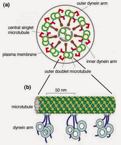
A cross section of an undulipodium axoneme looks like
this – although I’m not sure they’re color coded in the
cell. Notice how the inner are connects the nested tubule
of one doublet to the outside tubule of the next doublet.
The inner arms are responsible for the degree of bend,
the outer arms are involved in the rate of movement. There
are other proteins involved, but we aren’t getting that
detailed. Maybe you want to do that on your own.
The axoneme (axo = axis, and neme = thread) of the undulipodia has a very distinct structure which is best appreciated when you look at it in cross section. The long microtubles appear as small circles in the cross cut, but they are arranged very precisely (see picture). They come in doublets, and there are nine sets of them surrounding a central doublet (called 9 + 2 or 9(2) + 2). This is very different from the single hollow tube of the prokaryotic flagellum.

The way the axoneme is built is also the key to how it works. The different microtubule doublets are cross-linked by protein complexes called dynein arms. There are inner arms and outer arms. An inner arm connects one microtubule from one doublet to another microtubule of the adjacent doublet. When one doublet slides further out from the cell body and the connected adjacent doublet doesn’t, this creates stress and the whole thing must bend to maintain the connections.

So, walking proteins are how the undulipodia create their whip-like action. There is a walking system analogous to dynein arm movement that has developed in many animals. We have looked at muscle contraction before. Just like myosin heads walking along actin filaments that are anchored to the muscle cell membrane, the dynein arms on some doublets start to crawl up or down the adjacent microtubule of an undulipodia creating a bend and then they can reverse to whip back the other direction. This is very different from the spinning motor of the bacterial flagellum.


Pay attention to these cartoons, they show how the undulipodia
bend. ATP powers the sliding of the dynein arms. As they move
down one tubule, that filament moves up. If the filaments are
anchored in the membrane, as they are in undulipodia, the
movement creates tension and a bend. The cartoon on the
right is a good summary.
The motions the two different mechanisms produce is different too (a homophone). While the prokaryotic flagellum spins like a propeller, the sliding of the microtubules makes the undulipodium wave back and forth, like a field of wheat on a breezy day, or how a snake might thrash if you held his head down (don’t just hold his tail down, he’ll bite you).

The undulipodium motion often occurs in just one plane, back and forth instead of all around, but that doesn’t mean it has too limit itself to that. It can spin too; it just takes a very coordinated sliding back and forth of microtubules.

Another difference between prokaryotic flagella and eukaryotic undulipodia is in how they are powered. We saw that flagella in bacteria get their force from the spinning motor, and the motor gets its energy from an ion gradient across the membrane. But in eukaryotes, it was seen early on that if you strip the membrane off of an undulipodium, and added ATP, they start to move.

Yes, all undulipodia are held within the membrane. Some bacterial flagella are membrane covered, but all eukaryotic versions are sheathed in plasma membrane. But back to the ATP. Exposing the naked undulipodium to ATP, even on a dead cell, can initiate the dynein walking and microtubule sliding, so it is definitely ATP powered.

Also, this points out that the power for the bacteria movement comes from the motor in the base, but the eukaryotic movement is in the axoneme, not the base. The basal body that anchors the eukaryotic undulipodium into the cell membrane is amazing in its own way. The basal body is actually a centriole, the same structure that helps to move the chromosomes apart in the spindle apparatus during mitosis. We’ll come back to this double duty organelle in a a few weeks.


Chlamydomonas algae species have a double flagella
for swimming. A single stroke as shown above. The
power strokes on top is followed by the recovery stroke
below. Put all the numbers together and looks like
someone swimming underwater. The picture of the
organism is there because it’s always better to see the
real thing as compared to a cartoon.
Our two systems still look similar, but we see how they are not so similar in structure and action. Since eukaryotic cells are so much bigger than prokaryotes, the first flagella to be examined and found to have the 9(2) + 2 structure were in sperm tails.

But soon after that, the protozoans were discovered to have undulipodia as well. Organisms from the algae genus Chlamydomonas have two long undulipodia that they use for motility. Located at the front of the cell, their movement pulls the alga through the water. But protozoans are just as likely to have undulipodia that push them through water. They can work both ways.

Amazingly, when a Chlamydomonas finds itself out of water, the undulipodia resorb in short order. Nature hates to waste energy, so why maintain a boat motor if you’re not in the water. But place them back in a liquid environment and the two structures will reassemble with in an hour – with the same structure 9(2) + 2 and working the same exact way.

Of course, saying they all have the same 9(2) + 2 structure is an invitation to find exceptions, and science has found them. A 2006 studyfound that rabbits are quite the rule breakers. Sure, they have 9(2) + 2 axonemes, but they don’t stop there. Some rabbit embryo undulipodia show a 9(2) + 0 structure, where the central doublet is completely missing, yet the structure functions just as other motile undulipodia.

What’s more, rabbits can also have 9(2) + 4 axonemes, with double the number of central microtubules. Again, they function just fine. Is there a reason for these variations – maybe, but maybe they are just mutations that didn’t have a negative impact, so they were retained.


You know how annoying it is when you touch a cactus
and those little bristles get stuck in your skin? Well,
don’t touch a bristleworm. They’re lined with those painful
bristles – hence the name. Somebody studied these worms,
and found out they have parasitic protozoans in their gut.
Then someone studied those parasites and found that they
have sperm. And someone studied the sperm and found
that they have unique axoneme structures. I love science.
A couple of parasitic protozoans that live in bristle worm guts show differences in their axonemes. Lecudina tuzetae sperm tails have a 6(2) + 0 structure, while the Diplauxis hatti protozoan sperm has a 3(2) + 0 axoneme. Described in a 1980 paper, this is the simplest motile undulipodium known – as of now.

The undulipodium basal body (born as a centriole) can have exceptions as well. The vast majority have a structure of 9(3) + 0, where instead of doublets, they have triplets. This makes sense since they need to be strong to support the axoneme.

But diatoms, very small algae cells protected by a silica shell, can have sperm that look very different, according to a 2013 study. Their basal bodies have been observed to have doublet microtubules, and are very similar to the axoneme. Even weirder, a couple of insects feel the need to go big with their basal bodies. Acerentomonon microrhinus, a primitive hexapod insect has sperm tail basal bodies with 14 microtubule doublets, while Sciara coprophila, a fungus gnat (see picture), has up to 90 singlet microtubules in its sperm basal body.

We have talked a lot about sperm tails and protozoan motility structures, and these undulipodia look the most like bacterial flagella. But undulipodia come in a couple of flavors; those longer than 40 m or so are called flagella while the shorter ones are called cilia. See the naming problem and why Lynn Margulis came up with undulipodia?


This is a fungus gnat – it sounds like they eat fungus,
but nope. Only the larval form feeds; the adults never
eat. That’s OK, they don’t live very long at all. They
overwinter in their adult form because they are unique
in that they can both tolerate freezing weather and freeze
themselves without damage. It must be important, since
their sperm tails are loaded with 90 microtubules in the
basal body – everyone knows that massive numbers of
microtubules is the best way to avoid cold damage.
Both eukaryotic flagella and cilia have the microtubule and dynein arm structure, with centrioles for their basal bodies. There are exceptions to the cilia that we will look at in a few weeks, but the biggest difference between them, besides their length, is that cilia occur in groups, while flagella are usually found in ones or twos. You could say that cilia and flagella are like synonyms, they have almost the same meaning (and structure), although they are two different things.

Next week – Halloween is coming, so what better time to have a discussion of genetically modified foods and an 19th century teenage girl who wrote the best science fiction book ever.


Prensier, G., Vivier, E., Goldstein, S., & Schrevel, J. (1980). Motile flagellum with a "3 + 0" ultrastructure Science, 207 (4438), 1493-1494 DOI: 10.1126/science.7189065

Idei, M., Osada, K., Sato, S., Nakayama, T., Nagumo, T., & Mann, D. (2012). Sperm ultrastructure in the diatoms Melosira and Thalassiosira and the significance of the 9 + 0 configuration Protoplasma, 250 (4), 833-850 DOI: 10.1007/s00709-012-0465-8

Feistel K, & Blum M (2006). Three types of cilia including a novel 9+4 axoneme on the notochordal plate of the rabbit embryo. Developmental dynamics : an official publication of the American Association of Anatomists, 235 (12), 3348-58 PMID: 17061268


For more information or classroom activities, see:

Undulipodia –


Frankenstein Meets Genetic Modification

$
0
0
Biology concepts – Frankenstein, asystole, ethics, genetically modified organisms, genetically modified foods, synthetic biology, decomposers, electroconvulsive therapy


Mary Shelly was wedded to Percy and friend to Lord
Byron, one of the great poets of the early 19th century.
But she was a fair writer on her own. Note the bolts on the
monster's neck. These were added by make-up artist Jack
P. Pierce. He said they were electrodes, not bolts, even
though Mary Shelly never actually wrote that
the good doctor used electrodes on the body.
Can you think of anything scarier for Halloween than an irresponsible scientist letting his creation loose on the world? Now imagine that his creation is something that violates our sense of decency and our reverence for the dead. Well, that’s the story behind Mary Shelly’s Frankenstein.

Who's the victim of the story? Is it Dr. Frankenstein, who’s family is murdered or is it perhaps his monster, who was brought into the world and abandoned? He lives his second life shunned by all, misunderstood, lonely, unable to live with dignity or even die at all.

This is a big story for a 17 year old to pen. Yep, that’s how old Mary Shelly was when she wrote Frankenstein (published when she was 21 in 1818). Despite her age and inexperience, she rolled out one of the greatest novels ever. It was both romantic and a criticism of romance. It sparked the science fiction genre and was the beginning of horror stories.

The movies and stories about Frankenstein’s monster usually highlight the way in which the monster was created and his ugliness and hatred, but that isn’t what the book is about. It’s a story of responsibility in science and toward others.

The Age of Enlightenment had just ended when Frankenstein was written, and the Romantic period was in full bloom. A switch from science to emotion meant that the facts and discoveries about the world now needed to be examined, not just accepted. Here was Mary found her message – a person must be responsible for the things he/she creates – be they physical things, knowledge, or opinion.


Electrical impulses make muscles move. Adding salt to
freshly skinned frog legs is a lot like hitting the with a
mild jolt of electricity. This is like Galvani demonstrated
with the corpse of the murderer and the image Mary
Shelly evoked in her novella.
The science of the monster’s reanimation was not the focus, but Mary had good knowledge of the latest science of the day, and this is what informed her making of the monster. Sir Humphrey Davy of the Royal Institution of Science had just stated that chemistry would, eventually, control the conversion of dead matter into living matter.

This was combined with the advances in electricity at the time. Just before 1800, Luigi Galvani had published on the ability of electricity to excite the muscles of dead animals – the innate electrical force of living tissue came to be known as “galvanism.” In 1803, Galvani applied an electrical charge to the corpse of executed murderer Thomas Forster, and the body jolted and moved – a good visual for Mary.

So could a body be reanimated as Shelly relates in the novel? Nope…. at least, not yet. Let’s examine why.

Dr. Frankenstein uses a corpse, with some implied modifications through surgery. Not good. Immediately after death, cells that are starved for oxygen stop making ATP. ATP is required to maintain lipid membrane compartments and in general for the integrity of the cell. Once there is no oxygen and no flow of energy, the enzymes designed to break down wastes, toxins, and old organelles for recycling are released to the cytoplasm and start to destroy the cell.

Consider the process of rigor mortis. Muscle contraction requires ATP not to contract, but to release the contraction (see this post). With no ATP, the muscles become rigid in their contraction about 3-4 hours after death. Rigor lasts for about 12-20 hours, and is only released by the process of cell destruction that we described above.


Frankenstein’s monster better have been a very fresh
corpse. Decomposers like bacteria and fungi are already
in and on your body; it’s your immune system that keeps
them at bay. Once dead, we’re all just food for worms,
prokaryotes, protists , fungi, and of course buzzards.
Mary doesn’t describe anything to overcome this problem. But there are other problems as well. No life, no immune system. This is what keeps our fungal, bacterial, and protist flora in check. Without a working immune system, the microorganisms that are normally growing in and on us will be unchecked and start to grow and feed on the corpse. This is where they get their name, “decomposers.” Mary doesn’t mention anyway to overcome that problem either.

What about the electrical problem? We use electricity in our neural system and in our heart. Your brain is an electrochemical machine, using ions to generate and electrical current down axons. Electroconvulsive therapy (ECT) is useful in treating some forms of depression and schizophrenia and mania, but we don’t really know how it works yet.

Several hypotheses exist; none or all of which may be correct. ECT may alter neurotransmitter concentrations, which would change the degree to which impulses are transferred or suppressed from one neuron to another. It may work to prune back some neural connections in the brain, or it may work to stimulate hormone release that could alter the brain chemistry. A 2014 reviewprovides more information on the various theories of ECT mechanism.


From top to bottom we see a normal heart rhythm, and a
then a ventricular fibrillation that CAN be treated with
electric shock. Below that is a pulseless rhythm, which
looks normal but doesn’t move the heart, and then asystole,
with no pulse at all. The bottom two CANNOT be treated by
shocking the heart. That’s why they called it a defibrillator,
not a heart starter.
Electricity is used in the heart as well. We can modulate the rhythm of the heartbeat with a pacemaker, which is just a low voltage shocking device. When a person is dying from a poor heart rhythm (ventricular tachycardia or fibrillation), we can use a defibrillator to shock the heart back toward a normal rhythm.

Mary’s error: electrical shock won’t start a stopped heart (called asystole, a = not, and systole = contraction), despite what you’ve seen on the TV shows. For asystole, the treatment is CPR with a shockingly large dose of adrenaline every 5 minutes.

Dr. Frankenstein couldn’t have reanimated his self-digesting, microbiological dinner plate of a corpse with an electric shock, but if the muscles hadn’t gone into rigor yet, he might have been able to get a short slam dance out of him. But then, this isn’t really the point of the story.

Frankenstein’s monster was alive and wandering the world alone a mere 1/5 of the way through the story, so it’s really a story of how to deal with the products of science. Erasmus Darwin (Charles’ grandfather) had introduced the idea of mutations or “monstrosities” being passed on or inherited – so Mary now had the essence of the story. Who is the monstrosity - the monster or the scientist who creates and then abandons it?


Erasmus Darwin was Charles granddad. He was an
inventor, poet, natural philosopher and I hear a great
cook. One of his poems predicted the discovery of the
Big Bang, he also suggested the idea of natural
selection and mutation and sketched out a liquid
oxygen and hydrogen rocket.
One indication that a story is a classic is whether its themes are applicable in different eras. Frankenstein may be even more applicable to our times than it was to Mary’s. Current debates are boiling over concerning the uses and limitations of science.

The issue most often compared to Frankenstein’s monster is genetically modified organisms (GMOs). Have you heard the term, “Frankenfoods?” This is the name that opponents of GMOs and particularly GM foods use to taint the agricultural biotechnology industry.

The fear is that by tampering with nature and introducing genes into organisms, we are creating monsters that might have unexpected effects on us. It’s a good marketing campaign idea, and it has stuck in the minds of the public.

Europe and Russia have banned all GM foods out of fears that they may contain toxins or mutagens that would harm the consumers. One fear is that DNA from the genetically modified organisms would be transferred to the eater and combine with their own DNA. That is a scary sounding idea.

The problem is, you take up DNA from the food you eat every day, although not whole genes as the fear warrants. Digestion breaks down DNA, so we take up mostly nucleotides and short stretches of nucleic acid. No recorded evidence exists of uptake of an entire gene.


Dr. Frankenstein used all natural body parts, no artificial
sweeteners, additives or preservatives, and good old-
fashioned electricity. If he was sold in the market, the
monster could be labeled as organic! No genetic
modification here.
Websites and books talk about the dangers of GM foods, but the evidence hasn’t shown up in the scientific literature. What few papers have announced negative ramifications of GM foods have been retracted or have such vague conclusions as to apply to any food at all. I’ll give a typical example.

In 2012, a researcher named Seralini from the U. of Caen announced that an herbicide used with GM foods (glyphosate in Roundup) causes tumors. He didn’t just publish it, he had a press conference and concurrent release of his book on the subject and videos in three different languages. It turns out that he also had a company that was preparing to market a product as a “protectant” against glyphosate. The study was subsequently retracted, but a modified version with a conclusion that “more study is needed” was re-published in a lesser journal (see note below).

Other studies on the dangers of GM foods have been correlative, meaning that when you see “A”, you often see “B.” But that doesn’t mean that A causes B, or that B causes A. Remember this: correlation does not imply causation.

The truth is, we need more studies. There are real issues to be dealt with, such as - does introduction of a particular gene cause plant toxins to be increased – this could be bad for us. The idea is the same as in Mary Shelly book – we must be responsible for those things we make. No GMO or GM food should go to market without extensive testing.

The testing to date shows that there are no health risks associated with GM foods. Longitudinal studies from 2014, 2013, and 2012 of live stock feeds showed that animals fed GM crops over five generations showed no ill health effects and their meat was exactly like that of animals fed conventional feed. By the middle of 2013, over 600 studies showing that GM foods carried no health risks had been peer-reviewed and published. The key is always the same - responsible and thorough testing.


Synthetic biology has arrived. Vanilla is a very expensive
crop to produce. But a gene has been constructed and
vanillin is now produced in yeast. They ferment sugar and
produce vanillin. This is more natural than artificial vanilla,
and contains many of the metabolites that make vanilla
taste like vanilla.
The problem of hidden agendas like Seralina's does go both ways; a 2014 editorial on the safety of GM foods was written by a Monsanto employee, the company that markets GM corn and soybeans. Society must be diligent and demand topnotch, transparent, and responsible science. This was one of Shelly’s themes, Frankenstein conducted his work in private, with no comment from society about how or whether it should be done at all.

The next generation of people will have more issues to deal with, like synthetic biology (not merely taking a gene from one organism and putting it another, but constructing a gene or genes from nucleotides and then inserting them). How to ensure good use of science? - transparent methods and results, no hidden agendas, no jumping to conclusions, and a very science literate population that can judge and reason for themselves. And that’s why we learn biology.

Next week - Halloween is a time to focus on what's scary and what's dead. Can you actually be scared to death?


The retraction of the 2012 study of Seralini in Food and Chemical Toxicology can be found here. It was republished in modified form in the journal, Environmental Sciences Europe in 2014, but with no peer-review.


Goldstein, D. (2014). Tempest in a Tea Pot: How did the Public Conversation on Genetically Modified Crops Drift so far from the Facts? Journal of Medical Toxicology, 10 (2), 194-201 DOI: 10.1007/s13181-014-0402-7

Tufarelli V, & Laudadio V (2013). Genetically Modified Feeds in Poultry Diet: Safety, Performance and Product Quality. Critical reviews in food science and nutrition PMID: 24915369

Van Eenennaam AL, & Young AE (2014). Prevalence and impacts of genetically engineered feedstuffs on livestock populations. Journal of animal science, 92 (10), 4255-78 PMID: 25184846

Snell C, Bernheim A, Bergé JB, Kuntz M, Pascal G, Paris A, & Ricroch AE (2012). Assessment of the health impact of GM plant diets in long-term and multigenerational animal feeding trials: a literature review. Food and chemical toxicology : an international journal published for the British Industrial Biological Research Association, 50 (3-4), 1134-48 PMID: 22155268

McCall WV, Andrade C, & Sienaert P (2014). Searching for the mechanism(s) of ECT's therapeutic effect. The journal of ECT, 30 (2), 87-9 PMID: 24755719

 
For more information or classroom activities, see:

Genetically modified organisms/foods –


Death By Haunted House

$
0
0

Halloween is a time when fear is invited. The rush of
adrenaline in a controlled environment is life-
affirming. Not much else to comment on here,
except that he seems to have excellent oral hygiene
for a chainsaw-wielding maniac.
A big man with the chainsaw and the gaping wound on his face jumps out from around the corner and growls. You leap backward and scream, your heart pounding in your ears. You’re ready to either take that power tool and teach him a lesson or to run like the kid from Home Alone. Sure you're scared, but could it kill you?

Haunted houses are great examples of stimuli that induce the fight or flight response. The name suggests that two mechanisms are fighting it out, but there is really only one biologic pathway. Whether an animal tries to escape or tries to defend itself, its muscles and mind need to be ready.

In response to a threat, the brain triggers the release of epinephrine and cortisol from your adrenal glands into the blood. As a result, your heart beats faster and stronger, your blood vessels dilate to move more blood, and your lung vessels dilate to exchange more oxygen for carbon dioxide. Equally as important, your liver breaks down glycogen (a sugar storage molecule) to glucose and dumps it into your bloodstream.

All these processes work together to increase your alertness and increase the power of your muscles for a short time - like when mothers who lift cars off their small children. You are now ready to respond to the threat; however, there is an exception – you may do nothing at all.

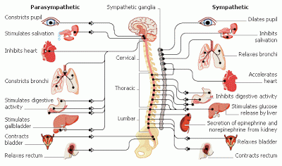
One of the major control mechanisms of the fight or flight response is the autonomic nervous system. This is part of the peripheral nervous system (PNS, outside the brain and spinal cord) and transmits information from the central nervous system to the rest of the body. The autonomic system controls involuntary movements and some of the functions of organs and organ systems.

Parts of the autonomic system acts like a teeter-totter, it's their relative balance that controls the outcomes. In the fight or flight response, the sympathetic system predominates and your heart rate increases and your blood vessels dilate.

The autonomic nervous system is divided into sympathetic
and parasympathetic. Much of the sympathetic innervation
comes from the thoracic and lumbar regions, while most
parasympathetic innervation is carried by the vagus nerve.
You can see that the two systems have largely opposite effects.
Butwhat if the parasympathetic systemgained an upper hand for a short time? The parasympathetic system controls what is sometimes called the rest and digest response – the opposite, get it? The heart slows, the blood vessels constrict in the muscles, blood moves from muscles to the gut, and glycogen is produced from glucose. Remember the old adage - don’t swim after your dine; eating puts you in a parasympathetic state of mind! (O.K., I just made it up)

Many people have had the experience of parasympathetic domination coincident to a threat, for some folks it proceeds long enough to have an observable result – they faint. The vagus nerve (a primarily parasympathetic cranial nerve) controls much of this response, so it may be called the vasovagal response. The parasympathetic-mediated reduction in blood oxygen and glucose do not spare the brain - and when your brain is starved of oxygen and glucose, you pass out. Fighting or fleeing is difficult when you are unconscious.

Lower animals will faint as well, but they have additional defenses along these lines. Mammals, amphibians, insects and even fish can be scared enough to fake death – ever hears of playin’ opossum?

There are overlapping mechanisms for feigned death, from tonic immobility (not moving) to thanatosis (thanat = death, and osis= condition of, playing dead). When opossums employ thanatosis, they fall down, stick their tongue out, and even emit a foul smelling odor from glands around their anus. One study in crickets showed that those who feigned death the longest were more likely to avoid being attacked, so this is definitely a survival adaptation – except for the opossums scared by cars and decide to play dead in the street.

Feigned death deters predation, so being scared ain’t all bad. Many predators won’t eat something that is already dead, so not moving could protect them from attack. Another theory is the clot formation hypothesis; it contends that slowing the heart and blood flow forces blood clots to form faster. This will reduce the amount of blood lost during an attack, improving chances for survival.

New evidence is suggesting that even humans undergo tonic immobility. Post-traumatic stress patients asked to relive their trauma show definite signs of tonic immobility, although first they show signs of "attentive immobility," which is more voluntary then the tonic form.

I highly recommend this new book for popular
biology and medicine readers. Zoobiquity explores
a powerful reality. No disease--whether physical or
psychiatric--is uniquely  human. We have much to
learn from animal patients and from the doctors who
care for them.  The impact on human medicine
will be significant.
We havediscovered one exception to the rule; instead of fight or flight, it is really fight, flight or faint – but can we take it further? Should it be fight, flight, faint, or fatality? The answer is yes, but it's very rare. Sometimes animals (including us) don’t just feign death when afraid – they actually die.

In their book, Zoobiquity, What animals can teach us about health and the science of healing, Barabara Natterson-Horowitz and Kathyrn Bowers talk about capture myopathy in animals. Small traps that limit movement, cause pain, or are associated with loud noises can cause spontaneous death in live-trapped animals. Several decades ago it was not unusual for 10% of trapped animals to die. In birds, the death rate often rose to 50%! More humane methods of live trapping have reduced the death rate, but point is made – these animals were scared to death.

A human analogy of capture myopathy may have been identified. People that have had a sudden emotional shock, perhaps the death of a loved one, some other tragic occurrence, or crippling fear can undergo something that looks a lot like a heart attack, even if they have no history of heart disease.

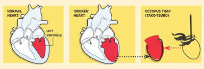
This sudden loss of heart rhythm has been called broken-heart syndrome, but is more accurately termed stress cardiomyopathy (SCM). In these cases, the heart actually changes shape! The part of the heart that pumps blood out to the body (left ventricle) balloons out and loses the ability to pump efficiently. Dramatically less efficient pumping leads to symptoms just like a heart attack.

In the normal left ventricle of the heart (left image), the muscle is
thick around the space (in red) and contracts strongly. In SCM, the
space is ballooned at the based (middle image) and the muscular wall
is thin, giving a weak contraction. The change in shape can be seen in
the superimposed images on the right, as is the octopus trap (tako-tsubo)
that the original Japanese describers thought the lesion looked like,
hence the early name takotsubo cardiomyopathy.
In most cases, SCM and the change in heart shape resolve after a time and there is little left to show they were present, but if they go too far for too long, they can cause death – called sudden cardiac death. There are many causes for sudden cardiac death, but emotion and fear are definitely among them.

I was wondering if there was a link between SCM in humans and capture myopathy in animals, so I asked Dr. Natterson-Horowitz. She told me that those studies have not been done yet; we don’t know if there is a heart shape change in captured animals. I think it would be hard to get approval for studies that would intentionally scare animals to death.

One interesting connection amongst fight or flight, capture myopathy, and SCM is the catecholamine dump involved. Epinephrine and norepinephrine control all three responses, and in humans they control even more. Recent evidence shows that catecholamines mediate the production of fear memories.

You remember fearful events more readily and more vividly as a survival adaptation. Strong memories help you to avoid dangerous situations in the future. In this way, your mind can affect how your body responds to a threat. We will see this again in just a bit.

All babies have an exaggerated startle reflex until
they are several months old, but in some cases it may
contribute to SIDS. An exaggerated startle can lead to
apnea (temporary breathing cessation) and this can be
compounded by a depressed heart rate if the baby is
sleeping on its stomach. Some clinicians also theorize
that swaddling may contribute by exaggerating the startle
due to confinement stress, but by far the greatest
association with SIDS is stomach sleeping.
However, dying or nearly dying from fright isn’t all in your head either; some conditions can predispose you to dying from a sudden shock. One unfortunate condition is called hyperekplexia, or startle disease of the newborn. Newborns with one or more of several mutations in the glycine receptor (an inhibitory receptor in the brain used in neuron signal transmission) can lead to these babies dying from loud noises or a sudden touch.

The startle reflex involves squinting to protect the eyes, raising the arms, hunching the body to protect the back of the neck, as well as inducing the fight or flight response. With the loss of inhibitory signaling, the signals that ramp up a startle response are unchecked and can lead to uncontrolled beating of the heart (ventricular fibrillation, VF) and sudden cardiac death.

Just as some cases of the fight or flight response going too far, the startle can sometimes lead to VF. A recent study has shown that the bigger the perceived threat, the bigger the startle reflex will be. Also, if there is a fearful environment prior to the threat, then the startle will be bigger. Once in a long while, it goes too far.

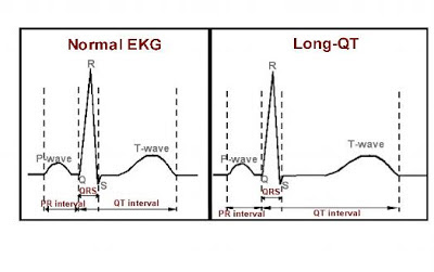
Similar to hyperekplexia, there is another condition that could lead to VF and death in the environment of fear. Long QT syndrome can either be inherited or acquired later in life, and affects the time between beats of the heart. In long QT, the interval is variable and longer, and can lead to inefficient beating and VF.

On echocardiogram tracings a heartbeat has a certain shape, and 
each point has a corresponding name which is represented 
by a letter. If the time between the Q point and the T point 
is too long, the heart rhythm is subject to disintegrating 
into chaos. In the 1990’s, the antihistamine Seldane was 
taken off the market due to QT interaction when it was 
given with the antibiotic erythromycin.
Highlightingour circle of fear and the body, evidence presented here and here suggest that SCM can cause acquired long QT syndrome. Dr. Natterson-Horowitz said today many patients with long QT may have implantable defibrillators. In earlier days, however, these patients were warned not to use alarm clocks or to jump into cold water – they could startle themselves to death.

Long agowe talked about premature burial. It would be easy to envision a person waking up inside a coffin, and then dying from the fright of being buried alive! Does this mean that you are putting yourself in peril every time you visit a haunted house at Halloween? Probably not, remember that deaths from fright are exceedingly rare. Maybe you could just feign death, and the horrible monster will leave you alone.

For the next couple weeks - back to the science of flagella. Undulipodia are present in many phylums, except for where they aren’t. On the other hand, some types of organisms don’t have undulipodia - except for those that do.


Greek, R. (2012). Zoobiquity: What Animals Can Teach Us About Health and the Science of Healing. By Barbara Natterson-Horowitz and Kathryn Bowers. Knopf Doubleday Publishing: New York, NY, USA, 2012; Hardback, 320 pp; $16.23; ISBN-10: 0307593487 Animals, 2 (4), 559-563 DOI: 10.3390/ani2040559

Volchan, E., Souza, G., Franklin, C., Norte, C., Rocha-Rego, V., Oliveira, J., David, I., Mendlowicz, M., Coutinho, E., Fiszman, A., Berger, W., Marques-Portella, C., & Figueira, I. (2011). Is there tonic immobility in humans? Biological evidence from victims of traumatic stress Biological Psychology, 88 (1), 13-19 DOI: 10.1016/j.biopsycho.2011.06.002


For more information or classroom activities, see:

Fight or flight –

Autonomic nervous system –

Thantosis/tonic immobility –

Stress cardiomyopathy –

Hyperekplexia –

Long QT syndrome -


Almost This Or Almost That? Must Be The Other

$
0
0

Biology concepts – Protista, taxonomy, phylum, kingdom, monophyletic, paraphyletic, cladistics, algae, diatom, dinoflagellate


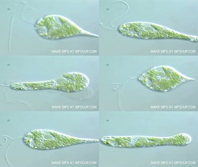
Euglena gracilis is an organism in the Kingdom Protista. It has
one long flagellar undulipodium, but it can also move by
amoeboid movement. It has chloroplasts and can do
photosynthesis, but it also can eat other organisms. Is it any
wonder that classifying protists is so hard?
Classifying living organisms is self-perpetuating job. Imagine if the dentist sold candy in his/her office, “Here’s your root canal and your Laffy Taffy.” Scientists try their best, but whenever you start sorting things out, you always have that pile left over that doesn’t seem to fit anywhere. So you have to rethink your categories and try again.

The best example of the inanity of classification is Kingdom Protista. The word means, “the very first,” probably because it is supposed that these were the first eukaryotes. How do we define the organisms of this Kingdom? The best we can manage is to say that they are the eukaryotes that aren't animals, plants, or fungi. Really, is that the best we can do?

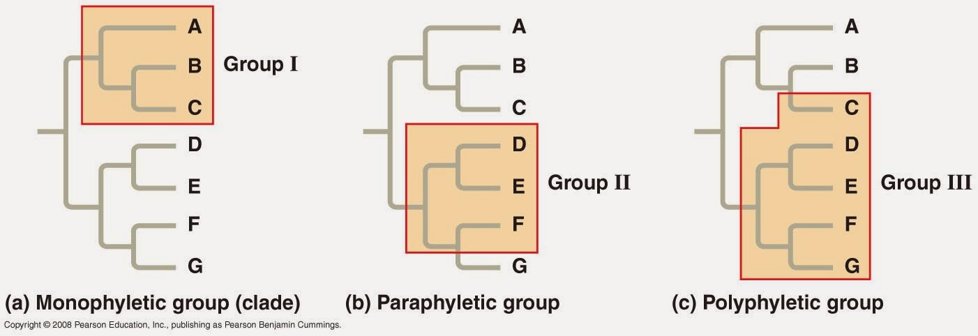
In a perfect system, all the organisms of one kingdom would be descended from a single common ancestor (be monophyletic, mono = one, and phulon = tribe). But it don’t work like that. And this is where Kingdom Protista serves as a good example.

There are protists that look a lot like animals, those that resemble plants, and those that share features of fungi. No way did they all come from a single ancestor. Protista is a paraphyletic(para = near) kingdom, the group may exclude a member with a common ancestor. As such, the protists are a catch-all, those that don’t fit in some other kingdom. Protists are like pornography – hard to define, but you know it when you see it.


Classification isn’t perfect, some groups come from
different ancestors. A shows a group (in yellow) that
is monophyletic, they all come from one ancestor.
The paraphyletic group (B) shows that some groups
can’t include all descendents of a common ancestor.
And if a group is made from descendents of different
ancestors, it is called polyphyletic (C).
We already said that some behave like animals, plants, or fungi. Some are unicellular, some are multicellular, and some can be either. Some do photosynthesis, while some eat other organisms. How can you break these up into phyla, classes, orders, families, genera, and species when they are all so different?

You might do it by common ancestor; let their genes do the talking. We are learning more and more about who begot who – this is the study of cladistics. But if you break down protists into their clades – they don’t seem to make sense. Organisms that look or act similar might be in different clades, with wildly different organisms linked close together.

Alternatively, you might divide them up based on the characteristics, as Linneaus did - the animal-like protists in one phylum, the plant-like protozoans in another. But this may separate genetically related organisms into very different phyla. Same problem. How about by the way they get around? Some use flagella-like undulipodia, some use undulipodia called cilia, some use cytoplasmic crawling called pseudopodia, and others are immotile. Again, disparate organisms may be lumped together just based on their preferred mode of travel.

The idea of the "phylum" is to place the organisms in categories so that they are “more related to each other than they are to any other group.” Wow, that sounds scientific. Related based on what? We just discussed motility, genetics, and physical characteristics or behaviors.


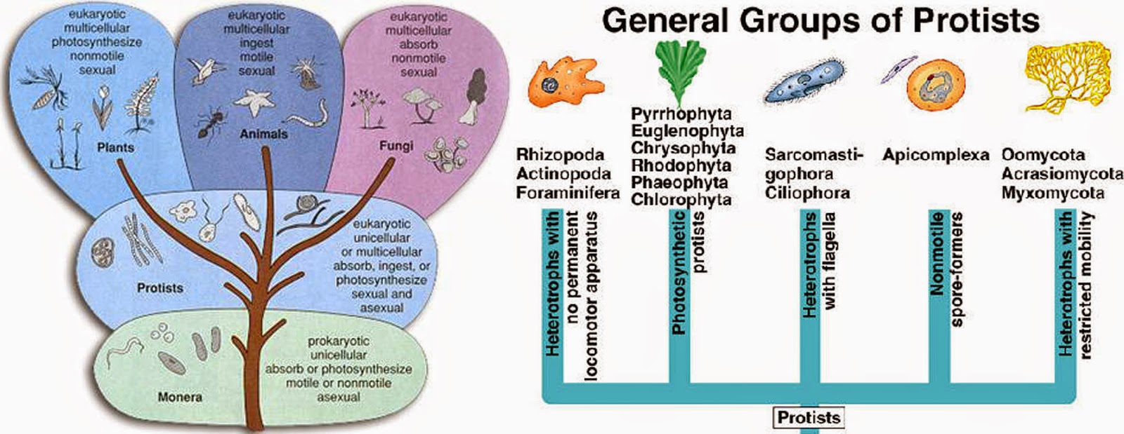
The Kingdom Protista sits between the modern plants,
animals, and fungi, and the ancient prokaryotes. As such,
they end up being a catch all group. The right image
shows how some people group the protists based on
undulipodia characteristics, not ancestry.
And this assumes that we even know how related they are to each other and to organisms outside each phylum. Genome studies haven’t even begun to get close to establishing the ancestral relationships between all the organisms.

So we guess. And then we change things as new information becomes available. The work never ends, and the students never get to just memorize the categories.

As of today, some scientists classify protists based on a combination of the characteristics above. In the system I like best, there are 15 phyla, and we can roughly divide them as we show below. But there are six different phyla just for the protists that perform photosynthesis! The reason I like this system best -it roughly mimics the way they use undulipodia. And this is what we’re interested in today.

Kingdom Protista contains the organisms that seem to have made the most obvious uses of undulipodia. Eukaryotic flagella and cilia abound, some protists have both, and some have them only part of the time. There are six phylums of plant-like protists. Many have flagella, none that I could find have cilia. Here are some examples:


Pyrrophyta organisms will bloom and then bioluminesce
in order to scare predators away. Movement in the water
causes vesicles in the dinoflagellate to rupture via action
potential and release the reagents to make light. It’s exactly
the same system that fireflies use.
Phylum Pyrrhophyta The dinoflagellates are in this phylum; they have two flagella, one from that side that beats and one on the posterior that whips more traditionally. Some species of this protist are responsible for the red tides that poison fish and can (and have) killed humans who eat the fish. Other dinoflagellates are bioluminescent and make the water appear to be on fire (hence the phylum pyrro = fire).
             
Phylum Euglenophyta This phylum includes the Euglena gracilis organism shown in the animation at the beginning of the post. These protists also have two flagella, but one of them is reduced and doesn’t stick out. They have an eyespot, perhaps the genesis of our own eye. The eyespot helps them to move away from strong light sources, sources that would overheat them.

Euglena are common model organisms, on this world and in (near) space. They traveled on the parabolic flights to have their flagellar motions studied in zero gravity. The 2010 publication that resulted from the experiments showed that the process of beating is regulated and physiologic, as the change from hypergravity to microgravity stopped the flagellum from moving. The opposite change in gravity reoriented the cells and they started swimming to the bottom of their tank again.

The remaining phyla of plant-like protists can be included in a supergroup called the Chromista(colored organisms).  In terms of their undulipodia, the chromists tendto have two flagella, one on each end. The forward flagellum is usually longer and has lateral growths called mastigonemes. The best description for this type of flagellum is that it looks like Christmas tinsel.  The back flagellum is shorter and smooth.


These are the phyla of the Chromista; the colored protists.
Problem is, not all of them are colored and some colored
protists aren’t included in this group. Top right and bottom
left are the chlorophyta, the green algae. These are the most
recognizable algae. The bottom right is the diatoms, they have
the most interesting shapes. Look them up.
The Chrysophytaare the golden algae and diatoms. The diatoms are only flagellated when undergoing sexual reproduction, and is just the male gametes that have the flagella, sounds like male gametes in mammals doesn’t it?
  
Green algae are the ones we recognize; they belong to the Chlorophyta phylum. These are the ancestors of the land plants, and some have flagella in all stages, while others only have flagellated gametes.  We will see soon how some land plants still have flagellated gametes.

Brown algae belong to the Phaeophyta phylum. They are exceptional amongst the protists because every organism in this phylum is multicellular. No brown algae live as individual cells. Kelp is an example of brown algae. Kelp forests are multicellular example of brown algae thalli, growing to 40-60 m (130-200 ft) in height! Kelp forests are some of the most productive ecosystems on earth.      
The gametes of the brown algae are flagellated like in most of the other chromists. A 2014 study has started to look at the flagella of the chromists, using brown algae as the model organism. The study found that the flagella have functions in motility, signal transduction, and even metabolic activities.  The two different types of flagella had common proteins and proteins specific to each form, for a total of 495 different proteins associated with flagellar function and structure. For instance, only the posterior flagellum has a protein that senses blue light, and may be used for steering the organism. 


The Rhodophyta are where we get food stuffs. On the left
represents agar that can be used to make things that are like
Jello, it fills the role of the gelatin. In the middle, agar is also
used as a polysaccharide source of nutrition for growing
bacteria in the lab. On the right, nori is a rhodophyta
seaweed used in sushi.
Finally,Phylum Rhodophyta is the last of the Chromists. They are known as red algae, but you may know this protist better as seaweed. We saved them for last because they are the biggest exception in the plant-like protists.

If you’ve eaten Japanese sushi rolls, then you’ve eaten red algae in the nori that the rice and fish are wrapped in. Nori is made from several species of red algae of the genus Porphyra. Not a sushi fan? How about ice cream? Carageenans that make ice cream smooth also come from red algae.

Ice cream is reason enough to love the red algae, but there’s more. A 2014 study indicates that one compound found in the Porphyra is a strong antibiotic. Studies of 1,8-dihydroxy-anthraquinone from this red algae genus can disrupt the cell wall of Staphylococcus aureus. This is hugely important, since many strains of S. aureus (like MRSA and VRSA) are now resistant to most existing antibiotics.

Rhodophyta algae are red because although they use some chlorophylls for photosynthesis, they also use phycoerythrins and phycocyanins. Interestingly, these are the same pigments that are present in the cyanobacteria. This suggests that there is an ancestral link. The link is supported by one other factoid. Both cyanobacteria and red algae lack undulipodia!


The seaweed Rhodophyta organisms often live in the tidal
pools. The spongy material in the stalks and “leaves” is the
agar and is related to the mucin product that attaches to the
male gametes as they are released. I couldn’t find a picture of
the gametes with their mucin tails, so this will have to do.
The male gametes of red algae are at a deficit; they don’t have flagella to swim toward the female eggs. They must relay on water movement to disperse them. An older study showed that when the male spermatia are released by the discharge from vesicles, the vesicle contents can hang on to the gametes and form mucin appendages. These are then more likely to be moved around by the water.

Whatever it is, the system seems to work. A 2014 study found that fertilization success was dependent on male organism biomass, but neared 100% when there were relatively few male gametes present. This was hypothesized to be possible because low tides in the tidal pools where the organisms live greatly increase the chances of male/female interaction and fertilization. Seaweed takes advantage of the moon’s effect on the tides to ensure reproductive success – who needs flagella!?

So far we have met protists that use flagella at some point in their life cycle (except for the red algae). Notice that none of them have used cilia. Next week, how about the animal-like protists? I bet there are some exceptions there as well.


Fu G, Nagasato C, Oka S, Cock JM, & Motomura T (2014). Proteomics Analysis of Heterogeneous Flagella in Brown Algae (Stramenopiles). Protist, 165 (5), 662-675 PMID: 25150613

Wei Y, Liu Q, Yu J, Feng Q, Zhao L, Song H, & Wang W (2014). Antibacterial mode of action of 1,8-dihydroxy-anthraquinone from Porphyra haitanensis against Staphylococcus aureus. Natural product research, 1-4 PMID: 25259418

Maggs CA, Fletcher HL, Fewer D, Loade L, Mineur F, & Johnson MP (2011). Speciation in red algae: members of the Ceramiales as model organisms. Integrative and comparative biology, 51 (3), 492-504 PMID: 21742776

Strauch SM, Richter P, Schuster M, & Häder DP (2010). The beating pattern of the flagellum of Euglena gracilis under altered gravity during parabolic flights. Journal of plant physiology, 167 (1), 41-6 PMID: 19679374


For more information or classroom activities, see:

Kingdom Protista –


Euglena –

Red tide –

Pyrrophyta –

Kelp –


Bacteria Are Intelligent Designers

$
0
0
Biology concepts – nature of science, flagella, intelligent design, irreducible complexity, motility, Gram+, Gram -, ion gradient

You don’t believe it now, but in the weeks ahead we’re going to discuss how bacterial motility, plant reproduction, intelligence, and the location of your heart are all related to whips and eyelashes. Sounds preposterous, but give me a few posts and a little leeway and you’ll be amazed.


Cheetahs can cover about 25 body lengths in a second, but
some Salmonella can move 60-80 of their own lengths in the
same time! See this post for finding out what the fastest
organisms are. Salmonella typhi is the bacterium that causes
typhoid fever and is spread in contaminated water or touch.
Mary Mallon was blamed for 51 cases of typhoid fever as a
carrier (no symptoms but still sheds bacteria). A 2013 study
shows that the bacteria turn on a fat regulator, PPAR-delta, in
macrophages which lets them live inside the cells forever.
Let’s get right to it. Bacteria are small, but they’re quick little devils. They have inboard motors – or are they outboard? – I can never keep those straight. This piece of machinery is so complex and fascinating that some people use it as a sign that someone or something had a hand in designing life on Earth.

The bacterial motor is called the flagellum, but it's so much more than just a way to get around, it’s often the means to saving their own lives. The word flagellum comes from the Latin word flagrum meaning whip, so you can see we are already starting to work on our challenge for these posts. Flagrum could also mean scourge, and this seems to be prophetic, since many flagella (the plural) we study have a hand in causing disease.

In typhoid fever, a potentially deadly disease that affects more than 20 million people each year, flagella are important not just for putting the bacterium, Salmonella enterica typhi, in the correct place to cause disease, but for attaching the bacterium to the gut wall and for invasion of the gut. A study in 1984 showed that even flagella that couldn’t move were still needed for S. typhi to produce disease. More about this in a couple of weeks.

A flagellum is very much like a boat propeller, it spins to produce force along its axis. This is possible because flagella aren’t perfectly straight. One of the main components of the flagellum is called the hook. This hook is located just outside the cell wall and lies in between the basal body and the filament. The basal body is the engine and is what attaches the flagellum to the cell, while the filament is the long whip like end that sticks out into the world.


A cartoon of the bacterial flagellum shows the structures,
filament, hook, and the entire lower part is the basal body
(motor). The filament is hollow and sends new flagellin
subunits up through it to be added on to the end. The electron
microscope image shows the basal body. It looks like art or
engineering.
The hook turns at about 90 degrees, but the degree of turn is different for different bacteria. This means that that filament, when spun by the motor in the basal body whips around in a circle, bigger at the bottom where the hook is located. So instead of rotating like a straight pencil and not generating any forward force, it spins like a propeller.

The basal body attaches the filament and hook to the cell, and is made up of several rings. In Gram+ bacteria there are two basal body rings that anchor the flagella apparatus, the M ring which attaches to the membrane and the P ring which is anchored in the peptidoglycan layer. In Gram- bacteria, the basal body is longer and has more rings since it must anchor the flagella into the LPS (the L ring) and the M ring has a buddy in the inner membrane called the S ring. All these rings support the rod, which is turned by the rotor and then spins the hook and the filament.

The filament is pretty cool. It’s either a left- or right-handed helix of subunits of a protein called flagellin. The filament is a prescribed length in each bacterium, but we aren’t exactly sure how the length is regulated. Scientists know that it grows faster at first and then slows down, but if broken it will start to grow again at the faster pace.


The filament of the bacterial flagella is capped by a small
protein called FLiD. This is an amazing protein that
regulates and mediates the assembly of the filament
subunits of flagellin at the tip of the growing filament.
The flagellin units are straight as they travel through the
middle of the filament, but their final shape is bent. The
FliD mediates this folding at the tip.
The amazing thing is that the filament grows from the tip, not the base where it attaches to the hook. The flagellin filament is hollow, and subunits of the protein travel from the cytoplasm up through the basal body and hook and then through the existing filament out to the end. Then they are attached to make the filament longer. That’s a pretty neat system because it alleviates the need for a way of exporting the parts, regulating their movement to the end of the filament and then attaching them. Sometimes, but only sometimes, evolution finds the simpler way to do something.

The energy for the motion of the flagellum comes from the movement of ions across the membrane of the cell. We have seen before how protons (or other ions) being pumped out and then allowed to enter through a pore can create the force needed to do work. That’s how ATP is made, how the neural action potential works, and how photosynthesis proceeds. But here, the proton motive force is used to spin the hook and the filament, driving the bacterium forward.

The flagella spin one way to move forward, but when they spin the other way, the bacterium just sort of tumbles around. We’ll talk more about this next time. We’re just now starting to learn how the motor can go from spinning counter clockwise (forward motion for a left handed filament) to spinning clockwise in no time whatsoever and without slowing down. Nothing looks very different in the basal body, the hook or the filament, but the direction of spin is reversed.


This is a complicated picture so stick with me. A) is the shape
of the FLiG protein from a certain bacterium. The end we are
interested in is red, it holds the charge for interacting with the
ion gradient across the membrane. B) shows the positive and
negative bubbles of charge in the helix. Below, see the ring of
FLiG proteins of the rotor. When spinning different directions,
the positive and negative bubbles are reversed, one shape
makes it go clockwise, the other, counterclockwise. It all has to
do with the pushing and pulling by same and opposite charges
as the ions pass through the membrane.
I can hear you thinking out there, “Well, just reverse the direction that the protons move, instead of outside to inside, go inside to outside.” Nope, when a flagellum switches direction, the protons keep moving the same direction. We do have information that one of the proteins that connect the motor (electrochemical gradient) to the physical turning (rotor), a protein called FLiG, can change shape.

Several studies have shown this change, and it is hypothesized that the change moves charged amino acids of FLiG around in relation to the cation gradient. By changing them, it changes the direction of the turning of the rotor (see the picture to the right). This might be akin to reversing the poles of a mag-lev train by flipping the electrical charge can make the train go the opposite direction.

Different bacteria have flagella that look similar but they have small differences. Nevertheless, it can be seen that this is a very complex machine for such a supposedly “primitive” domain of organisms. We have to remember that bacteria have been here the longest; they must be doing something right. There are over 40 genes that are required to build a flagellum, and they all fit together just so.

This complexity and order leads some people to declare that flagella couldn’t have evolved on their own. The concept is called irreducible complexity. People who support the idea of intelligent design (ID) say that some biologic components are so complex and have so many working parts that they could not arise through a series of mutations.

All the parts of a flagellum must be present for it to work (therefore they say it is irreducible) and must be assembled all at once which suggests it could not be random (complex in ID means improbably occurs by chance). Therefore, a flagellum could not have evolved over time and, ipso facto, it must have been designed as one unit by someone or something.

ID proponents haven’t always focused on the flagellum. They first talked of the blood coagulation cascade as irreducibly complex, but then it was shown that portions of the cascade were not necessary for function – whales don’t have factor XII and jawless fishes only use about half the proteins that vertebrates use. It was also shown how the cascade evolved over time.
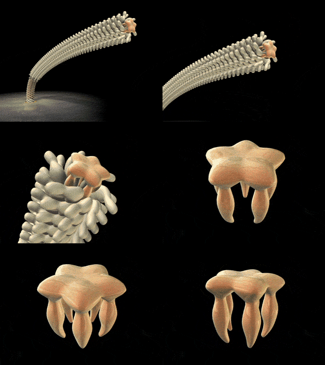

A vibrio bacterium can make two different flagella types,
signified by the two sides of the dotted line above. The ions are
different that run the gradient, and the genes are different for
the motor/rotor. Are there two different irreducibly complex
flagella or did one modify into the other – then they aren’t
“complex.” Vibrio vulnificus is shown on the bottom. It has been
unusually numerous this summer (2014) and causes a disease
that looks like flesh eating disease, but isn’t.
Over the years, ID has proposed that the eye, the immune system, the flagellum and the eukaryotic cilia and its production system were irreducibly complex.  But each time, the ideas of specified, irreducible and complex (must have all come together at once) have been refuted for each example.

For the bacterial flagellum, arguments against ID include the facts that different bacteria use different systems, although they are all variations on a theme. One exception is the Vibrio. They use two different kinds of flagella on the same cells, each needing its own genes. Likewise some bacteria don’t use protons for the gradients, they use Na+ ions. The bacterium Vibrio parahemolyticus is an exception in both cases.

It uses a single flagellum at its end (polar) to swim in liquid water, but many flagella all around its cell when in something thicker. The polar flagellum uses Na+ ions to drive the rotor, while the lateral ones use protons. The genes are different for each flagellar type and mutations in one don’t hurt the other.


The top cartoon shows that when a gene duplicates (and they
do, often) one copy can drift and acquire mutations without
hurting the cell. This can lead to better function or new
function. Over generations, one set of genes for a function
can be replaced with another set – this would hardly be
called irreducibly complex. On the bottom, you see the type III
secretory system for injecting bacterial toxins on the left and
the flagellum on the right. They are very similar, so why is the
flagellum irreducibly complex and the not the type III system?
Lastly, only some Vibrio and other bacteria have a protein sheath over their flagellar filaments. These protein sheaths cover the filament and aid in sensing changes in chemicals outside the cell. So which flagellar type is irreducibly complex and which is not?

Spirochetes don’t even have flagella that protrude from the cell, they’re located between the inner and out membranes (endoflagella). This is a different system and again argues against irreducible complexity in flagella, unless different systems were designed differently. More about spirochete motion next week.

Please read more about ID and decide for yourself if it holds up to the tenets of science - that something that is true must be observable, repeatable, and able to be refuted if incorrect. Irreducible complexity is refutable, and has been for every example proffered by ID. But the conclusion that ID draws – that a designer must be involved, is a belief not a hypothesis – you can’t refute a belief, it doesn’t rely on observable evidence, therefore ID is not science. It doesn't make it wrong - it just makes it faith, not science.

Next week, let’s look at the different ways flagella help bacteria move, and some exceptions in bacterial motility.


Eisele NA, Ruby T, Jacobson A, Manzanillo PS, Cox JS, Lam L, Mukundan L, Chawla A, & Monack DM (2013). Salmonella require the fatty acid regulator PPARδ for the establishment of a metabolic environment essential for long-term persistence. Cell host & microbe, 14 (2), 171-82 PMID: 23954156

Lee LK, Ginsburg MA, Crovace C, Donohoe M, & Stock D (2010). Structure of the torque ring of the flagellar motor and the molecular basis for rotational switching. Nature, 466 (7309), 996-1000 PMID: 20676082

Minamino T, Imada K, Kinoshita M, Nakamura S, Morimoto YV, & Namba K (2011). Structural insight into the rotational switching mechanism of the bacterial flagellar motor. PLoS biology, 9 (5) PMID: 21572987

Carsiotis M, Weinstein DL, Karch H, Holder IA, & O'Brien AD (1984). Flagella of Salmonella typhimurium are a virulence factor in infected C57BL/6J mice. Infection and immunity, 46 (3), 814-8 PMID: 6389363


For more information or classroom activities, see:
Bacterial flagella –
   You must be careful to vet the source of material on flagella, much “science” is actually put out by Intelligent Design proponents, masking it as science.

Intelligent design –

Typhoid fever and Typhoid Mary –
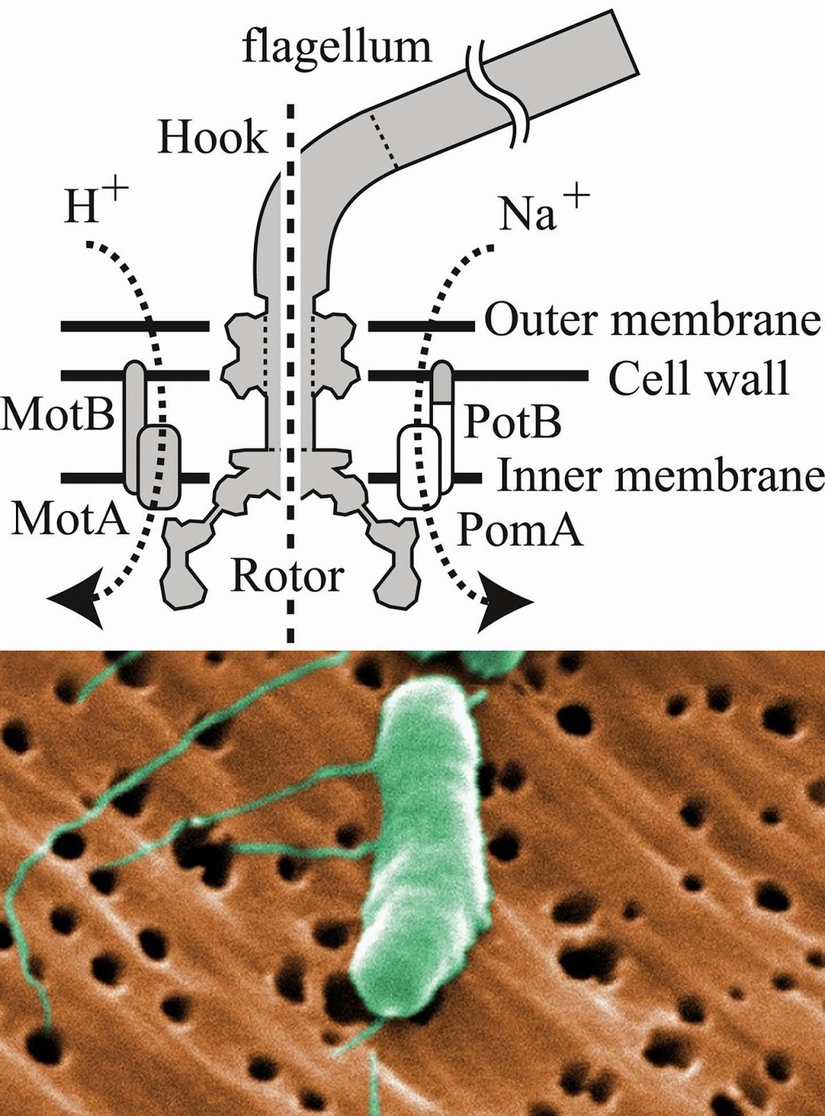
Vibrio bacteria -


Doing More With Less

$
0
0
Biology concepts – protists, complexity, undulipodia, flagella, cilia, amoebas,


Emotions are one of the things that make humans so complex.
Memories attached to associations, stimulated by
individualized brain chemistry makes it so you can’t predict
how any one person might feel about a particular stimulus.
But perhaps we are not so complex. A new study suggests that
there are really only four human emotions, happy, sad, afraid,
and mad. The other two commonly held states, disgusted and
surprised are just sides of mad and afraid, respectively. Read
the study and feel…… something.
Are humans the most complex animals? Humans have cells, tissues, organs, and organ systems that allow us to do things that no other organism can do – like invent doughnuts.

Indeed, this makes us complex and hard to understand, especially when we mumble. But on the other hand, wouldn’t it be more amazing if an organism could do complex things withoutthe benefit of all that organization and without all those trillions of cells doing different jobs?

What if an organism did many complex things but was only made of one cell? I think this blog has shown on many occasions that bacteria are capable of some pretty astounding feats, and they don’t even have a nucleus! True, they don’t have structures as complex as ours, most of their behaviors are responses to chemical signals from other cells, and they can’t make doughnuts.

But there are other single celled organisms that might match us for complexity, or even exceed our level of complexity, and they do it all within the confines of a single cell. Of course I’m talking about the ciliate protists. If don't know them, stick around and meet them and their kin. Sometimes, less is more.

We have been talking about the undulipodia in the last few weeks, and our last story started to describe the great catch-all kingdom, the protists. They use cilia and eukaryotic flagella (these being the undulupodia), but this is just one characteristic that can be used to separate them into groups.

The last post talked about the plant-like protists and how they can use flagella to either move around or to have their gamete cells move around. Today let’s discuss the animal-like protists; they use undulipodia in more ways.

There are six phylums of animal-like protists, just like there were six phylums of plant-like protists – but I think that was just a happy accident. The animal-like protists have more diversity amongst their phyla than did the plant-like protists; some use flagella, some use cilia, some use neither. Each phylum is amazing, but we’ll save the most complex – or is that most simple – for last.


The radiolaria are counted amongst the actinopoda. They
have intricate mineral skeletons. What you can’t appreciate from
these photomicrographs is that also have an inner skeleton that
divides them into an endoplasm and ectoplasm. Some house
algae in there ectoplasm to harvest their photosynthetically-
produced carbohydrates.
We start with the Phylum Actinopoda– their pictures are very impressive. No, I don’t mean that they are good photographers. Seeing them shows you how delicate and complex they are. The have silica (glass-like) coverings that protect them from the outside world. Though many of these organisms are among the zooplankton(zoo = animal-like, and plankton = drifter) that are in the oceans; remember that plankton don’t have to be microorganisms; many species of jellyfish are zooplanktonic as well.

The radiolaria are amongst the actinopoda and are quite complex. They're one celled, but the cell has two parts, an endoplasm that contains the nucleus and organelles, and the ectoplasm, that has frothy bubbles to control their buoyancy.

Radiolaria are floaters, which would suggest that they don’t use undulipodia for motility. In fact, the only time that flagella have been observed in radiolarians is in some swarmer cells. Scientists think these represent sexual reproductive gametes that might be released from a swelling in the adult cell. Sexual reproduction has not been confirmed; scientists must be too embarrassed to ask them about it.

The swarmers are very small, as a new study shows and can move in the water column. This may be why we often find radiolarian DNA at depths where they don’t live – it’s their swarmers. And they do seem to get around. A Russian cosmonaut just reported having sampled the windows on the space station and found plankton! They think they have escaped the atmosphere on the wind. If true, that certainly changes our post about life moving from Earth to space.

Phylum Foraminifera– these organisms have tests, shells of calcium carbonate with little holes in them from which they stick out a pseudopod (we'll see more of this below) and walk.  The foarminifera look like and are closely related to the actinopoda. Some wonder if they shouldn’t be lumped together, but we all know that arguments about protist classification are the rule, not the exception. Like actinopoda, they only show flagella on their gametes, and like actinopoda, the gametes are biflagellated.


The foraminifera are protists with calcium carbonate shells. I
wonder if the RAF pilots returning during WWII knew that the
White Cliffs of Dover are chalk formed from these protists.
Likewise, the limestone quarries in Bloomington , IN where they
filmed Breaking Away in 1979 are also made from the tests
of these protists.
While radiolarian zooplantokton skeletons are responsible for a lot of the sediment at the bottom of the ocean and over time – forming rock called radiolarite, the tests of foraminifera organisms go to form limestone and chalk.

Oil spills and other such disasters are having an effect on the ability of foraminifera to maintain their calcium tests, and this affects us beyond just having some pretty cliffs to look at. We may be using foraminifera in the future to repair bone injuries.

A 2014 study showed that using foraminifera exoskeletons is a good way to promote bone growth in skull defects in rats. The hope is that we can use these for bone grafts and for bone repair in the future. As long as we don’t destroy all the formanifera.

Phylum Apicomplexa– We could talk about the best known apicomplexans for years and just touch the surface of their biology. Why do we know so much about them? Because they kill us. Plasmodium falciparum is an apicomplexan – it causes malaria. Toxoplasma gondiiis an apicomplexan; it causes toxoplasmosis that can chew holes in your brain and kill you as well.

Apicomplexans are immotile, a lot like the planktonic foraminifera and actinopoda. But in this case they're usually carried around from place to place inside a living host. The vast majority of them are parasites. Since they get carried around, they don’t need flagella to move, but some of the species have gametes that have three flagella on their back ends. Even though I’m breaking my parasitology friend Bill’s heart, I am going to leave the apicomplexa here and move on.  Don’t worry Bill, we’ll come back to them soon.

Phylum Rhizopoda - These are the amoebas; most don’t have cilia or flagella. They move by pseudopodia (pseudo = false, podia = feet), oozing their membrane and cytoplasm in one direction and then pulling the rest along. Of course, this means they need a surface to move across, you don’t use pseudopodia to move in water. But this phylum also includes the ameoboflagellates,


Naegleria fowleri lives in warm waters. It enters the body
through the nose and travels straight to the brain. The
feeding structures look like a clown face, which makes it
double frightening for my daughter. The infection is almost
always fatal. Must be a sad clown.
Amoeboflagelleates are the exception, and can switch back and forth from amoeba form to flagellated form, depending on their environment. When in a liquid environment, amoeboflagellates use flagella to move about. But when in dry environment or a surface, the resorb their undulipodia and move by amoeboid mechanism. One amoeboflagellate in particular gets my attention.

Naegleria fowleriis the brain-eating protozoan that is transmitted through contaminated drinking or swimming water. It may not happen often, but I hate to think about something swimming (or would they crawl?) around my brain and feasting. Primary amoebic meningioencephalitis from N. fowleriis fatal in about 95% of cases, and though it is rare worldwide, most cases occur in the U.S.

A 2014 study in Arizona found that N. fowleri in five of 33 lakes studied. That’s scary enough, but the researchers also found that the protozoa were present in the cooler months, when it had been supposed that the cooler water temperatures were lethal to the organism. I am not retiring to Arizona.


Choanoflagellates look so much like the choanocyte cell
type in sponges. They are both collared and have a
flagellum sticking out. Since sponges are the basal phylum
of kingdom Animalia, it is believed that choanocyte protists
are the ancestor to all animal cells. But a new study says don’t
be so hasty. They resemble each other, but it could be parallel
evolution not straight descent.
Phylum Sarcomastigophora – this group of protists includes the most animal-like organisms that aren’t officially animals. The trichonympha have over 1000 flagella, but more species are like the choanoflagellates (means collared flagellar cells). These are believed to be the ancestor of all animal cells. They are very similar to the choanocytes of sponges, the most basal of the animals. However, a 2014 study warns against assuming that they are the same. The bending of the modified cilium that marks the choanocyte functions differently in the protists as compared to the sponges. The two diverged more than 600 million years ago, so similarities are there, but so are differences.

Phylum Ciliophora– The ciliates are those “less is more” organisms we described at the beginning of this post. These organisms have cilia all over their bodies, and they use them for feeding, motility (swimming and crawling), attachment and for sensing chemicals and perhaps sensing mates. These are all very complex behaviors for single-celled organisms. But wait, they do more.


Vorticella is one of the ciliates. It uses it’s cilia to shovel prey into
its oral groove, but the adult doesn’t use cilia to move. They
anchor to one spot and duck predators. The myoneme doesn’t
spring them up, it coils them down when something tries to eat them.
Some modified cilia funnel food into an oral groove, something that looks supiciously like a mouth to you and me. This is just the start of how they look like animals, they also get rid of waste in just one place, like our backside.

They have a complex cytoskeleton, made of connections between all those cilia. They have contractile vacuoles that control their water content. They even carry spears called trichocysts for defense against predators. These are like the cnidocytes of the jellyfish, and are a trick that we, with all our complexity, can't come close to matching

So do they go beyond us, even though they only have one cell? Yep, you have seen anything yet. Ciliates have up to six life cycle stages, they all have two nuclei, and they go through an insanely complex sexual reproduction that uses seven different sexes – with all our complexity, we only manage two - or are there more? That could be a good series of posts as well.

Ciliates are free living and cause us no problem. But in a great show of disrespect to their inferiors, Balantidium coli can be a horrible problem for us. It's the exception among ciliates in that it causes a disease, Balantidiasis, in primates. The organism usually lives in the guts of pigs, but if it gets into our gut, and we then become immunocompromised – watch out.


This is B. coli, the only know ciliate to cause disease in mammals.
You see all the cilia, that is how it moves around. It is transmitted
via the oral-fecal route, but might also be transmitted by
undercooked pork. Some people say fecal-oral, but that sounds
more gross to me.
Balantidiasis is characterized by explosive diarrhea, as much as one explosion every 20 minutes. Don’t think that "explosion" is too extreme a word. In some cases, the colon can literally have a hole blown in it (perforated) by the action of this ciliate. All that is bad enough, but a 2013 case study showed that B. coli infected an immunocompetent man’s spine, led to a pinching of the cervical nerves, and caused a temporary quadriplegia! Ciliates are complex, yes – and apparently one of them is mean.

We haven’t talked about the fungus-like protists. We’ll include them when we talka bout the undulipodia of the plants and fungi. Some plants cells have flagella for movement. Is my salad going to crawl off my plate?

But next week we'll start some Thanksgiving posts. Should we really be eating goat for Thanksgiving?


Jack, R., Garrod, O., & Schyns, P. (2014). Dynamic Facial Expressions of Emotion Transmit an Evolving Hierarchy of Signals over Time Current Biology, 24 (2), 187-192 DOI: 10.1016/j.cub.2013.11.064
  
Dhawan S, Jain D, & Mehta VS (2013). Balantidium coli: an unrecognized cause of vertebral osteomyelitis and myelopathy. Journal of neurosurgery. Spine, 18 (3), 310-3 PMID: 23259539

Chou J, Hao J, Kuroda S, Ben-Nissan B, Milthopre B, & Otsuka M (2014). Bone regeneration of calvarial defect using marine calcareous-derived beta-tricalcium phosphate macrospheres. Journal of tissue engineering, 5 PMID: 24808939

Yuasa T, & Takahashi O (2014). Ultrastructural morphology of the reproductive swarmers of Sphaerozoum punctatum (Huxley) from the East China Sea. European journal of protistology, 50 (2), 194-204 PMID: 24447698

Sifuentes LY, Choate BL, Gerba CP, & Bright KR (2014). The occurrence of Naegleria fowleri in recreational waters in Arizona. Journal of environmental science and health. Part A, Toxic/hazardous substances & environmental engineering, 49 (11), 1322-30 PMID: 24967566

Mah JL, Christensen-Dalsgaard KK, & Leys SP (2014). Choanoflagellate and choanocyte collar-flagellar systems and the assumption of homology. Evolution & development, 16 (1), 25-37 PMID: 24393465


For more information or classroom activities, see:

Plankton –

N. fowleri –

Ciliates –


A Goat For Thanksgiving

$
0
0
Biology concepts – cornucopia, goat, nutrition, sustainability, browser/grazer, Cassandra hypothesis


The image on top is the traditional cornucopia, filled with
foods or riches. The bottom image is the version form the
Hunger Games movies, filled with survival gear and weaponry.
Boy, did they go the other way with that idea. I prefer a
different Horn of Plenty, the Dizzie Gillespie album from 1953.
Thanksgiving is a traditional time to remember the work of planting and tending, and to be grateful for the harvest. In a larger sense, it’s a time to be grateful for life’s unending bounty, both the good and the challenging.

As a symbol of the gifts of the Earth, the ancient Greek cornucopia (comes to us as the Latin cornu= horn, and copiae = plenty) has been adopted as a symbol of this holiday in the USA. Not every country has a specific date set aside for celebrating the harvest, but almost every culture has a version of the cornucopia.

Looking at the horn of plenty in a little more detail, we will see that it fits more nicely with the American holiday tradition, and perhaps the American future.

From Greek mythology, there are a few stories about how the horn of plenty came to be. My favorite is that when Zeus was born, he had to be hidden from his father Chronos. Dad had a nasty habit of eating his young, so Zeus was whisked away to a mountain cave on Crete where he was suckled by Almathea, a lesser god that took the form of a goat.

Zeus, being the he-man god that he was, accidentally broke off one of Almathea’s horns while playing. Of course he blamed it on his sister ….. wait, that’s what used to happen at my house. No, Zeus felt bad about his rude feat of strength, so he enchanted the horn so that it would provide Almathea with whatever she desired. I imagine that for a goat that would be anything edible, and goats will eat almost anything.

Other versions of the myth say that Almathea was a nymph that fed goat’s milk to Zeus and he subsequently blessed one of the goat’s horns to provide her with unlimited bounty as a way of thanking her – but either way it didn’t end well for for the goat. A teenage Zeus showed his appreciation by killing and skinning the goat. The skin was used to make his shield, the aegis, and is shown in many depictions of Zeus. But the horn was still around, spewing forth sustenance for the holder.


This is from the 1555-1556 painting, Infancy of Jupiter, by
Giorgio Vasari. It shows the god being suckled by the divine
goat Amalthea. Notice we said Jupiter, not Zeus. Roman and
Greek myths often stole from one another, the same stories
can be found in each traditions; only the names have been
changed to protect the copyright.
This brings us to the goat horn and the American Thanksgiving. The goat just may have been a part of the first Thanksgiving, and it may have implications for future Thanksgivings, speaking biologically.

The first Thanksgiving wasn’t too much of feast; it looked nothing like the table we look over as we watch football and stuff ourselves. The pilgrims had it rough, but they were thankful for the harvest that they did manage and the wisdom that the native Americans had passed on to them about finding food in their new home.

They might have had some turkey, but they certainly didn’t have a big steak. The pilgrims had not brought any cows with them on their journey, and they weren’t native to this country. They may have had some pork, as they did have a few pigs, but they may also have had another meat – goat.

It's true, goats are not native to the Americas. No, the Rocky Mountain goats (Oreamnos americanus) that grace the crags and cliff faces so majestically are not really goats, they’re actually members of the antelope family. And it turns out that they aren’t even native to the Rocky Mountains, at least not the lower Rockies. They were originally found in Alaska, and were introduced to Colorado, Montana, and South Dakota much later.


Check out the nutritional values for goat meat. If you, unlike
most Americans, can stomach the idea, you should be adding
it to your diet in place of some other red meat.
The pilgrims had at least some goats with them in 1621, they were remarked upon in one person’s journal. They certainly used them for milk and cheese at the first Thanksgiving, but they may have eaten goat meat as well. Does that sound weird to you?

True American’s don’t eat goat to any great degree. But in this instance, we’re the exception. Over 70% of all the red meat consumed worldwide is goat! I don’t remember trying it, but I am sure going to try and procure some now – not just for the novelty of it, goat meat (sometimes called chevon) is really good for you.

Chevon has greater vitamin and lower fat content than other red meats, even fewer calories and fat than chicken. People around the world haven't necessarily known about the nutritional value of goat meat for centuries; goats lived there and they ate them. Even south of the border in Mexico and Latin America they eat lots of goat (and I don’t just mean the cryptozoologic chupacabra).

All across Asia they eat goat, especially northern China, but as many of my Chinese friends say, the only thing with four legs the Chinese won’t eat is the kitchen table. Andrew Zimmern, he of the bizarre foods shows, says that goat is like soccer - it’s popular everywhere but the US.

Despite the fact that we don’t eat it nowadays, our story tells us that goat might have a bit more to do with Thanksgiving that one might think. Is there more – you bet. Along with their meager harvest and maybe some roast goat, the pilgrims enjoyed native American foods, things that were grown, hunted and/or gathered.


The black trumpet mushroom lives in Europe across to Asia.
Avery closely related species lives in North America, still
called the black trumpet or horn of plenty. Recent studies
show that in America, the black trumpet lives in a symbiotic
relationship with several pine trees. This is interesting, since
goats are now being fed pine bark to make their meat taste
better. Everything is connected.
In southern Virginia, this would certainly have included many different species of mushroom. And wouldn’t you know it, one of the most bountiful mushrooms in this region is named Craterellus cornucopioides, or Craterellus fallax. Also called the black trumpet or the horn of plenty mushroom.

I have read that native American Indians didn’t eat a lot of mushrooms, but they did use them for medicines and as symbols. But the European pilgrims were certainly mushroom eaters, so it is likely they made good use of the horn of plenty mushrooms. I can see a big bowl of mushroom stuffing gracing the tables of the first Thanksgiving.

Maybe the Indians were on to something when they used the mushrooms in their medicine. Recent studies of the black trumpet mushrooms show have medicinal effects. A 2012 study indicated that several mushrooms related to, and including the black trumpet mushroom have the ability to regulate blood sugar levels so as to prevent hyperglycemia. I know my diabetic wife would be interested in that, even if she doesn’t like to eat mushrooms.

In addition to this, the same study showed that the mushrooms also have antioxidant activities. They could scavenge iron ions that can do damage to cells as well as oxygen radicals that can devastate cellular function, see this post for a discussion of oxygen radicals and antioxidants.

A newer 2014 study show that the black trumpet has significant anti-inflammatory properties. An alcohol extract of the black trumpet was able to prevent inflammatory cytokine and nitric oxide production in macrophages that were stimulated with LPS, a potent inflammatory agent.

We have seen that goat plays a role in the American Thanksgiving, perhaps in the symbol we use for the harvest, perhaps as a meat source, and even perhaps in the mushroom stuffing. Now we can see how it may represent the future as well.


Evolution is amazing. Some ruminants evolved as grazers, while
others became browsers. That way, they can live in the same
areas and they both still have enough to eat. Goats are browsers,
so they have to digest things that even cows can’t digest.
Goats are browsers, not grazers. This means that they don’t eat grasses down to the roots; they also feed on what ever is around. They eat bark, twigs, leaves, license plates, homework, just about anything laying around. As such, they have a smaller effect on the environment than do grazing livestock, like bovines. Cows can strip a pasture clear of grass and its roots, and this takes time to replenish.

So goats are better for the land, you can raise more goats on the same amount of land as cows, and you don’t need to grow as much grain for them to eat. The commercials should really be saying, “Eat More Goat.” Goat numbers in the US have doubled in the past few years, but they still pale in comparison to those of bovines. Given the nutritional and environmental advantages of goats, maybe they could use a marketing slogan – “Goat, the other green meat.” Maybe not.

All this environmental talk leads us into the last discussion for our Thanksgiving post. Is the horn of plenty really a good symbol for the Earth today? Are the resources of Earth limitless and will keep pouring forth no matter what we do to our home?

This is the issue in an argument about population growth referred to as the Cornucopia vs. Cassandra hypothesis. The cornucopia position is one that says that the Earth will be able to sustain human population no matter how much it grows, while the Cassandra hypothesis states that the Earth’s resources are limited and that uncontrolled population growth is untenable.


Alan AtKisson wrote Believing Cassandra in 1999. He
explains how working for sustainability is necessary but
possible. Global sustainability is explained via anecdote and
good science, unfortunately, his method for reaching
sustainability is abbreviated ISIS, not so good for late 2014.
I recommend the book highly.
Cassandra was another character from mythology. Apollo gave her the power of prophecy in hopes becoming her boyfriend, but she spurned his advances and he took revenge. He left her with the power to predict the future, but cursed her so that no one would believe her predictions. Ouch, that “hell hath no fury like a woman scorned” line might have to be amended to include Greek gods.

The argument seems odd to me. Of course the Earth can take what ever we throw at it, but we can’t. Population growth and climate change are untenable only to us. Life will change and go on, but we won’t be here to see it.

The key is to manage the growth so that we don’t destroy the Earth for ourselves and for the rest of the living organisms that we depend on. And we depend on them all in one way or another. Maybe we should give thanks that we still have time to embrace the goat. OK, that sounds bad too.

Next week - does Thanksgiving turkey really put you to sleep?


O'Callaghan YC, O'Brien NM, Kenny O, Harrington T, Brunton N, & Smyth TJ (2014). Anti-Inflammatory Effects of Wild Irish Mushroom Extracts in RAW264.7 Mouse Macrophage Cells. Journal of medicinal food PMID: 25136763

Liu YT, Sun J, Luo ZY, Rao SQ, Su YJ, Xu RR, & Yang YJ (2012). Chemical composition of five wild edible mushrooms collected from Southwest China and their antihyperglycemic and antioxidant activity. Food and chemical toxicology : an international journal published for the British Industrial Biological Research Association, 50 (5), 1238-44 PMID: 22300772


For more information or classroom activities, see:

Cornucopia in myth –

First Thanksgiving meal –

Browser versus grazer –

Cassandra and population growth –


A Meal More Powerful Than The NFL

$
0
0
Biology concepts – genetic code, neurotransmitters

A turkey dinner with all the fixins can lead to a
satisfying nap. But the meal usually takes a little
longer than this to have an effect. This fellow might
be more affected by last night’s activities than today’s
meal.
Turkeydinner at Thanksgiving brings the family together, celebrates the bountiful harvest, and puts you to sleep just as the NFL games are ready to start. Many people think that if you eat less turkey and fill up on the other goodies you can escape the post-Thanksgiving meal sleepiness. Other people look forward to eating seconds and thirds and then stretching out on the couch for a long nap, forcing Aunt Ethel to sit in the chair with the spring that surprises you every once in a while.

The culprit, or the hero, in this eat and sleep saga is said to be the tryptophan in the turkey. Other people think that it is simply how much you eat, not the turkey's tryptophan, but it isn’t quite that simple. What is tryptophan, and is it indeed responsible for the snoring that follows Thanksgiving dinner?  Some background will help.

Tryptophan is an amino acid, one of the twenty standard building blocks of proteins. Each amino acid has a similar basic structure, as shown in the picture below. The central carbon has an amino group (NH3) on one side and a carboxylic acid (COO-) moiety on the other; hence the name – amino acid. The third side group is a simple hydrogen (H), while the fourth side (R) refers to any of several different side groups and is what makes one amino acid different from one another.

Tryptophan is an aromatic amino acid, meaning that its side group contains a six-sided carbon ring structure (each corner represents a carbon). It also has a second ring group of four carbons and a nitrogen. As such, it is the largest and most massive of all the standard amino acids. However, tryptophan is the least abundant amino acid in plant and animal proteins; it accounts for only 1-1.5% of the total number of amino acids in proteins.

Amino acids are the building blocks of proteins. The NH3
is the amino part and the COO is the acid part. The R is
different for each amino acid. On the left, you see that
tryptophan’s R group is a big structure with two different
rings (each angle where two lines meet stands for a carbon,
they just don’t write in each “C”). Two lines means a double
bond. In producing the protein, the COO of the last amino acid
added gets connected to the NH3 of the next amino acid to be
connected. Which amino acid it is determine by the mRNA
and the genetic code.

Tryptophan’slarge structure and intricate rings make it costly to produce in terms of ATP invested. In fact, it takes so much energy to make that we have stopped making tryptophan all together. Tryptophan is abundant in a number of food sources commonly available to humans, so over evolutionary time we have turned it into an essential amino acid. True, it is essential for life, but here the word “essential” means that we MUST get it from our diet, we cannot produce it ourselves.

Of the 20 standard amino acids, 10 are essential in humans (9 that we must eat and 1 that we make from an essential amino acid), but bacteria make them all just fine - although the parents of newborns may wish it wasn’t so. Gut bacteria make tryptophan or use the tryptophan we eat. They transform it into molecules they need to survive, but the byproducts of these reactions are skatole and indole – these are the precious little molecules that give dirty diapers that wonderful smell!

Tryptophan is different from many other amino acids in another way as well; it gets no respect from the genetic code. Each amino acid is coded for by a group of three RNA bases, together called a codon. Since there are four different bases in mRNAs (A, C, G, and U – remember that T is used in DNA but not RNA), then there are 64 different codons (4 x 4 x 4). This is more than the 20 amino acids that the codons code for, so most amino acids have two or three codons that signals that they should be added to the growing peptide. But tryptophan is encoded by only one codon (UGG).

It may make sense that an amino acid that is not used often in proteins might rate only one codon, but the amino acid methionine is used much more often than tryptophan, and it's only coded for by one codon as well (AUG). You know nature must have a reason why tryptophan has a single codon, we just don't know it yet.

The genetic code is how mRNA codons (3 bases sequences)
get translated into a signal to build proteins from specific amino
acids. The first base of the codon is represented by the biggest
letters (ACGU), the middle base is the middle size letters, while
the third position (wobble position) is usually where you see an
amino acid coded for by more than one codon. For instance,
serine is coded for by UCU, UCC, UCA, or UCG. But tryptophan is
only coded for by UGG. Three codons signal the protein to stop
growing, called stop codons (UAG, UAA, and UGA).
Eventhough it is used sparingly in proteins, tryptophan is an essential amino acid - don’t eat enough of it and you die. This is because tryptophan’s most essential functions have nothing to do with protein synthesis or structure – tryptophan is important to your brain function. The crucial neurotransmitter, serotonin, is synthesized only from tryptophan.

It takes two enzymes to turn tryptophan into serotonin (also called 5-HT).  First is tryptophan hydroxylase; hydroxylase means it splits water, here it adds an OH to tryptophan. Next, the amino acid decarboxylase removes a carboxylic acid (COOH), producing serotonin.

Amongst the many functions of serotonin are a few that are not brain related. Serotonin is released by enterochromaffin cells that line your gut to tell your gut to move. The movement helps push the food along your digestive tract, but serves a protective function.

If you eat something toxic, the enterochromaffin cells produce more serotonin – your gut moves much faster, and you get diarrhea. If even more serotonin is made and released, it moves through the bloodstream to your stomach and esophagus and causes you to vomit.

But it is in the CNS that serotonin has its significant activities. As a neurotransmitter, it is responsible for controlling how electric messages are passed from one neuron to another. When serotonin is released in the synapse (the gap between the upstream and downstream neurons) and is taken up by adjacent neurons, it produces a sense of well-being.

Where one neuron ends and others begin there is
a gap called the synaptic cleft. Different types of
neurons use different neurotransmitters, of which
serotonin is one. It is released into the synapse, and
adjacent neurons with serotonin receptors can be
stimulated to conduct a nerve impulse. The serotonin
is broken down in the synapse by MAO’s and taken
back up to produce more serotonin.
It isn’t surprising that depressed individuals often have low blood levels of tryptophan, as well as reduced serotonin. Classic treatments for depression include increased tryptophan intake, monoamine oxidase (MAO) inhibitors, and serotonin reuptake inhibitors (SSRI). With more tryptophan, you make more serotonin – problem solved. On the other hand, MAO’s break down serotonin, so their inhibitors enhance the action of tryptophan. SSRI’s prevent the reuptake, this leaves serotonin in the synapse longer. Both types of drugs make tryptophan more likely to be taken up by downstream neurons.

Unfortunate, but interesting, is the study showing that the suicidal thoughts that sometimes accompany anti-depressant therapies (TESI – treatment enhances suicidal ideation) use may be related to polymorphisms in one form of the tryptophan hydroxylase enzyme that starts the serotonin production from tryptophan.

When non-suicidal patients were compared to those with TESI or those who were suicidal without treatment, a pattern emerged. Only those with TESI showed a polymorphism pattern in the tryptophan hydroxlyase 2 (TPH2) gene. This polymorphism had previously been associated with suicide victims and major depressive disorder. It seems that a slight alteration in function of TPH2 due to a single nucleotide change can contribute to the genetic background of treatment induced suicidal thoughts.

The feeling of general well being induced by serotonin also participates in the sleep/wake cycle. So is tryptophan – through serotonin – responsible for the post-Thanksgiving nap? Well… yes and no, it's an accomplice in a larger conspiracy.

Serotonin is use to produce the hormone melatonin, and melatonin promotes sleep, so you could say turkey dinner promotes sleep. But turkey doesn’t have that much tryptophan! Tofu has much more tryptophan than turkey, but you don’t get a post-Chinese takeout urge to sleep, so what gives?

Melatonin is made from serotonin in the pineal
gland. Sunlight stimulates the suprachiasmatic
nucleus (SCN) which inhibits the pineal from
making melatonin. As the sun goes down,
inhibition is reduced, more melatonin is made
and released from the pineal, and sleep is
promoted.
The melatonin effect has to do more with how much of everything else you eat at Thanksgiving dinner, especially carbohydrates. Here is how it works – eating lots of carbohydrates causes a release of insulin into the blood (to reduced blood glucose levels). Another function of insulin is to promote the uptake of some amino acids (but not tryptophan) into muscle cells. This leaves the blood higher in tryptophan as compared to other amino acids than it would normally be.

The brain takes in amino acids through a neutral amino acid transporter, which now finds more tryptophan than other neutral amino acids, so the brain level of tryptophan goes up. More tryptophan in the brain, more serotonin – more serotonin, more melatonin. More melatonin = nap time! So if you want to avoid the post-Thanksgiving nap, eat the turkey and skip the mashed potatoes.

You didn’t know how much tryptophan controlled your daily life, did you? Well, there’s more. Tryptophan is also important in synthesizing niacin, a.k.a. vitamin B3 or nicotinic acid. Niacin is important in production of NAD/NADH for energy metabolism, for production of steroid hormones and balance of lipid forms in the blood, and as an anti-convulsant.

The tryptophan-niacin connection is made stronger by recent evidence that high dietarytryptophan can prevent epileptic seizures in mice. In this study, a whey protein called alpha-lactoalbumin (ALAC) was found to have much tryptophan, much higher levels than in most proteins. Feeding epileptic mice ALAC resulted in reduced numbers of seizures.

So even if you don’t want to sleep or think happy thoughts, you still need to eat food that contain tryptophan or niacin. And many of those foods are plants, because plants use tryptophan to control their own activities. Tryptophan is easily converted to auxins, a type of plant hormone. Auxins are responsible for several different plant behaviors, namely the falling leaves in autumn and ripe fruits all year long.

Here is an interesting attempt to get kids to read
history. During the spring, captive warriors were
killed by cutting out their hearts, then their skin was
flayed off their body, and the priests would wear them
around for 20 days. This was meant to celebrate the
god who sacrificed himself to allow a new growing
season to begin. This time period corresponds
 to when they would have had the lowest amount of
 tryptophan in their daily die. No - I wouldn't want
to be an Aztec sacrifice!
Having dietary choices for tryptophan is good, and plants provide our major source. However, cooking grains and corn reduces usable tryptophan and niacin levels dramatically, so poorer environments where corn is the staple food need also to have additional dietary sources of tryptophan. A deficiency of this amino acid leads to some disturbing conditions. Low tryptophan leads to low serotonin levels and agitation, insomnia, and depression. A study in the Archivesof General Psychiatry stated that chronically low levels of tryptophan led to relapses of purging behaviors in bulimics.

More amazingly, studies in the 1970’s to 1990’s suggest that low tryptophan levels can lead to increases in religious fanaticism. Several studies from a single author correlate the Aztec human sacrificial ceremonies to the times of year when their diets depended more on foods that had less tryptophan. Think of all the lives that could have been saved by tofu!

But turkey is more than just tryptophan. You have to love an animal that has caruncles, a wattle, and a snood! What's a snood? Come back next week.


Musil, R., Zill, P., Seemüller, F., Bondy, B., Meyer, S., Spellmann, I., Bender, W., Adli, M., Heuser, I., Fisher, R., Gaebel, W., Maier, W., Rietschel, M., Rujescu, D., Schennach, R., Möller, H., & Riedel, M. (2012). Genetics of emergent suicidality during antidepressive treatment—Data from a naturalistic study on a large sample of inpatients with a major depressive episode European Neuropsychopharmacology DOI: 10.1016/j.euroneuro.2012.08.009


Russo, E., Scicchitano, F., Citraro, R., Aiello, R., Camastra, C., Mainardi, P., Chimirri, S., Perucca, E., Donato, G., & De Sarro, G. (2012). Protective activity of α-lactoalbumin (ALAC), a whey protein rich in tryptophan, in rodent models of epileptogenesis Neuroscience, 226, 282-288 DOI: 10.1016/j.neuroscience.2012.09.021

For more information or classroom activities, see:

Genetic code –


Neurotransmitters –
http://science.education.nih.gov/supplements/nih2/addiction/activities/activities_toc.htm


As A Bird - It's No Turkey

$
0
0
Mr. Carlson and Herb Tarlek had to deal with the
aftermath of bombing Cincinnati with live turkeys.
The line about turkeys being able to fly is one of the
most famous in TV history. But he should have at least
questioned whether they could fly, there are more than
50 species of flightless bird alive as we speak.

In a famous 1978 episode of the TV sitcom, WKRP In Cincinnati, station manager Arthur Carlson releases turkeys from a helicopter to a waiting crowd below as part of a holiday publicity stunt. The birds crashed to the ground (off camera), as intrepid reporter Less Nessman described the carnage. You can find the entire episode here; it’s as funny now as it ever was.

This comedy had a 1940’s parallel in real life, when the town of Yellville, Arkansas dropped Thanksgiving turkeys off the courthouse roof for several years in succession, and then from low flying planes. They didn’t seem to have any qualms about the flight problems of the domesticated turkey.

In contrast, the North and South American wild turkeys would have survived the stunts. In the genus Meleagris, there are several species of wild turkey, and they can and do fly short distances. In fact, they spend their nights perched in the low branches of trees from Maine to Peru.

The Aztecs introduced the Spanish to Southern Mexican turkeys (Meleagris gallopavo gallopavo), who took them back to Europe in the 1520’s. The Spanish trade capital at the time was Turkey, and from there turkeys spread all across the continent by the 1550’s. Therefore, the English called them turkeys, because they thought the birds originated in Turkey.

The Meleagris g. gallopavabirds brought back to Europe were domesticated and became the eating turkeys of today. They were bred for large breast muscles, and being raised in domestication caused them to lose much of their flying musculature; the domesticated turkey is flightless and mimics a bowling ball when released from a helicopter.

Here’s proof for you city folks that turkeys can fly.
Their perch is bird-like, drawing up one leg. This might
be to conserve heat, or to change their outline and
make them look like a plant in order to avoid predation.
Truly, that is one hypothesis… I mean it….really I do.
In animals, only birds have a vertical extension (keel) on their breastbone to allow for attachment of the large breast muscles required for flight. Birds are also the only animals to have fused collarbones, called a furcula. This bone attaches to the muscles important in the down stroke of wings, and also helps to pump air into the lungs - we know this structure as the wishbone. The furcula is more massive in the middle, and can flex and act like a spring during flight.

We use the furcula as a sign of good luck, but for domesticated turkeys it is just an unfortunate reminder that they used to have a fighting chance at avoiding a gravy bath. It's ironic that breeding to increase the size of their flight muscles is exactly why the domesticated turkey can’t fly.

Over many generations, the domesticated turkey’s muscles have become too big to allow it to fly and its legs have becomes shorter, so it has a hard time running. In fact, they are so large and cumbersome that they can’t even mate; they are inseminated artificially in order to breed them further. Many have been bred for white plumage, so that the small pin feathers left after plucking are harder to see.

But the noble turkey (Benjamin Franklin suggested the turkey as our national bird) can trace its line back to about 1100 CE, with the Spanish entering the picture about 400 years later. But new evidence suggests turkeys were raised in captivity much earlier than either of these estimates.

A recentstudy based on excavations of Mayan ruins shows that as early as 300 BCE there were male, female, and juvenile M. gallopava within the settlements, and some reduced flight morphology suggests that they had begun to be domesticated by that time. What is more, the native turkeys in southern Mexico were M. ocellata, not M. gallopava (from northern Mexico and America), suggesting that trade in the animals with the north had already commenced by this time period.

All this traveling suggests that by the time the pilgrims landed in Massachusetts they were already familiar with the turkey, and its inclusion in the first Thanksgiving feast was probably not a surprise to them. There is no evidence that turkey was the served at the first Thanksgiving, but it makes sense; both cultures were familiar with the bird. American Indians even had tribes named for turkeys and believed that their feathers had mystical powers; Central American Indians had turkey gods.

I find it a little odd that the turkey was revered as a god, considering its looks – that is truly a face only a mother could love. It has appendages and little growths everywhere. If a turkey spins its head around when startled, it could slap itself silly! But as nature proves again and again, everything has a purpose – or did.

You can see the differences between wild turkeys andthose bred 
for lots of meat. True, the domesticated version is puffed up in
a display, but he is much bigger weighing twice as much as 
the wild version on average (16-50 lb.s/7.25-22.5 kg for domestic).

The fleshy appendage around the head a throat of many bird species is called the wattle. In turkeys, the wattle hangs from under the beak and down the throat, but in pheasants it is located around the eye and cheek. Another name for this structure is the dewlap, and many animals have these. Even your grandmother might have a dewlap under her chin or upper arms!

The wattle is a mark of sexual dimorphism in many birds (di = two, and morph = shape). The males and females look different in species that are sexually dimorphic. It is hypothesized that birds’ wattles are a form of ornament for mate selection. A male with a larger wattle may be seen as more fit and a may have more reproductive success. The hypothesis states that a large ornament is energetically costly, so only the strongest, most disease resistant males will be able to survive the cost of a large ornament and still live to reproduce.

In terms of the female turkey, picking a male with a bigger wattle would be the same as picking a male with stronger genes. Indeed, a 2010 study inpheasants showed that there were different immune genotypes (MHC, major histocompatibility complex) associated with wattle size. The functional difference between the different MHC genotypes is not known, but they did show a significant difference in the genotypes of males with larger wattles, and those are the more highly preferred mates, so it may also represent stronger MHC types.

But domesticated turkeys don’t worry about selecting mates or appearing healthy, all decisions are made by the breeder, so why do they still have wattles? It may be because their breeding is anything but true natural selection, but it may also be that the wattle has another function. Being highly vascularized (having many blood vessels), the wattle can release body heat by placing a large amount of blood close to the surface, thereby acting as a physiologic control.

Use this picture to memorize the parts of the turkey’s
head.  Cousin Eddie asked Clark to save him the neck
in National Lampoon’s Christmas Vacation, but I doubt
that anyone ever specifically for the snood! There are
no snood recipes - believe me, I looked.
On top of a turkey’s head and down its wattle are smooth surfaced growths called caruncles. At the base of the wattle are larger growths called the major caruncles (not very imaginative). The exact function of the caruncles is not known, but they are significantly larger on males than on females, so sexual ornamentation might be one of their functions. Together, the wattle and caruncles are also a mood detector. When threatened or ready to mate, the wattle on a tom turkey will turn bright red.

Thestrangest part of a turkey’s head is the snood. The English word “snood” was around long before the English were aware of turkeys. It referred to a decorative hair net or bag worn by women on the back of the head to confine their hair. The resulting mass of hair does look something like the snood that hangs over a turkey’s beak, and this might be where the name came from (see below).

While many animals have wattles, and several different kinds of fowl have caruncles, the turkey is the exception in that it is the only animal with a snood. Its functions may be similar to those of the wattle and caruncles, as they are much larger in males than in females. The snood length in males is linked to testosterone levels, and males are more likely to dominate or steal food from shorter snooded (just made up that word) males than long snooded (there it is again) ones.

I haven’t found any documentation that the turkey
snood is named after the hair snood, but it makes sense.
The snood as a garment makes a comeback every 100
years or so, now they are all the rage in McDonalds and
abattoirs (slaughterhouse) –turkeys with snoods are
processed by workers wearing snoods!

But turkeys are rarely served with the head intact, and eating them is what we are most interested in at Thanksgiving. Like chickens, turkeys have both dark and white meat. The difference in color is due to the makeup of the muscles and how they store and use energy.

The red meat of mammals and the dark meat of birds are similar in that they contain high amounts of myoglobin. The muscles that have myoglobin are for prolonged use; muscles used most of the time require lots of oxygen to make lots of ATP. Myoglobin is to muscles cells what hemoglobin is to red blood cells; it is a molecule that binds and holds oxygen. In the muscle cell, the myoglobin will release the oxygen as needed to allow the muscle to make more ATP and then use that ATP for contraction.

Myoglobinis highly pigmented, so the muscles look darker (redder). When denatured by temperature, the myoglobin turns a tan to dark brown color, giving the cooked meat its look.

Myoglobin is structurally similar to hemoglobin, in that it
looks like one of hemoglobin’s subunits. Each subunit in
hemoglobin can carry one oxygen molecule, but act in
cooperative behavior; the first one is hard to bind, the
second is easier and so on. Myoglobin stores oxygen within
the muscle cell. The more you exercise that muscle, the
more myoglobin it will produce.

White meat, on the other hand, has much less myoglobin. Why? The muscles with white meat (like flight muscles) need energy in short bursts, perhaps to evade predation. To do this, they need less oxygen most of the time, but need lots of glucose some of the time. Therefore, they have less myoglobin but more glycogen (a storage form of glucose) so they can react quickly, rather than waiting for the blood to bring more glucose. The glycogen makes the cooked meat look white and glossy.

So, we have a big bird (did you know that Big Bird’s costume is made of turkey feathers painted yellow?) providing us with a big meal on a big holiday. Next week, we look at three stories of fungus-like protists. Can you believe that they are responsible for Irish priests in America and video game theory, and that they can be ranchers with hired cowhands?


Baratti, M., Ammannati, M., Magnelli, C., Massolo, A., & Dessì-Fulgheri, F. (2010). Are large wattles related to particular MHC genotypes in the male pheasant? Genetica, 138 (6), 657-665 DOI: 10.1007/s10709-010-9440-5

Thornton, E., Emery, K., Steadman, D., Speller, C., Matheny, R., & Yang, D. (2012). Earliest Mexican Turkeys (Meleagris gallopavo) in the Maya Region: Implications for Pre-Hispanic Animal Trade and the Timing of Turkey Domestication PLoS ONE, 7 (8) DOI: 10.1371/journal.pone.0042630
For more information or classroom activities, see:


Sexual dimorphism –

Myoglobin –
http://www.getbodysmart.com/ap/respiratorysystem/physiology/gases/myoglobin/animation.html


How Slime Molds Our World

$
0
0
Biology concepts – Protista, fungus-like protists, penicillin, undulipodia, serendipity, potato famine, networks, co-evolution, slime mold


It’s one thing for Dr. Fleming to have discovered pencillin by
accident. It’s another to admit to everyone – most people
would just say, “I meant to do that.” Fleming was great at
serendipity; he discovered human lysozyme when some
snot dripped from his nose when he had a cold onto a
bacteria filled agar plate and they died.
Have you ever had the fortunate experience of looking for one thing and finding something better? Your find wasn’t what your were looking for, or even meaning to find, but there it is. Now you’ve got something you can really use. And you didn’t even know it was out there. I think they call it serendipity.

Serendipity plays a crucial role in doing science, and there are myriad examples of how scientists have discovered one thing while looking for something else. Penicillin is a good example. Alexander Fleming had an awful time keeping extraneous organisms from growing on his bacterial agar plates. It was his prepared mind that recognized that the mold (penicillium) growing on one of them had cleared the bacteria away in a wide circle.

Today we have an example of serendipity in story-telling. I set out to continue our story of undulipodia(flagella and cilia) use in the different types of organisms. We had used the undulipodia to talk about how hard it is to classify protists, first animal-like protists, then plant-like organisms, and today the fungus-ish protists.

However, in terms of undulipodia, the fungal-like protists are pretty much all the same. They all use flagella to propel their gamete cells. It’s interesting that some fungal cells are motile, but it’s not the most interesting thing in their story. In looking at the three phyla of fungus-like protists, I found that the biology of each is amazing – see if you agree.

Phylum Acraisomycota– As unappealing as it may sound, this phylum consists of the cellular slime molds. These protists spend much of their life as individual cells, moving around via amoeboid motion through the soil, looking for decaying organic material and bacteria to eat. But when hunger, they become ranchers. Yee-ha!


When food is scarce, the amoebas of D. discoidium will
join together into a grex, or sometimes called a slug. They
remain individual cells, unlike acellular slime molds that
join together as a single huge cell. This video is sped up considerably.
When food is scarce, acraisomycota cells join together, both emotionally and physically. Dictyostelium discoideum is a cellular slime mold that has been used in research labs for years and is a well-studied example of the slime mold development.

When about 100,000 cells join together to form a grex or a slug, they all move as one to try and find food, and to produce reproductive bodies so that their progeny will be protected, waiting as spores for better conditions. Without the benefit of speech or sign language, the single cells will start to take on different jobs, including building stalks that stick up into the air with fruiting bodies on top.

Inside the fruiting bodies are the spores, the progeny cells inside protective cellulose spore coats. They are environmentally resistant in this form and can wait until the conditions are right to become amoeboid cells of the next generation.


After forming a slug, fruiting bodies will be formed on top
of D. discoidium stalks. Remember that the individual cells
are organisms, but they act like a multicellular organism.
Now it gets interesting. Some researchers noticed (serendipity?) that some of the fruiting body spores contained bacteria, but only some of them. Were they a contaminant? When the scientists killed off the bacteria nothing bad happened, but they noticed that the spores of the progeny that had contained bacteria also sported some bacteria in their newly formed spores.

Their 2011 paper showed that some of the dictyostelium cells were not eating all the bacteria available; they were letting them reproduce and then storing them in their spores. When those spores were blown or carried to new locations and germinated, the bacteria would grow as well, becoming food for the cells. In turn, their progeny would be gathered into the next generation of spores.

Protist ranchers are amazing enough, but there’s more. The ranchers hire cowhands to protect their herd. The spores contain not only the food bacteria, but some other bacteria as well. These other bacteria (of the family Burkholderia) secrete chemicals that keep the non-ranching clones of dictyostelium from rustling the bacterial cattle. Not only are some of the protists ranchers, they protect what they raise using armed guards! It only took us several hundred million years to catch up to them.

Phylum Myxomycota - These are the acellular slime molds. The individuals cells don’t join together just when food is scarce, they spend most of their lives all packed together.


This is P. polycephalum, or dog vomit mold. I bet you have
seen it before and walked FAR around it. When grown
in high moisture and food, it is slimy, when it dries out, it
forms a crust over the top to protect the huge
plasmodium below.
With acellular slime molds, the individuals actually merge together as one cell, not one multicellular organism. This plasmodium is a single cell with thousands of nuclei, reaching sizes of 0.3 meters (1 ft. or more), so these are called the plasmodial slime molds.

A good, if not so pleasantly named, example of the myxomycota is the dog vomit mold (Physarum polycephalum). It’s named that because it often looks like that. But the skills of this slimy mass help it to overcome the poor name and the yellow gooeyness (gooiness?). Acellular slime molds are math geniuses – and they are going to help you avoid your math homework by playing on the internet.

It all started when a 2000 paper showed that dog vomit mold can find its way through a maze. With food at the opposite end of a labyrinth, P. polycephalum will consistently find the shortest path to the food.  The mold grows toward food in such a way as to be most efficient.

Here, efficient means using the least amount resources and the quickest route, ie. the shortest path. It creates a redundant system as well – more than one way to get to the target in case the primary path is disrupted by a misplaced footstep or a carelessly discarded whoopie pie wrapper (I must be hungry).

Finding fast, short, efficient, and redundant paths involves high level math - very high level math. P. polycephalum doesn’t have a calculator, or even a brain, but we're learning a lot from this mathlete.

In an amazing 2010 study, pieces of food were placed on an agar plate in the relative locations of Japanese towns around Tokyo. A small amount of P. polycephalum was placed where Tokyo would be, and it was allowed to grow toward the food. The result – the mold recapitulated the Tokyo rail system map!!! Five dollars worth of agar and 48 hours achieved the same design result as years of time and hundreds of millions of Japanese yen – maybe just a bit embarrassing?


The networks formed by dog vomit mold become well
defined as they are reinforced. The fronts are looking for
food, and many are resorbed to reveal only the most efficient
pathways once food isfound (watch the left side).
All this math is important because many of the things you care about use this slime mold derived math to build virtual ….stuff. For example, the internet gets you pages of information by bouncing the information around networks. The more efficient that bouncing is, the faster your page loads.

Graphics programs use slime mold math to build geometric shapes which become smooth and realistic surfaces and moving objects in your video game. Dog vomit mold can even be used to model the movements of characters within the games. Who knew that Assassin’s Creed involved so much math?

Phylum Oomycota - These are the water molds or downy mildews. We have talked about them before in terms of their presence in your bathtub and shower, but they have more stories to tell. For instance, they were responsible for the number of Irish Catholic priests in America.

One of the most amazing things about this phylum is that you pronounce both of the first two O’s – say “Oh! Oh! Mycota” real fast. The name means “egg fungi,” and as with the two other phyla, they used to be mistaken for fungi. However, these molds grow in long filaments, not as slime molds.


This what happens to potatoes contaminated with the P.
infestans parasitic oomycete. The potatoes rot in the ground,
so when you finish growing them all summer, you have
nothing to harvest. It isn’t the plague, but it still killed a
million people.
The oomycota were originally mistaken for fungi because many feed on decaying material, but some other species are parasitic. They cause damage to crops and fisheries. One species attacks potatoes.

You can directly relate the number of Irish priests in America to an oomycota called Phytophthora infestans. The land in Ireland in the 1700-1800’s was particularly fertile; they were the breadbasket of the UK. This meant that they grew a lot of potatoes. True, the English land owners took most of the crop, but the Irish that worked the land benefited as well by having more food than most other people in their sociopolitical group could manage.

Because they had more food, they had better overall health. Better health led to, amongst other things, more children. The population growth in Ireland was much higher than in other parts of the UK. Then a ship arrived with P. infestans in 1845 and the Great Famine followed in its wake.

P. infestans wasn’t a problem for the potatoes growing the Americas because the parasite and the potato had co-evolved, every mutation that made the protist more dangerous to the potato was countered by a potato mutation to increase their defense. This was possible because, as we have discussed before, the potato is one of the crops native only to the Americas.

Historically, a ship from South America Andes has been blamed for bringing the ill-fated protozoan to Ireland, but 2014 research on genetics shows that the particular P. infestans that went to Ireland probably developed in central Mexico.


This what happens to potatoes contaminated with the P.
infestans parasitic oomycete. The potatoes rot in the ground,
so when you finish growing them all summer, you have
nothing to harvest. It isn’t the plague, but it still killed a
million people.
Regardless of where it came from, the European potato cultivars had not been pressured to develop defenses against P. infestans, and they rotted in the ground in a disease called late blight of potato. More than one million people died in 1847. To survive, millions left Ireland. Many came to America.

With their new land, the Irish adopted a new attitude. So many children had been lost to the potato blight that they began to rethink the idea of large families. They looked to the teachings of economist Thomas Malthus for ways to have fewer children and still remain true to their Catholic beliefs.

Malthus said that people could reduce their population growth by marrying later, by going into public service, or by joining the clergy. So many Irish boys became priests or policemen. By the 1870’s, over 80% of priests ordained in America were from Irish families. A protozoan parasite led directly to Barry Fitzgerald's and Bing Crosby's characters in Going My Way.

Do you agree that those are some amazing stories? If you didn’t already love biology, I bet you do now. Let’s bring Christmas into this lovefest next week. Your evergreen Christmas tree actually fights off the Sun in winter time; it could die otherwise.


Goss, E., Tabima, J., Cooke, D., Restrepo, S., Fry, W., Forbes, G., Fieland, V., Cardenas, M., & Grunwald, N. (2014). The Irish potato famine pathogen Phytophthora infestans originated in central Mexico rather than the Andes Proceedings of the National Academy of Sciences, 111 (24), 8791-8796 DOI: 10.1073/pnas.1401884111
 
Tero, A., Takagi, S., Saigusa, T., Ito, K., Bebber, D., Fricker, M., Yumiki, K., Kobayashi, R., & Nakagaki, T. (2010). Rules for Biologically Inspired Adaptive Network Design Science, 327 (5964), 439-442 DOI: 10.1126/science.1177894
 
Toshiyuki Nakagaki, Hiroyasu Yamada & Ágota Tóth (2000). Intelligence: Maze-solving by an amoeboid organism Nature, 407 (470)

Brock, D., Read, S., Bozhchenko, A., Queller, D., & Strassmann, J. (2013). Social amoeba farmers carry defensive symbionts to protect and privatize their crops Nature Communications, 4 DOI: 10.1038/ncomms3385

Brock, D., Douglas, T., Queller, D., & Strassmann, J. (2011). Primitive agriculture in a social amoeba Nature, 469 (7330), 393-396 DOI: 10.1038/nature09668


For more information or classroom activities, see:

Serendipity in science –

Slime molds –

Potato famine –


Christmas Trees Have Trouble Seeing The Light

$
0
0
Biology concepts – photoprotection, photosynthesis, non-photochemical quenching, reaction center, yule, evergreen, chlorophyll


Yule was/is a pagan celebration in midwinter. Krampus
was the spirit who came during Yule to punish children
who had misbehaved. Yule celebrations used evergreens
(note his headdress) and this has continued in the
modern Christmas celebration, but the Krampus became
paired with good Saint Nicholas, so they kind of went
the other way with that one.
Christmas trees are pagan holdovers from when early Christianity adopted December 25th as the date of the holiday. Pagan religions used evergreens as a reminder that the Earth would bloom with life again even in the winter when the sun was scarce. The joke is on them though – biology shows us that evergreens have to protect themselves from the life giving sun.

The Romans decorated their houses with evergreen boughs in December, and in particular, the Germanic and Norse pagans celebrated the jexla or jol (respectively) festivals in late December until early January. In English, this became Yule, or Yuletide (yule time).

Evergreens, as the name suggests, stay green all winter; they don’t drop the majority of their leaves (needles) and they keep chlorophyll in their leaves all year round. Chlorophyll is green, so there you have it - evergreen.

Chlorophyll is energetically costly to make and maintain, so if there isn’t enough sunlight to make photosynthesis a net gain (once you subtract the energy needed to make and maintain leaves and chlorophyll); a good strategy might be to just drop them and start over again in the spring.

So one reason for being an evergreen would be that there is enough sun and moisture in a certain location to warrant chlorophyll production and maintenance year round. But there might be a more pressing reason for keeping leaves all year round - nutrients.

When deciduous trees lose their leaves, there are letting a lot of nutrients blow away with the wind – usually into my yard. This is costly, especially if you grow in nutrient poor soil like many evergreens do. In cold climates, leaf litter doesn’t decay fast enough to allow nutrients back into the soil, so holding on to your nitrogen is a good idea. Conifer needles tend to be lower in nitrogen than deciduous leaves, so they hold on to their nutrients better.


Conifer forests are often quite bare on the ground. This is
for a few reasons. One, the ground is usually nutrient poor
and fewer things can grow there. Two, the needles cover
the ground and reduce the light for seedlings. And three,
needles are acidic and make the ground acidic, making it
even harder for other things to grow. Fewer competitors
makes it good for the conifers.
In addition, being able to grow in acidic soil or soil that has less nitrogen and phosphorous is an evolutionary strategy for the evergreens; if fewer plants can grow there, than then will have fewer competitors for what nutrients there are and for sunlight. Some evergreen litter is designed to make soil acidic, so that fewer competitors will try and put down roots.

The result of all this is that evergreens, conifer trees and some bushes, stay green year round. But they’re just fighting to stay alive, not growing year round. It turns out that evergreens spend a lot of energy to avoid sun damage caused by the very mechanisms that allow them to gather sunlight light year round. In winter:chlorophyll + sunlight = death. Just like vampires, they have to protect themselves from the suns rays, or they burn up.

The truth is, evergreen trees in cold climates do very little photosynthesis in the winter, even if there is sunshine. They'll make carbohydrates from sunlight, water, and CO2 (the three ingredients needed for photosynthesis) when they’re each available, but available is a relative term.

Air is always available, so CO2 isn’t problem. The sun is at a lower angle in the winter, but it isn’t cloudy and gray everywinter day; it just seems that way. So sunshine is available at least part of the time - usually daytime.


The large diameter tubes are vessels, while the narrower
ones are tracheids. Conifers only have tracheids, no vessels.
This is good for growing in cold weather environments.
When water freezes in the vessels, the width promotes gas
bubble formation. This will lock the vessel and no water can
ever be transported again. The narrow tracheids of conifers
prevent gas bubble formation.
The problem is water. Sure it’s there, but it may be solid. If the weather is cold enough to freeze the water on and in the shallow soil, it may also be cold enough to freeze the water in the tree trunk and leaves. Any photosynthetic plant, including trees, needs to split water into hydrogen and oxygen during photosynthesis. If they don’t have a source of water, then they can’t perform that little miracle that is the source of all life on Earth.

Even temperatures nearfreezing can slow down water movement in plants, and since cold air holds less water (humidity), more water can be lost from leaves - even the wax covered, thick leaves of conifers. The end result is that photosynthesis is just not feasible during most points of the winter.

Yet the evergreens don’t drop their leaves and keep their chlorophyll in their chloroplasts. That means that every time the sun comes out, some of the energy of the rays are caught and transferred to the photosynthetic reaction centers, including chlorophyll. If photosynthesis can’t be completed because of a lack of available water, then what happens to all that energy? It is free to bounce around and damage plant tissues, usually in the form of reactive oxygen species (ROS, see this post). Enough damage and the plant will lose its ability to function and die.

To avoid the irony and embarrassment of becoming a dead midwinter symbol of life, evolution has provided plants with certain photoprotectivemechanisms. Not just evergreen plants, but all plants. It turns out that photosynthesis pathways are saturable, only so much sunlight can be used to produce energy and then carbohydrates.

The sun is unwilling to play the game; once the saturation limit is reached, it just keeps on shining. On bright summer days, just about any plant is susceptible to damage from excess energy absorption in chloroplasts. Some plants have elaborate mechanisms to change the angle of their leaves so that they receive less sunlight.


Some plants, like this Oxalis triagulariscan quickly change the
angle of their leaves so that low levels of sun can be maximized
or that high levels of sun can be avoided. Evergreens can’t do t
his, so they need more mechanisms of photoprotection
Evergreens, especially conifers, can’t regulate the amount of light that shines on them, so they need additional photoprotective mechanisms. Plants of the genus Taxus, like English yew, move their chloroplasts (discussed in this post) instead of their needles.

According to a 2007 study from Japan, yew cell chloroplasts congregate in the center of the cell volume in response to low temperatures, whereas in summer they can be found along the edges closest to where the light comes in. In this way, many chloroplasts can be shielded from sunlight in the winter, so less energy will be harvested. Pretty smart.

For many evergreen plants, their chlorophyll is doomed to harvest sunlight all winter without being able to use it for photosynthesis, so what can they do with it - other than just let it damage them until they die?

It is important that the plant dissipates the light energy before it reaches the reaction center. Chlorophyll isn’t the molecule that changes sun energy to chemicoelectrical energy. Chlorophyll is the pigment that absorbs the energy. That energy can be transferred to an adjacent chlorophyll molecule and so on until it reaches the reaction center. Here, the accumulated the energy is used to split a water molecule and two electrons move into the electron transport chain. It's the reaction center that actually transduces (changes) the energy from light to chemical form.


The photosystem is made up of the reactions center and the
surrounding light harvesting complex (LHC). The LHC is made
up of many chlorophyll molecules that gather light energy and
bounce it around toward the reaction center. The reaction center
has many proteins that work together to transduce the light
energy into chemical energy by splitting water.
If the electrons are generated by the combined work of the protein complexes of the reaction center, then damage can be done because they can’t go on to fix CO2 and turn it into carbohydrate. Those electrons are free to attack any nearby proteins or lipids and break them down. In particular, they can attach themselves to oxygen and create ROS. These molecules are just itching to react with something, anything, and this leads to damage to many structures of the cell.

Plants can try to stop this by producing more antioxidants, which can absorb the electrons from ROS molecules. They might be destroyed in the process, but at least they aren’t allowing the ROS to damage something important. Evergreens ramp up antioxidant molecule production (especially glutathione and alpha-tocopherol) in the winter to prevent ROS damage.

Notice that we said above that the reaction center splits a water to generate the free electrons. Didn’t we also say that in winter, freezing conditions wouldn't allow for available water? This true, but damage to the reaction center and/or chlorophyll is possible BEFORE the point where water would be split. This energy has to be dealt with as well.

To dissipate the energy before it damages the chlorophyll or the reaction center, plants use a technique called non-photochemical quenching(NPQ). Demonstrated in a classic 1987 study, the physical positions of proteins in the photosystem (chlorophylls + reaction center) can be shifted during NPQ so that they create energy traps. In these traps, different pigments, called carotenoids (see this post for plant pigments), can accept the energy of the light.

When the energy hits a carotenoid pigment called violaxanthin, it converts it to another pigment, zeaxanthin. Zeaxanthin can’t passed energy along to the reaction center, but is good at giving it up as heat. When sunlight can be used for photosynthesis, zeaxanthin is turned back to violaxanthin and the photosystem redistributes itself so that light energy can be focused to the reaction center. This is called the xanthophyll cycle.


Cadmium is used in batteries, paint pigments, metal plating
and in the production of other metals, like copper and zinc.
Long-term exposure can lead to kidney damage, but a new
studyshows the problem may be worse than that. The
experiments showed that cadmium can interrupt non-
photochemical quenching in barley. This can lead to damage
and reduced barley harvests. Barley is used in making beer –
something must be done!
The zeaxanthin NPQ mechanism (truthfully, it's much more detailed than we have talked about here) is best for quick changes in sunlight level, However, the cycle can be disconnected in winter so that zeaxanthin is maintained for a long time. In addition, a 1995 study showed that some evergreens will prevent damage by inactivated or down-regulating (making less of) proteins of the reaction center, so that the high energy electrons won’t be generated.

This would presumably create more oxygen radicals from the chlorophyll since the energy can’t be transferred to the reaction center, but since zeaxanthin is being kept all winter, that energy is dissipated too. It sounds like a lot of work, but it requires much less energy than dropping leaves in the Fall and then re-growing them in Spring. You wonder why all plants aren’t evergreens – because then we wouldn’t have a special symbol to cram presents during the holidays.

So instead of being a vampire that has to stay out of the sun, the evergreen is more like a superhero that can overcome the power of the sun – his power of photoprotection saves Christmas for us. But maybe not - next week we’ll talk about how many different ways your Christmas evergreens can kill you.


Lysenko EA, Klaus AA, Pshybytko NL, & Kusnetsov VV (2014). Cadmium accumulation in chloroplasts and its impact on chloroplastic processes in barley and maize. Photosynthesis research PMID: 25315190

Demmig, B., Winter, K., Kruger, A., & Czygan, F. (1987). Photoinhibition and Zeaxanthin Formation in Intact Leaves : A Possible Role of the Xanthophyll Cycle in the Dissipation of Excess Light Energy PLANT PHYSIOLOGY, 84 (2), 218-224 DOI: 10.1104/pp.84.2.218

Ottander C, Campbell D, Öquist G (1995). Seasonal changes in photosystem II organization and pigment composition in Pinus sylvestris. Planta, 197, 176-183.

Christmas Greenery - Friend Or Foe?

$
0
0

Biology concepts – toxin, botany, cancer chemotherapies, pregnancy, evergreen


Noche de Rábanos (Night of the Radishes) is celebrated in
Oaxaca, Mexico on December 23. The townspeople carve
radishes into shapes, characters or scenes and then they are
judged.  It began as a suggestion by a couple of monks to
bring people in to the market to buy the produce that the
farmers had raised, so it’s a Christmas plant tradition that
really has little to do with Christmas.
In the middle of Northern hemisphere’s winter we use plants to help celebrate Christmas, but the practice is much older than Christmas. Decorating houses with evergreen boughs was related to the pagan tradition of the circle of the seasons, a guarantee that life would again return to the land.

Different regions used different evergreens, based on their folklore and what was locally grown. The poinsettia (Euphorbia pulcherrima) wasn’t considered a winter flower at first. It is native to southern Mexico and Central America, where it carried no real significance in holiday traditions, but was used by the Aztecs for making red dye and for treating fevers (using just the sap).

The first US ambassador to Mexico, Joel Poinsett, brought them back to the states and distributed them to botanical gardens. In the 1950’s, the Tonight Show was one of the first shows to be broadcast in color, and growers offered them free poinsettia plants as set decoration near Christmas. This, with additional donations for magazine layouts and Bob Hope specials created a huge market for the flower, which is now the most popular potted plant in America.

No matter what plants your corner of the world has chosen to include in their winter holiday tradition, they bring joy and warmth and yes, a sense of returning spring. ….. And many of them can kill you. To be fair, science is finding ways that they can save us as well. Let’s look at the major Christmas evergreens and how they can ruin or save your holiday.

Christmas trees– Several species of pines are used in the United States and Europe as Christmas trees. While not lethal, my pet peeve is the itchy rash I get on my arms while putting the tree in its stand and decorating it.


Many types of firs, pines, and spruces are used for Christmas,
but I think the bonsai Christmas has been severely underused.
Think of the budget savings, not many presents will fit under
that tree. And if it catches on fire, it might not even set off
your smoke detector.
It’s called irritant contact dermatitis (ICD) and can come from the wood or needles. Many people have trouble with the sawdust from cutting pine trees. I know it won’t kill me, but since I have few allergies and therefore am not used to being itchy, this bugs me.

Many people tout the health benefits of drinking tea made from pine needles, but this could get you into trouble. Many evergreen tree species (Ponderosa pine, Lodgepole pine, the cypresses, junipers) have high levels of a chemical called isocupressic acid (ICA).

ICA causes spontaneous abortions in cattle, and perhaps humans. A 2002 study showed that progesterone levels were affected by ICA. Progesterone is important for maintaining a pregnancy through the third trimester, ie. stopping premature labor. Progesterone relaxes the uterine muscles to prevent contractions; giving high risk women progesterone gel has been show to reduce premature delivery by 45%. You can see why ICA might be a problem.

A very recent study showed that ICA inhibits the transcription of two enzymes called StAR and P450scc. And a 2005 study links these two enzymes progesterone production. This is compelling evidence that eating pine needles that contain ICA suppresses the body’s work to prevent early labor.

On the other hand, pine needle tea might just save your life. A 2014 study showed that extracts from the Taiwan white pine (Pinus morrisonicola) needle can affect some cancer cells, specifically glioblastoma cells (cancer of the support cells of the brain).


Not all Christmas trees are evergreens. The New Zealand
Christmas tree (Metrosideros excelsa, or pohutukawa) turns
red around Christmas. A 2010 study indicated that extracts
from this tree were lethal to tuberculosis organisms as well
as Staph. aureus….. you know, that bacteria that is becoming
resistant to so many antibiotics.
One chemical in the pine needles, chrysin, could not only kill cancer cells on its own, but also prevented and reversed the molecular events in glioblastoma cells that had become insensitive to the cancer drugs used to treat gliobastoma. And lucky for us, Taiwan white pine needles are very low in ICA , so we can use it to treat cattle that are pregnant and have brain cancer…. and people too.

Speaking of cancer drugs, a different kind of evergreen tree is the source of one of the most powerful cancer chemotherapeutics we have. In many parts of Britain and Europe, yew trees were used as Christmas trees instead of pines or firs. Taxol itself is found in greatest quantity in bark of the slow growing Pacific Yew.

As you have probably guessed, some people like to make tea from the bark of the Pacific Yew, so it care needs to be taken here as well. Remember that cancer drugs kill cells that are reproducing. In the case of taxol, the chemical binds to parts of the cell that help line up and pull apart the chromosomes during mitosis (called microtubules).

Since normal cells use microtubules in exactly the same way as cancer cells do for cell division, taxol inhibits their function as well. This is why people lose their hair during many cancer treatments. Cancer drug therapy is balanced on a knife edge; hopefully it kills the fast dividing cancer cells just a little bit better and faster than it kills the cells that we need, like stomach lining cells, bone marrow cells, and hair follicles.

Mistletoe – prior to the advent of the Christmas tradition in Britain, kissing bunches were popular. An evergreen would be hung in a circular hoop and couple would kiss beneath it. In some traditions, the white berries of the mistletoe (Viscum album) also were used. Each time a kiss was taken, a berry had to be pulled; when the berries were gone, the kissing was over.


On the left is the older kissing hoop, or kissing bough. The
purpose of the apples eludes me, but they may be there to keep
the hot wax from dripping on your head. The right side is our
more modern mistletoe bough. It still has the white berries, but
we don’t pick them off with each kiss anymore.
The berries of mistletoe are poisonous to humans, so I hope that the young men who had to pick the berry after a kiss didn’t just pop it in their mouth in an attempt to impress the girl or make her laugh. Both the leaves and the berries contain toxins that would require a call to Poison Control if ingested.

Different species have different cocktails of toxins, but most, including phoratoxin and tyramine, will bring you (before they kill you) nausea, vomiting, blurred vision, and dangerously low blood pressure. Some companies tout European mistletoe extract as a natural antihypertensive treatment, but safe dosages have not been worked out, so I would caution against it as an herbal medicine not taken under a doctor’s supervision.

ON THE OTHER HAND, some extracts from some species of mistletoe might save your life. A chemical called viscothionin from a Korean species of mistletoe has been shown in a 2014 series of experiments to improve liver function in people that have fatty buildups in their liver tissue. Another 2014 study showed similar benefits to the liver and other tissues in rats that had low levels of estrogen, implying that it could be of benefit to post-menopausal women.


Pancreatic cancer is staged from 1 to 4, with 1 being the least
aggressive or least progressed. Notice that the survival
timess are in MONTHS, not years. No wonder we need to
find additional drugs for this cancer and why people are looking
for drugs to make people’s live easier while they suffer with
this cancer.
In a more philosophically complicated study, mistletoe extract was given to patients with advanced, terminal pancreatic cancer. Only patients who had refused further treatment were included in the study, to see if the mistletoe extract could improve their quality of life in the time they had remaining.

It did, as witnessed by increased body weight, increased appetite, less pain, better sleep, and less fatigue during the day. Great you say, but it also prolonged their life. Again you say great….. but I wonder if is this what all the patients wanted? Improved quality of life is amazing, but if they live longer, it just means living a little longer with a horribly painful cancer. A study of the ethics of this treatment should be warranted as well.

IvyHedera helix is scientific name for English ivy, used for centuries in Christmas decorations and symbols. One of the most popular Christmas carols remains, The Holy and the Ivy, but we will focus on just the ivy here.

As an evergreen, ivy was used as a symbol of everlasting life, but there was a time when Christians in England outlawed the use of ivy as a Christmas decoration. Some thought that since it grows in the shade, it could represent the sin that takes place outside of the light of day; secrecy and debauchery must have been on their minds.


English churches used to decorate the outside with 
ivy for Christmas, but only the outside. And of, 
course this during the period when they believe
that ivy was the houseplant of the devil.
I’m not sure why they would decorate with ivy, 
most of them are covered with it anyway.
In keeping with the evil associated with ivy, all parts of the plant are toxic. Cats and horses are especially susceptible to the toxins ivy contains, including facarinol. Ingestion can lead to convulsions, breathing problems, paralysis and coma. At least in humans it takes a pretty large dose to bring on big trouble.

However, many people develop a contact dermatitis reaction to ivy. Weeping blisters are common and severe itching accompany even the slightest contact. Heaven forbid the kind of itching that might accompany the eating of ivy! A 2010 review concluded that ivy should be one of the standard botanical allergens to be tested for.

ON THE OTHER HAND, ivy extracts might save your life. The same toxin that makes you sick, falcarinol, inhibits breast cancer cells from becoming resistant to cancer drugs (2014 study). Falcarinol stops the action of proteins in the cancer cell that work to pump cancer drugs back out. Called efflux pumps, cancer cells with increased pump activity keep pumping the cancer drugs out of the cell. Ivy extract stops the efflux pump from being produced so the cancer drug can do its job.


The poinsettia has undergone many hybridizations over the years
and has been selectively bred to keep its colored bracts (leaves)
for much longer. But the purpose of the blue poinsettia escapes
me. Is it supposed to look cold? My wife says it doesn’t have to
have a purpose, it’s just pretty.
According to another recent study, a toxin from ivy leaves, hederagenin, induces colon cancer cells to kill themselves (apoptosis). I think the take home message here is that Christmas evergreens are pretty and can be meaningful, but don’t add them to the holiday meal unless instructed by a doctor.

And as for poinsettia, they have a bad reputation for being poisonous, but a child would have to eat about 500 leaves for them to be in big trouble. And they taste so bad, that no kid would eat more than one. It’s strange how some have the reputation and other that are more dangerous are not thought of as toxic.

Next week, myrrh was one of the original Christmas gifts. It had many functions in ancient times, but now we know WHY it's such a great gift.


Liu, B., Zhou, J., Li, Y., Zou, X., Wu, J., Gu, J., Yuan, J., Zhao, B., Feng, L., Jia, X., & Wang, R. (2014). Hederagenin from the leaves of ivy (Hedera helix L.) induces apoptosis in human LoVo colon cells through the mitochondrial pathway BMC Complementary and Alternative Medicine, 14 (1) DOI: 10.1186/1472-6882-14-412

Tröger W, Galun D, Reif M, Schumann A, Stanković N, & Milićević M (2014). Quality of life of patients with advanced pancreatic cancer during treatment with mistletoe: a randomized controlled trial. Deutsches Arzteblatt international, 111 (29-30) PMID: 25142075

Tsui, K., Wang, J., Wu, L., & Chiu, C. (2012). Molecular Mechanism of Isocupressic Acid Supresses MA-10 Cell Steroidogenesis Evidence-Based Complementary and Alternative Medicine, 2012, 1-12 DOI: 10.1155/2012/190107

Earl, E., Altaf, M., Murikoli, R., Swift, S., & O'Toole, R. (2010). Native New Zealand plants with inhibitory activity towards Mycobacterium tuberculosis BMC Complementary and Alternative Medicine, 10 (1) DOI: 10.1186/1472-6882-10-25


Since most of this post is about how certain plants can be poisonous, I have decided not to include links for more information on how these things can kill you. If you must know, you’ll have to look them up yourself.


One Myrrh-aculous Christmas Gift

$
0
0

Biology concepts – synergism, multidrug resistant cancers

The Commiphora myrrha is the classic source for myrrh
resin. It is a short tree that grows in low moisture and
poor soil areas. Its branches are very thorny; some
propose that the crown of thorns Jesus is said to have
worn was made of myrrh twigs.
The three original Christmas gifts are usually listed as gold, frankincense, and myrrh, but why that order? Some say it is because you give gold to a king, frankincense was used by priests, and myrrh was used to anoint the newly dead.

The order goes along with representations of how he was born (as a king), how he lived (as a preacher), and how he died. But I think that sells myrrh short. True, it was used in consecrating and embalming dead bodies, but it is so much more. As with gold and frankincense, there is “myrrh” here than meets the eye.

Likefrankincense, myrrh is a resin from a tree that grows in the Middle East, in this case Yemen, Somalia, Eritrea, and Ethiopia. Frankincense and myrrh trees even come from the same family, the Bursceraceae. Being deciduous trees, both frankincense and myrrh are exceptions to the rule that coniferous trees are more likely to be resin producers.

Myrrh resin is an oleo-gum-resin, since it is has essential oils (oleo) and long polysaccharides (gums), as well as resins. It is more complex than frankincense, containing over 300 individual secondary metabolites and other compounds. Being a more complex substance, it might follow that myrrh would have more uses than frankincense, both in ancient times and now. And here is an instance in biology when the logical answer is the correct answer. In addition to being used as incense in rituals and perfumes, it had other mystical properties. It was so prized that it was often worth more than gold.

Greek soldiers always carried myrrh in their travel kits because it was a potent antibacterial and anti-inflammatory agent. Being soldiers, they were likely to be wounded, and those wounds would get infected and swell. If they died, it's good that they had myrrh, because it was also used as an embalming agent and to consecrate the dead bodies.

In Greek mythology, Myrrha was a young lady who
committed an awful no-no, and was chased across
the desert by her father. The gods took pity on her
and turned her into a tree so she wouldn’t have to
run anymore. The myrrh resin that drips from the
tree is said to be her tears. But I don’t get the part
where she gives birth to Adonis while she is still a
 tree – family trees aren’t supposed to be literal.
In fact, the Egyptians were some of the first to use myrrh in this way. Combined with natron, a form of salt from the desert, they would stuff the bodies of the dead to pull out the water. This was a big part of the mummification process. The myrrh was there to prevent rotting and to help with the smell.

Myrrh smells good, but tastes horrible. In fact, the name myrrh originally came from the Aramaic word for bitter. To this day, the bitter taste of myrrh oil or powdered myrrh has limited it use in medicines. A recent study fiddled with making emulsions of myrrh in water in order to cover the taste, or adding fat-soluble compounds and using it as a suppository (there is usually good uptake of drugs from the south end of the gastrointestinal tract).

But the ancients still consumed myrrh despite the taste. It is said that someone gave Jesus myrrh dissolved in wine as a painkiller while he was on the cross. Others mixed it with red raspberry leaves to soothe a sore throat. Pliny the Elder wrote of using myrrh to kill bugs in wine and wine bottles before bottling the drink for transport and sale.

Though myrrh has been used for centuries, we have just now started to explain how myrrh functions in these capacities. For example, it is now known that compounds in myrrh called terpenes can interact with opioid receptors in the brain. This is how they act as painkillers.

Myrrh and frankincense components are also being tested in combination as antimicrobial agents. Oils of myrrh alone can kill or slow down some microorganisms; so can oils of frankincense. But adding them together has been shown to be a case of 1+1=3.

This is a demonstration of the concept of synergism. Let’s say that one antimicrobial drug can kill or stop X number of organisms when given at a certain dose. It is often the case that as you increase the dose, you will kill or stop more organisms – up to a point. Almost any drug becomes toxic when you ingest a lot of it. The lowest amount you can give to do the job is the miminal effective dose, and the most you can give is the maximum recommended safe dose.

To get a bigger bang for your buck, sometimes you can add a second drug to the regimen. Drug 1 inhibits or kills X number of organisms and drug 2 affects Y number of organisms. Often, giving drug 1 and 2 together will then inhibit or kill X+Y organisms. This is an additive effect. Drugs with additive effects often work on different targets; they are like eating a foot-long hotdog from both ends. The hotdog goes away twice as fast because the two mouths aren’t competing for the same part of the hotdog.

The white dots are paper soaked in two different antibiotics.
They are put on a plate of bacteria (the hazy diagonal lines).
As the drugs diffuse out, they kill the bacteria (darker, clear
areas, but their concentrations go down the farther they
travel. But look between them, the area where they both are
low concentrations is a bigger cleared area (between red
lines). This is synergistic action.
Everyonce in a while, using drug 1 and drug 2 together gives you a bigger effect, greater than X+Y; this shows synergy. Synergistic effects are the exception, they don’t come around often and a researcher is lucky to find them. Synergism in drug activity can mediated by different mechanisms, but it may be caused by the second drug turning off the fall-back pathway a cell may use when the primary pathway is affected by the first drug - there are many redundant pathways in cells.

Synergism and additive effects are examples of pharmacodynamic effects; basically, how the drugs work on cells. We will later see how some drugs have pharmacokinetic effects on each other.

When a groupin South Africa tested two myrrh oils in combinations with three frankincense oils, they found that a combination of B. papyrifera and C. myrrha oils were synergistic in controlling both Cryptococcus neoformans, a fungus, and Pseudomonas aeruginosa, a gram negative bacterium.

The anti-inflammatory mechanisms of myrrh are just being worked out as well. Recent studies from South Korea indicate that myrrh stops the inflammatory process by inhibiting the production of molecules that promote inflammation. Their 2011 studyindicates that myrrh turns off the enzymes that produce nitric oxide, prostaglandins, and some inflammatory cytokines (messengers that have many effects) when inflammation was stimulated by LPS, a cell wall component of many bacteria called lipopolysaccarhide, also called endotoxin. LPS is responsible for things like septic shock and necrotizing enterocolitis.

Rheumatoid arthritis (arthus = joint, and itis =
inflammation of) is mediated by an autoimmune
process that brings much inflammation. Myrrh
has been used for hundreds of years as an anti-
inflammatory drug, but we are just now figuring
out why it works.
They added work in 2012 that shows that myrrh is very good at controlling inflammation after a rupture of the large bowel (which usually causes peritonitis and is very dangerous). This is probably due to its ability to stop inflammation induced by the LPS of the gut bacteria and its ability to kill the organisms as well. Those wise men were really quite wise – they didn’t know why myrrh worked, but they knew it worked, and that was enough.

But even they did not suspect all the wonders of myrrh. It is with cancer that one myrrh component is turning out to be a gift. There are several species of myrrh trees, and a couple, C. mukul and C. molmol, contain a compound called guggulsterone (I love saying that name out loud – go ahead, it’s fun). Guggulsterone is not necessarily toxic to cancer cells by itself, but it may solve a big problem that currently affects many cancer treatments.

We talked a while ago about how bacteria have pumps to kick antibiotics out of their cell, and thereby prevent their action. Cancer cells also have a pump to do this to many cancer chemotherapeutic drugs. The most common of these drug pumps is a membrane channel protein called P-glycoprotein (P-gp). This protein is present in some normal types of cells, working to pump out toxic compounds, like in liver cells and skin cells. This means that cancer drugs on these types of cancers have a hard time staying in the cells.

P-gp pumps cancer drugs back out of cells with
the help of changing ATP to ADP. This can lead to
drug resistant cancers. We are looking for inhibitors
that might block the action of P-gp by taking away
its ATP or by competing with the drug for the pump,
so less drug is pumped out.
Other cells can up-regulate the production of P-gp once they start to receive the cancer drugs. Either way, it leads to multidrug resistant (MDR) cancers – a serious problem. Many attempts have been made to develop P-gp inhibitors, but most have been either ineffective or toxic.

Enter guggulsterone (let’s call it GGS for short) – new research shows that this compound from myrrh can reverse MDR in several types of cancer. The mechanism is just now being uncovered; GGS can act as a competitive inhibitor of P-gp, meaning that it is pumped out just like the cancer drugs. But the more time P-gp spends pumping out GGS, the less time it is pumping out cancer drug, so it becomes more effective. It does not appear that GGS stops production of P-gp or other actors in this play, it just keeps them busy – but it does it without being toxic. This is a pharmacokinetic effect, one drug (GSS) has an effect on how another drug (cancer drug) is acted on by the cells, in this case by keep the drug in the cancer cell much longer.

In the cases of pancreatic cancer and gall bladder cancer, very new studies show that GGS in combination with the cancer drug gemcitabine, works much better than the drug alone. The combination causes higher levels of apoptosis in these cancers, perhaps through the action of keeping more drug in the cancer cells, but GGS may have other cytotoxic effects as well.

Osteoporosis leads to less dense bones, which can alter posture 
and lead to bone breaks. It looks like the guggulsterone in 
myrrh can prevent bone resorption after menopause. It may 
even increase density and be a treatment for bone breaks.
And this is the most amazing part, even though it may be inducing damage in some cells, a new use for GGS is to prevent damage to heart muscle cells (cardiomyocytes). The cancer drug doxorubicin (DOX) is a very good cancer killer, but its use is limited because it damages the cardiomyocytes. GGS has recently been found to protect cardiomyocytes from DOX damage by preventing the up-regulation of many pro-apoptotic proteins. But GGS helps kill cancer cells by promoting apoptosis – what gives? Become a biologist and find out, it can be your gift to the rest of us.

Next week – the biology of New Years’ resolutions!


Xu, H., Xu, L., Li, L., Fu, J., & Mao, X. (2012). Reversion of P-glycoprotein-mediated multidrug resistance by guggulsterone in multidrug-resistant human cancer cell lines European Journal of Pharmacology, 694 (1-3), 39-44 DOI: 10.1016/j.ejphar.2012.06.046

Wang, W., Uen, Y., Chang, M., Cheah, K., Li, J., Yu, W., Lee, K., Choy, C., & Hu, C. (2012). Protective effect of guggulsterone against cardiomyocyte injury induced by doxorubicin in vitro BMC Complementary and Alternative Medicine, 12 (1) DOI: 10.1186/1472-6882-12-138

de Rapper, S., Van Vuuren, S., Kamatou, G., Viljoen, A., & Dagne, E. (2012). The additive and synergistic antimicrobial effects of select frankincense and myrrh oils - a combination from the pharaonic pharmacopoeia Letters in Applied Microbiology, 54 (4), 352-358 DOI: 10.1111/j.1472-765X.2012.03216.x


For more information or classroom activities, see:

Myrrh –

Additive and synergistic effects in pharmacology –

Multidrug resistance in cancer –
http://mayoresearch.mayo.edu/mayo/research/chang_lab/

It May Be A New Year, But It’s The Same Old Brain

$
0
0
Biology concepts – learning, habit, long term potentiation, neural plasticity

50% of Americans will make at least one New Year
resolution, but a quarter of them won’t even make it
one week before relapsing. However, those who write
down a resolution are much more likely to make
changes than those who don’t make a specific
demand of themselves.
I swear, this year I’m going to get these posts written a month in advance. Really, I mean it this time. I know I said the same thing last year, but this time I’ve got a plan in place –- yeah, sure. Biology is stacked against me here; making new good habits is definitely an exception. Our brains function to make it hard to change our behaviors – but it is possible.

First things first, I am not a neurologist. I don’t even play one on TV, but we’re going to delve into some neuroanatomy and neurochemistry here. I’ll try to keep it from making your brain hurt.

Before diving into the gooey mess inside our skulls, we need to know that keeping a resolution means creating a new habit, or breaking an old habit and replacing it with a new one. But, what is a habit anyway?

A habit (from old French meaning “to hold” or “customary practice”) is an extreme form of learning, ingrained to such an extent that we do not think consciously about performing the behavior. But we still have the ability to turn the behavior on or off consciously. This is what separates a habit from an addiction. A poor man’s definition – if you have to decide to do it, it’s not a habit, and if you can’t decide not to do it, it’s an addiction.

William James was trained as a physician, but was
the first professor to start offering psychology classes
at the college level. His brother was novelist Henry
James, who wrote about the social corruption of
England versus the brash selfishness of America. His
father was a theologian who worried about the moral
evil have thinking about oneself, and Sigmund Freud was
a family friend. No wonder William went into psychology.
The philosopher and psychologist William James said, “99% of our behavior is purely automatic ….. all of our life is nothing but a mass of habits.” This is mostly true, we need to save our thinking for things that are important and undetermined, not for everyday things for which we can easily predict the outcomes and do not threaten our existence. You don’t think about putting one foot in front of the other when you walk, you look for the bus that may stop you dead in your tracks.

Habits are important, they keep us safe and alive for the most part. Good habits aren’t easy to make, while bad habits seem so simple. Bad habits are rewarded at more primitive levels of the brain, and the rewards are more tangible and shorter term. Good choices may be their own reward, but in terms of our brains, they aren’t as strong as a big ice cream sundae.

Rewards reinforce our habits and learning in a chemical sense as well. The reward centers of the brain release a neurotransmitter called dopamine, and we will see below that dopaminergic neurons are very important in learning, memory and making habits.

We need to know how our brains make habits if we want to increase our chances of keeping our resolutions. First comes intent and motivation, then comes learning, then comes making the learned behavior an unconscious act. As it turns out, there are brain centers for all these things, and they're all tangled together.

Dopaminergic neurons release, and may respond to, dopamine. They are involved in reward, learning, and in reinforcing learning to make habits. Dopaminergic neurons are located in many parts of the brain and a new study shows just how important they are in forming habits.

To help uncover the mechanisms of habit making, a mouse model has been developed that can’t form strong habits. A certain receptor was eliminated from dopaminergic neurons, and then the mice were taught new conditioned behaviors, like stepping on a lever to give them food. They could learn that the lever motion provided food, but they stopped after a while. Normal mice will learn the habit, and just keep stepping on the lever to get more and more food.

NMDA receptors contribute to LTP by allowing calcium
into the cell. This stimulates a retrograde signal that
causes the presynaptic neuron to release even more
glutamate. This stimulates more NMDA action and even
more calcium influx. This loop can literally remain
turned on for months!
Thereceptors in question work with dopaminergic neurons to reinforce signals and strengthen nerve firing. They are called NMDA receptors, and they respond to glutamate, an amino acid and important neurotransmitter. In the synapses (gaps, Greek; syn = together, and haptein = junction) between neurons, NMDA receptors bind glutamate and then allow sodium and calcium into the downstream neuron. These work in different ways to make the firing of the neuron stronger. Calcium in particular can keep the upstream neuron firing and keep stimulating the down-stream neuron. This leads to long-term potentiation (LTP).

LTP results in repeated firing of those neurons, from minutes to months in duration. Every time they fire, that individual pathway gets strengthened. This is the key to learning, called neural plasticity. When neural pathways are repeatedly used, they become strengthened and a behavior is learned or remembered. If they are not used, the connections fade away. Dopaminergic neurons are especially important because they can generate LTP through NMDA receptors but can use additional mechanisms as well.

Many parts of the brain are involved in habit formation, like those that link intent with action. Peter Hall at University of Waterloo near Toronto has been looking at intent and brain function, specifically, a portion of the brain called the superior prefrontal cortex (SPFC), located just behind that place on our forehead where you smack yourself when you do something stupid.

Some people have better SPFC function than others, and they find it easier to act on intentions and make behavior match intention. But good habits can increase SPFC function – see the end of the post.

Adolescent brains are maturing at an astonishing rate
during the teen years, but the maturation is uneven. This
means that they often revert to the more primitive,
emotional brain for decision making. The emotional brain
includes the reward center, so teens are more likely to make
habits based on short-term rewards. Good school work and
behavior habits are tough to develop in these befuddled brains.
Theprefrontal cortex is more than just the SPFC. A 2009 study showed that the ventromedial prefrontal cortex is important in self-control, while the dorsolateral prefrontal cortexis important in meeting goals. And we all know that we need some hefty self-control to keep resolutions.

The entire prefrontal cortex is a big player here, as this is the seat of the executive function, those functions of the brain that control and manage other thinking; like planning, problem solving, resisting immediate reward, and mental flexibility. It boils down to this: the PFC is the chief weigher of risk vs. reward and is the boss decision maker – although he often listens to the primitive brain that, “wants what it wants when it wants it.”

The signaling from the PFC communicates with other brain areas that are needed for habit formation. These include the nucleus accumbens and the ventral tegmental area that are deeper and older. These just happen to be those reward centers we talked about that reinforce actions based on the pleasure they bring.

Dopaminergic signaling in the nucleus accumbens has a lot to do with LTP and plasticity. A 2012 study shows that dopamine in the nucleus accumbens works to reinforce strong signals while inhibiting weak ones. So burgeoning habits get reinforced and become strong habits, while changing habits is difficult because the signals to do so are inhibited. Plasticity isn’t an easy thing to induce.

For every resolution you make, there is an unconscious
resolution not to change. One reason that habits
(good or bad) are hard to break is because they have been
successful to this point; you aren’t dead yet. Changing a
habit means a journey into the unknown, and change is
evolutionarily dangerous; why change what has hasn’t hurt
you yet? This is why bad habits that take a long time to
manifest are so insidious – like a chain-smoking 2 yr. old.
Anotherreason habits are hard to break is the reinforcers; those things that trigger the behavior are a part of our everyday lives. You need to stay away from these reinforcers (temptations might be a better word) because your brain remembers those reinforcers for a long time. It stores the contexts in which the habits are triggered and can bring back the behavior of the context is encountered again. It takes time for plasticity to weaken these pathways.

It takes willpower to keep yourself out of those situations where bad habits are reinforced. It turns out that your willpower is a real thing, requiring energy to work and it can actually tire out. First proposed by RoyBaumeister in 1998, he showed that when people are asked to employ willpower to resist a temptation, it became harder for them to resist a later temptation. We all know this is true.

In addition, it seems that people with the best self-control use their willpower less often. A 2012 study of Wilhelm Hofmann from U. Chicago showed that people should set up their environments to minimize their temptations, so their willpower was energized for when it was really needed. If you want to stop gambling, don’t go to the track – duh!

Let’s put together all we have learned and get some tips from the experts (Peter Hall at University of Waterloo, B.J. Fogg at Stanford, and others) on how to keep your resolutions.

Exercise affects habit formation. A 2012 study from Brazil
shows that running rats on treadmills induced plasticity
in the habit formation portions of the brain. Proteins and
genes that control the formation and function of synapses
were affected in the striatum – which includes the
dopaminergic neurons of the ventral tegmental area.
1) Make your goal something concrete, you can’t resolve an abstraction.

2) Focus on tiny habits that can be implemented in small doses until you can build it up to something bigger. Don’t say you will learn to play the banjo – say you will learn to play one chord. Then do it over and over.

3) Don’t just say you have intent, make the implementation concrete as well. Where and when will you practice that chord on your banjo?

4) Place your new behavior directly after a good behavior that is already a habit – you will be less likely to avoid it.

5) Reward yourself – even just a nice thought about your ability to meet your goal for that day. It will help reinforce the pathways.

6) Limit your temptations, this will help degrade the pathways that lead to the behavior you wish to change and reinforce the new pathways.

7) Get some exercise– superior prefrontal cortex function in making habits and good executive function improves with physical exercise.

Next week we go back to the undulipodia. Fungi can teach us alot about evolution by looking at which ones have flagella. And one is killing off all our frogs.


Wang, L., Li, F., Wang, D., Xie, K., Wang, D., Shen, X., & Tsien, J. (2011). NMDA Receptors in Dopaminergic Neurons Are Crucial for Habit Learning Neuron, 72 (6), 1055-1066 DOI: 10.1016/j.neuron.2011.10.019

Wang, W., Dever, D., Lowe, J., Storey, G., Bhansali, A., Eck, E., Nitulescu, I., Weimer, J., & Bamford, N. (2012). Regulation of prefrontal excitatory neurotransmission by dopamine in the nucleus accumbens core The Journal of Physiology, 590 (16), 3743-3769 DOI: 10.1113/jphysiol.2012.235200


For more information, see:

NMDA receptors –

Long-term potentiation –

Neural plasticity -
http://www.google.com/url?sa=t&rct=j&q=&esrc=s&source=web&cd=10&ved=0CG8QFjAJ&url=http%3A%2F%2Fwww.acnp.org%2Fasset.axd%3Fid%3D852ca1c4-ece9-4f2b-988d-bd6b5222e5ac&ei=9Ty-UKeYM9S80QHLtYHgBQ&usg=AFQjCNER4QfEVPqNhq6jrFAXfcQE4DVN_A


The Fungus And The Frog

$
0
0
Biology concepts – common descent, evolution, direct descent, fungi, undulipodia, amphibians, phylogenetics

THIS IS NOT HOW EVOLUTION OCCURS!! What this
animation implies is that one type of animal became
another type of animal. It shows a chimp becoming a
human. If so, how come there are still chimps? This
suggests direct descent with adaptation and this is a
fallacy. What is correct is that the animals shown did all
share a common ancestor at some point.

The first life on Earth is a mystery to us. Our best guess right now states that whatever it was, it showed up about 3.5-3.7 billion years ago. It was a cell, let’s call him Luca (last universal common ancestor). Luca had DNA, or maybe just RNA. He had ways to harness and use energy for his purposes, to adapt to changes in his environment, and to reproduce. Luca possessed all seven of the characteristics of life.  

Every living thing you see around you, and all the living things you can’t see around you, are descended from Luca. But here is the part that’s a little harder to understand – it hasn’t been a straight line from Luca through all other life forms to you.

It’s the difference between common descent and direct descent. Just because humans and pine trees have a common ancestor, it doesn’t mean that we are direct descendents of the same organism.

At some point, about 3.5 billion years ago, archaeal prokaryotes were recognizable. Luca gave rise to archaea and also to bacteria, but we can’t say that bacteria descended directly from archaea. There are as many differences between archaea and bacteria as there are between you and a pine tree, maybe more. All we can say is that they diverged from a common ancestor, which is why they have somecommon characteristics (from a common ancestor), and manydifferent characteristics (because they diverged).


This is a much better representation of evolution
through common descent and adaptation. We may not
know what exact organism was the last common ancestor
between any two branches since only the fossil record only
represents about 1% of species that lived.
Bacteria were, and are, just about perfect organisms. They were bacteria then, and they are bacteria now - why mess with perfection. They have diverged into many types of bacteria, and those types are diverging even today, but they have stayed as bacteria.

Archaea, as opposed to bacteria, diverged again and again, giving rise to the rest of the kingdoms we know today – protists, plants, fungi, and animals. The question is, why did they diverge so much, while bacteria stayed so much the same?

I don’t know the answer, but a good theory is the development of organelles. We have talked before about how an archaea swallowed a bacteria, but didn’t destroy it, and how that the eukaroytic nucleus. Another instance in this endosymbiosis theoryformed the mitochondrion. The more complex something is, the more chance for it to respond to changes in the environment – more adaptation could have led to more divergence.

Through billions of years of struggle and adaptation, the eukaryotic kingdoms emerged – but again, you can’t say that one descended directly from another. Contrast evolution to the alphabet. C follows B, which follows A. Humans like to think linearly. But consider that there may be millions of A’s. Must they all become B’s and then C’s?

No, millions of A’s may breed and remain A’s over the years, while what became a B breeds and stays a B. Maybe later on a couple of A’s breed and have a little baby that looks like neither an A nor a B. Now we have C. B and C or both diverged from A, but B and C are different from one another. They are linked by common, but not direct, descent.


This is a phylogenetic map of the life on Earth. Notice that
we don’t know just what the last common ancestor was, but
you see that eukaryotes diverged from archaea and that
animals, plants, and fungi diverged from protists
(microsporidia, flagellates, ciliates, slime molds) rather
recently. Trees may look a bit different based on what DNA
target gene is being compared.
Scientists use changes in DNA over time to track just when two lines of organisms diverged from their last common ancestor. They can plot this out by time and distance, forming a diagram. This is the study of phylogeny (from Greek phylos = race, and genesis = birth of) and its tools are phylogenetics. Because the diagrams can look like trees, with the common ancestor as the base of the trunk, they are called phylogenetic trees.

A long time ago, a few archaeal descendents had diverged enough to be called protists. They were eukaryotic now, with organelles and special functions, and were starting to think about working together as multicellular organisms. The protists of that timewere direct descendents of archaea, but the different protists we know today (see our last few posts here, here, and here) aren’t necessarily directly descended from one another or even from the same archaea.

Some protists diverged from one another. Many stayed as protists, even though they might have evolved to different orders or families over the years. However, others became animals, fungi, or the plants. Our last common ancestor with plants was a protist of some sort, so we have a common ancestor with a pine tree, we just can’t draw a direct line through humans and a pine tree to get to that ancestor.

Let’s use undulipodia(eukaryotic flagella and cilia) to illustrate, or confuse, the situation. We saw that protists – most all of the different protists, use flagella or cilia in one or several ways. Flagella and cilia are characteristics that have been retained in the various descendents of archaea…. But didn’t we also say that prokaryotic flagella and eukaryotic undulopdia are completely different in structure and genes? Yes we did - so what gives.


This is a typical fungus that someone might think of. This is the
basidiomycete mushroom, Phallus indusiatus. Fungi can range
from single cell yeasts (yeah beer!) to hyphal forms that make
your tongue fuzzy, to mushrooms that can take up thousands of
acres. By the way, in New Guinea they worship P. indusiatus as
sacred and that lacy web is called an indusium.
The archaea that diverged to become eukaryotes lost their flagella at some point, and then very quickly evolved them right back again. The structure of the new flagella were different; made from different proteins and having a different structure, but they did about the same job(s).  This re-evolving of a flagellum (and cilium) must have occurred pretty quickly, because all the kingdoms that descended from those early eukaryotes (protists, plants, fungi, animals) have them to some degree or another.

Yet, if you look for undulipodia in the fungal species on Earth today, you’ll find them in only one of the five phyla. Again, what gives? Didn’t we just say that fungi descended from undulipodia containing ancestors?

Four of the phylums of fungus are grouped by characteristics, both genetic and life cycle – the Chytridiomycetes, the Zygomycetes, the Ascomycetes and the Basidiomycetes. The fifth phylum is called the imperfect fungi because we haven’t found a life cycle in them that we can place into one of the other four phyla. But when we do, as we have for some individuals imperfect fungi, they usually fall into the ascomycete or basidiomycete phylum.

However, only chytrid fungi have flagella. Every species in every other phylum of fungi has lost their undulipodia. If you are thinking in terms of phylogenetics – what does this suggest to you?

Yes, very good – the chytrids are probably the common ancestor for all the other fungi. Those that didn’t diverge enough and stayed chytrids retained their undulipodia, but at some point, others lost their flagella and kept on changing into all the other phyla of fungi over time.


Here is a phylogenetic tree of the fungi, showing that flagella
were lost after the other phyla diverged from the
Chytridiomycetes. On the bottom right is a microscopic image
of the mature sporangium of a chytrid fungus. The
zoospores are inside the sporangium. The dark filaments are
rhizobia, not flagella.
You could do the genetics studies to find out which phylum diverged first, and then which one diverged from that and so on, but it would be a waste of time – it’s already been done. The picture to the right shows the phylogenetic tree for four of the phyla of fungi (minus the bothersome imperfect fungi).

What is so different about the chytrids that they kept their flagella, while the others were suited to live without them? There must be something about the chytrids life cycle and/or environment that required the retention of their eukaryotic flagella. Well, they all live in water, maybe they need flagella to swim around. But look at the picture of the chytrid on the right, I don’t see a flagellum anywhere there.

The chytridiomycete name comes from the Greek chytridion, which means little pot. The pot holds all their zoospores, their reproductive form. It is the zoospores that have the flagella, so they can swim away and establish themselves somewhere with less competition. But that isn’t a good enough explanation. Many water borne fungi come from the basidiomycete or ascomycete phyla, and they don’t have flagella in any of their life cycle stages. Hmmm.

We have tens of thousands of extant (living today) fungal species, and only about 750 or so have any undulipodia. This makes the chytrids exceptional, and is directly related to their being the common ancestor for all the other fungi. If undulipodia are so important that animals, protists, some fungi, and even some plants have them, then why is it that so many fungi seem to get along fine without them? Maybe they’re the exception.

Let’s tell one story of how the chytrid flagella are important. A species of chytrid, called Batrachochytrium dendrobatidis,is responsible for perhaps the largest vertebrate mass extinction in the history of the Earth - and it's going on right now. Most chytrids are sabrophyitc, meaning they eat dead tissue, but a few are parasitic, mostly on plants.


Although the chytrid infection is a large reason for amphibian
collapse, there are others. Mutagens in pollution are responsible
frogs with extra or missing limbs. You can imagine how that
might interfere with long life and mating. It turns out that
amphibians, being in and out of water, are especially vulnerable
to environmental changes.
B. dendrobatidis, on the other hand, is the only known chytrid parasite of living animals - amphibians specifically. The fungi embed themselves in the keratinized skin of amphibians that find themselves in chytrid-contaminated water and then proceed to digest their skin. As many as 6000 species may be vulnerable to this fungus, and several hundred have gone extinct because of it.

You may have heard of the frightening loss of amphibian diversity on the past decades. The reasons for this are known to be several, including toxic pollutants, climate change, and habitat destruction. But B. dendrobatidis has played a significant role as well. Fortunately, a 2011 study showed that a water flea has a voracious appetite for our frog foe and might be used as a control measure. Unfortunately, a 2013 study showed that chytrid-susceptible amphibians may be succumbing to at least two different, and probably more, species of Batrachohytria. So the problem is probably more complex than we thought.

And yes, you read correctly a couple of paragraphs above. Some plants have cells that have flagella. There are moving plants cells! Those will be our exceptions for next week.


Martel, A., Spitzen-van der Sluijs, A., Blooi, M., Bert, W., Ducatelle, R., Fisher, M., Woeltjes, A., Bosman, W., Chiers, K., Bossuyt, F., & Pasmans, F. (2013). Batrachochytrium salamandrivorans sp. nov. causes lethal chytridiomycosis in amphibians Proceedings of the National Academy of Sciences, 110 (38), 15325-15329 DOI: 10.1073/pnas.1307356110

Buck, J., Truong, L., & Blaustein, A. (2011). Predation by zooplankton on Batrachochytrium dendrobatidis: biological control of the deadly amphibian chytrid fungus? Biodiversity and Conservation, 20 (14), 3549-3553 DOI: 10.1007/s10531-011-0147-4


For more information or classroom activities, see:

Common descent –

Fungi –

Amphibian collapse –

Phylogenetics –


Everybody In The Gene Pool - Plants That Swim

$
0
0
Biology concepts – botany, taxonomy, alternation of generations, cycad, gametophyte, sporophyte, gametes, motility, ginkgo, archegonium, antheridium


In the LOTR The Two Towers we find the tree herders
that can move on their own despite being plants. Today’s
exception is a version of this, if on a much smaller scale.
There are a few types of trees that have motile parts; they
don’t rely on wind, gravity, insects or anything else to
move from one place to another.
Plants are divided up into many categories, and few people agree completely on the groupings. I’ve got a new grouping – swimming plants versus non-swimming plants! It’s as good as anyone else has come up with – let’s investigate the plants that can move on their own.

Plants are most often called by their binomial system names (genus, species), but this is just naming, not categorizing in a large way. Animals, protists, fungi, etc. are all divided into different phyla based on their similarities ad differences. But for some reason (I hope there’s a reason) botany uses divisions instead of phyla.

There are 10-12 divisions of plants (we’ll use 10), covering everything from the mosses to the flowers. Compare that to 21 phyla of animals and it seems like the plants will be easier to classify – but not so fast.

Group plants according to various characteristics and you start to muddy the waters. You might divide them up according to whether they have vascular tissue or not. Non-vascular plants are short because they don’t have vessels to move water very high – these are the Anthocerotophyta (hornworts), the Bryophyta (mosses), and the Marchantiophyta(liverworts). Together they are called the Bryophytes.

The vascular plants are all seven of the other divisions – the Lycopodiophyta (the spikemosses and clubmosses), Pteridophyta (ferns and horsetails), Coniferophyta (conifers), Cycadophyta (cycads), Ginkgophyta (just one species, Ginkgo biloba), the Gnetophyta (a weird group), and Angiospermae (flowering plants).  The conifers, cycads, ginkgo and gnetophytes are often grouped together as the gymnosperms – you’ve probably heard of them.

On the other hand, you might divide the plants up into the seed plants and non-seed plants. In that case, you lump the club mosses and the ferns with the bryophytes, since they all reproduce using spores, not seeds.


Every plant alternates between versions of itself that are
diploid (sporophyte) and haploid (gametophyte). Different
types of plants spend different amounts of time as one or
the other. Mosses and other bryophytes are almost always
in their gametophyte state, while trees are sporophytes with
microgametophytes (antheridium and archegonium) located
in their flowers only.
Lest you think that all those divisions that are seed plants are the same, you can divide them all differently based on whether their seeds are naked (the gymnosperms) or those with fruit (parts of the ovary or accessory organs that overgrow – all in the angiosperms).  Or you could divide them on the basis of how many leaves the embryonic plants have; monocots have one while dicots have two. Angiosperms play by these rules, but gymnosperm seeds don’t, their embryos may have none, one, two, or dozens of cotyledons (embryonic leaves).

Here’s one classification method you may not have heard before – gametophyte dominant vs. sporophyte dominant plants. This has to do with the cycle of life of plants.

Every plant has two lives. Part of its life is spent as a haploid gametophyte (produces haploid gametes) while another plant of the species is a diploid sporophyte.  The sporophyte produces spores that grow into the gametophyte, then the gametophyte produces gametes that join together during fertilization to become a new sporophyte.

Some plant types (like bryophytes) exist mostly in the gametophyte stage and are therefore called gametophyte dominant. Other plants (like trees and flowers) spend all there time as sporophytes and only small parts of them become gametophytes (like pollen or cones).

One way you shouldn’t classify plants is based on their movement. Sure, some plants can grow in a certain direction, toward or away from some stimulus (tropisms, see this post), but plants aren’t motile. They don’t pick up and move themselves from one place to another under their own power.


The Himalayan Balsam is an invasive viny flower that has
become a problem in Europe and is invading the US as well.
Their seed pods contain fins under great strain when fill
with water. The slightest provocation will cause them to
explode, sending seeds more than 25 ft (7.5 m).
Plants also have many ways of moving their seeds and this is sort of a way of plants moving, but I think it’s cheating a bit. Seed dispersal mechanisms can rely on the wind (maples, dandelions) or water (cranberries, coconuts). They can use animals that eat them (many), or just grab ride on them (devil’s claws), or they can burst out (peas) or be shaken out (poppies) and use gravity. But this isn’t really a plant moving by it’s own power.

Plants disperse seeds as new plants, but they also disperse their male gametes in order to find the egg on another plant of their species. You have to get the pollen of seed plants (containing male gametes) or the male gametes themselves to the egg. Like seeds, pollen grains can be moved by insect, by wind, by rain, etc. These are the ways most plants get their male gametes to the egg in order to create a new plant, either in a seed or without a seed.

But there are exceptions, and this is weird exception. Some plants have male gametes that are motile. They swim to the egg! No big deal for animals, they pretty much all have motile male gametes (we’ll look at the exceptions to that), but it’s quite the stunner in plants.


Who knew SpongeBob cartoons were science lesson. Plankton
is green, a phytoplankton – an algae to be precise. Those two
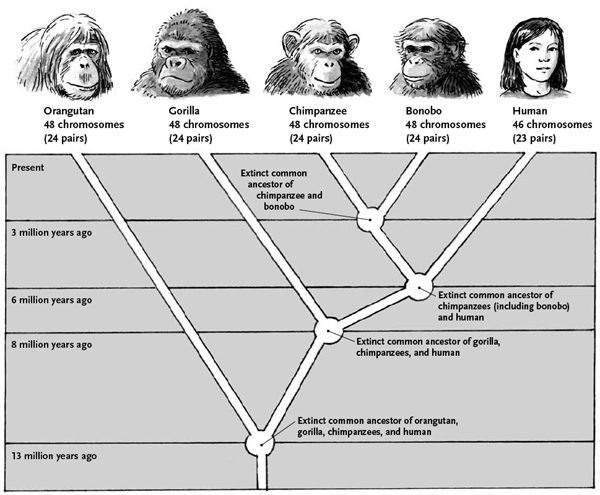
antennae? Probably his two flagella, the way he would swim
around. The feet and the single eye-not so scientific.
Algae are probably the ancestors of all land plants, and we know their gametes have flagella for swimming from our post on them. But some land plants retain this method of male gamete dispersal, but they do include some weird twists. The plants that have motile male gametes cross many of the classes that we described above. There are some seed plants and some spore plants, some vascular and some non-vascular, some are gametophyte dominant and a few are sporophyte dominant – but no flowering plants do this.
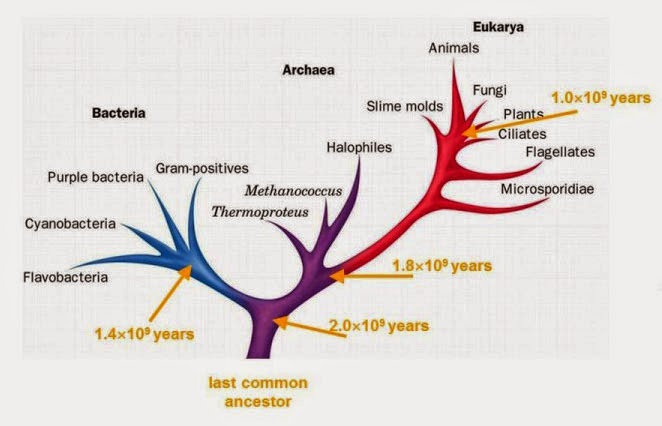
Bryophyte males gametes are swimmers. The mosses, liverworts and hornworts  live close to the ground and must have standing water for the make gamete to reach the female gametophyte and egg. The haploid gametophytes are the moss that we usually see. The antheridium grow on the top of the male plants to produce male gametes, while the archegonium on the female plant tip produces the ovule with the egg. When the water is high enough the antheridium releases the male gametes and they swim to the egg using two flagella. The sporophyte (diploid) plant grows from the top of the female gametophyte.

Ferns, horsetails, and club mosses are taller than mosses because they are vascular, but they still require water for their male gametes to swim to the egg. The gametpophyte is a heart shaped leaf that lies near the ground. At one spot the archegonium grows the egg, while the male gametes in the antheridium grow nearby. Water resting on the leaf allows the male gametes to swim across the leaf to the egg (or from leaf to leaf). The sporophyte grows from the heart-shaped gametophyte and is the fern we usually think of.


Cycads (left) are gymnosperms whose trunk are formed from
the bases of the leaves as they grow and are lost. There are
about 300 species of cycads known, with several added every
year. The Ginkgo biloba is the only extant species of the
division. Because its wood is insect resistant, some trees may
be over 2500 years old.
Angiosperms and other seed plants use pollen to send the male gametes to the ovule. When the pollen reaches the archegonium, a tube grows into the ovule and to the egg. The male gamete cells are carried along by the pollen tube right to the front door of the egg. This is when fertilization occurs.  But not allseed plants work this way. A few of the gymnosperms still use a modified version of swimming to the egg.

Cycads (about 300 species) and the lone extant ginkgo, Ginkgo biloba, do have pollen grains that represent the male gametophyte plant. They get blow or carried to the female gametophyte cone and then it gets weird.

The ovule produces a drop of liquid that sticks into the air. The pollen gets caught in this drop and then the drop and the pollen are pulled back into the ovule. The pollen tube grows into the female reproductive organ, but not right to the egg. When the pollen tube reaches the entrance of the archegonium, it ruptures and the gametes are released into a watery fluid that surrounds the eggs.


Cycad and ginkgo male gametes move on their own, the only
exceptions in the seed plants. They have hundreds to thousands
of cilia, as opposed to flagella in bryophyte gametes, which
pull the cell forward. Ginkgo male gametes are huge (0.3 mm),
larger than an entire Wolffia globosa plant.
The male gametes have about a thousand of cilia (not flagella) that pull the cell through the watery environment inside the ovule toward the egg. Fusion and fertilization occurs when the male gametes find the egg – as always. The cycad and ginkgo male gametes swim, but they swim in the indoor pool, not out in the old swimmin’ hole like the bryophytes.

The male gametes of cycads and ferns are very different, from where they swim, to their relative sizes – ginkgo male gametes are HUGE compared to those of ferns, to the use of thousands of cilia as opposed to a couple of flagella.  However, research shows that they are remarkable similar in structure and function.

A 2006 studylooked at the proteins involved in gamete movement in ferns and ginkgo. Their results indicated that most of the proteins in both were homologous enough that it suggested a direct descent from bryophyte to gymnosperm, not a case of parallel evolution.


In Guam, there is a neurologic disease that looks a lot like
Alzheimer’s. Research in 2004 found that it was actually
coming from cycad trees. Here’s how it happens. Cyanobacteria
live in the tree roots and put the toxin BMAA into the tree
tissues. Bats eat the fruits and people eat the bats. Than BMAA in
the brain causes disease.
One more cycad exception while we’re here. The cycads can do something that I thought was reserved only for philodendrons and a few relatives. These thermogenic plants can raise the temperature of their male cones by several degrees when the pollen is mature. A 2013 study showed that they can raise the temperature of male cones 2-15 degrees above ambient temperature.

This is believed to attract more insects as pollen distributors, and the researchers did find that more insects visited the plant when the temperature was increased. The mechanism may involve volatilizing more attracting chemicals though the added heat, which would then attract more pollinators (usually weevils).  Pretty advanced for a plant with a so-called primitive reproduction mechanism.

Next week – the base of the undulipodia has a special story all its own. Is it another instance of bacteria evolving into one of our organelles? And it has two very different jobs – which came first?


Suinyuy, T., Donaldson, J., & Johnson, S. (2013). Patterns of odour emission, thermogenesis and pollinator activity in cones of an African cycad: what mechanisms apply? Annals of Botany, 112 (5), 891-902 DOI: 10.1093/aob/mct159

Vaughn, K., & Renzaglia, K. (2006). Structural and immunocytochemical characterization of the Ginkgo biloba L. sperm motility apparatus Protoplasma, 227 (2-4), 165-173 DOI: 10.1007/s00709-005-0141-3

Murch, S., Cox, P., & Banack, S. (2004). A mechanism for slow release of biomagnified cyanobacterial neurotoxins and neurodegenerative disease in Guam Proceedings of the National Academy of Sciences, 101 (33), 12228-12231 DOI: 10.1073/pnas.0404926101


For more information or classroom activities, see:

Alternation of generations –

Seed dispersal –

Pollen –

Cycads –

Ginkgo biloba –

Evolving A Second Job

$
0
0

Biology concepts – protein moonlighting, undulipodia, evolution, basal body, centriole, GAPDH, intraflagellar transport


Today’s post is on a multitasking cell structure. This
would make Alton Brown proud, since he hates tools
that do only one thing. The University of Miami of
Florida football team runs through fire extinguisher
blasts when they enter the stadium – maybe Alton
can find a second use for his.
Alton Brown from Food Network hates a unitasker. He wants all his kitchen tools to have more than one function – I least I think it’s just his kitchen tools. But he might just as well be talking about biology. Nature hates a unitasker, that’s why some many things in our cells have multiple jobs.

This phenomenon is called protein moonlighting. The re-evaluation of the human genome (about 19,000 genes) suggests that many proteins have more than one distinct function. This would allow for a relatively small number of genes to provide a large functional proteome (the total number of protein functions).  As such, a 2014 study is showing the importance of moonlighting proteins in health and biology.

There are rules for a protein to have a legitimate second job. It’s only moonlighting if the two functions are unrelated,  the functions can't be carried out by two different domains of the protein either. This would suggest a gene fusion event. The two functions must be independent, so ablating one doesn’t affect the other.

There are hundreds of examples of moonlighting proteins in the literature now, and more are sure to follow. There are examples aplenty within the glycolysis pathway; you know, the breakdown of sugar for energy. No fewer than seven of the ten glycolytic enzymes are known to have other jobs.


Crystallin proteins (alpha and beta) make up the
majority of the lens and cornea of the eye. They are
transparent, but they do more than that. Recent
studies show that they have enzymatic activity in other
tissues. Aldehyde dehydrogenase and transketolase are
enzymes that turn out to be moonlight crystallins.
The king of the moonlighting proteins is glyceraldehyde -3-phosphate dehydrogenase (GAPDH). Sure, it's one of the enzymes that breaks glucose down to pyruvate, but it does so much more – like helping to maintain the ends of our chromosomes (telomeres), working to move tRNAs out of the nucleus, controlling the expression of some genes, especially those involved with gamma-interferon, repairing our DNA when it is damaged. And apparently GAPDH is crucial for helping cells to bring in particles from outside (endocytosis). That’s a full day.

But as amazing as GAPDH is, today’s example of a multitasker is even more rare, in that the moonlighter is a complete structure, made of many proteins, and has two distinctly different jobs. What’s more, each function has its own exceptions. What's our structure of interest? The basal body, or perhaps I should call it the centriole.

Let’s talk about the basal body first. This is the base of the eukaryotic undulipodia (cilia and flagella). These moving tails come in two parts; the basal body and the axoneme. We talked at length about the axoneme a few months ago, with its nine doublet microtubules surrounding two singlet microtubules (9[2]+2, see picture below). Undulipodia movement, as opposed to the motor driven prokaryotic flagella, is achieved by sliding the different doublets forward and back past one another.

But in those previous posts we didn’t talk much about the basal body. It too is a ring of microtubules, although these are shorter polymers that in the axoneme. Instead of doublets, there are almost always nine sets of triplet microtubules, and there are no center microtubules (9[3]+0). Of course, there were a couple of exceptions, and we talked about them.


The basal body has a 9(3) + 0 structure, while the
axoneme is 9(2) + 0. While this cartoon shows the
complexity of the axoneme, our post today highlights
the complexity of the basal body. One thing this cartoon
does show, the axoneme is sheathed in the plasma
membrane, it isn’t a protein structure sticking out through
the membrane.
The basal body is about 100 nm wide and 150 nm long, and serves as the base of the flagellum or cilium. If a cell has hundreds of cilia, like the male gametes of the cycads we talked about last week, then it has hundreds of basal bodies as well – one per cilium.

The basal body serves as the nucleation site for mictrotubule growth into the axoneme. It’s like a skyscraper, the microtubule girders are built vertically on the basal body foundation; only here, the basal body sparks a self-assembly of the microtubules. You don’t need fearless guys climbing the beams to build an axoneme.

If we turn our attention to the centriole, we find that it’s used in mitosis. When a cell divides, each progeny cell receives one centrosome. Don’t confuse the centrosome with a centriole or a centromere (a near center point of a chromosome, it holds the two chromatids together). The centrosome is more of an area, it contains two centrioles, a mother and a daughter, and the pericentriolar matrix (PCM) amorphous group of proteins that help the centrioles do their job.

Each centriole is a complex microtubule formation in a 9(3)+0 arrangement, about 120 nm wide and 175 nm long. This is exactly the structure of the basal body – they’re the same thing! Well, almost.... a centriole has to mature into a basal body.

During S phase of the cell cycle (when the chromosome are replicated) the centrosome will duplicate. Each centriole grows another one from its side, at a right angle. The mother is a mother again, and the daughter becomes a mother for the first time. The two mother/daughter pairs then gather their own PCMs and move to the sides of the nucleus. When mitosis time comes, microtubules grow toward the chromsosomes, but from the mother centriole only. This is the spindle and will help pull the chromatids apart during cell division.


Start at the top left. During the cycle, a pair of centrioles
will separate and each will grow another from the proximal
end. The daughter centrioles then have to mature, with
proteins added toward the distal end. During mitosis, they
two pairs separate and form the spindle body, which pulls
the chromatids apart.
If you remove the centrioles after S phase, most cells can still go through mitosis just fine. In fact, there many cell types that don’t have centrioles at all (higher plants, some protists, most fungi). Even in cells that are supposed to have centrosomes, destroying centrioles with a laser after S phase doesn’t always affect mitosis negatively.

A commentary published in 2010 talks about how many cell types, including some oocytes can undergo meiosis without centrioles, while centriole numbers than are re-established after fertilization. However, in other tissues, loss of centrioles leads to genetic instability over time, even if the spindle will develop without the centrosome. To this point, we still haven’t resolved the issue of whether centrioles are necessary for proper cell division.

In the vast majority of cases, centrioles come from centrioles. One serves as a template to form the second. The process through which this occurs has just begun to be revealed in the past few years. There is a linking fiber from the proximal end of the mother centriole that acts as a seed point to start aggregation of the daughter centriole at a 90˚ angle to the first. 

The process is very complicated, but is carried out spontaneously, without specific gene products to guide it. It was first believed that if centrioles were lost, then they could not be regained. Of course, they also thought that centrioles had their own DNA. Now we know that in cases where centrioles are lost, they can form de novo, and function just fine in mitosis or as basal bodies. In several studies, removal of centrioles or cells without centrioles to begin with allows for new centrioles being formed from aggregated microtubules.


Intraflagellar transport is the method by which the
axoneme grows, adding tubulin monomers to the end.
The proteins are carried up the axoneme by walking
proteins. These are important not for just cilia assembly,
but also for cell signaling and sensing that occurs in
the undulipodia.
After centriole duplication by prescribed pathways, the basal body matures. A 2011 review shows that there are many proteins involved in the process. First the distal end of the centriole is capped, then it migrates to the cell membrane and docks. A transition zone is produced that allows for selective movement of molecules up and down the inside of the axoneme (intraflagellar transport, IFT).

Finally, there is attachment of accessory bodies like rootlets to anchor it to the membrane, transition fibers to move to the axoneme and distal appendages. Only after all this maturation of the basal body can the axoneme be built by IFT. This is how the process happens in all species EXCEPT fruit fly male gametes and the microbe Plasmodium yoelii, where the axoneme grows BEFORE plasma membrane docking of the basal body. 

What’s more, you can always reverse it and go from basal body back to centriole, so their functions must somewhat overlap. The basal body and centriole both serve as microtubule organizing centers (MTOCs). So what makes it a moonlighting structure?


Prokaryotic flagella just whip around in a circle, but
eukaryotic undulipodia have more of a beating motion.
The side view shows how the flow is one direction, while
the top view indicates that the motion is circular, but not
like a propeller.
The basal body does a different job by controling the direction of movement of the cilium or flagellum! The prokaryotic flagellar motor spins the prokaryotic flagella as we have described, but eukaryotic cilia or flagella beat, rather than flop around, and the direction in which they beat is important. Cilia on a cell beat in concert in one direction, and the basal body is asymmetrical enough to drive this directional beating. Believe it or not, this is controlled by a protein called disheveled.

That’s a little weird, but what’s really weird is the kids. In a 1991 paper, part of the oviduct of a quail was reversed, so that the cilia beat toward the ovary instead of toward the uterus. When cells divided through embryo and chick development, the cilia of the progeny cells had the same orientation as those in the parent! They continued to beat in the wrong direction. Since basal bodies are duplicated from basal bodies, the mother basal bodies gave rise to daughters that beat the same direction.

The evidence presented shows that we have an ancient, yet complex, structure that has a couple of important jobs. The question for evolutionary biologists is which came first, the basal body or the centriole? The last common eukaryotic ancestor (LECA), the cell from which all eukaryotic cells descend, had undulipodia, so the basal body is an extremely old structure. But all eukaryotic cells undergo mitosis or meiosis, so the centriole must be important too.

Notice the cilia that are the upper most in the animation.
They show best the coordinated beating that pushes fluid
in one direction.

Cells without either centrioles and basal bodies suggest that basal body function came first; since you can’t have undulipodia without basal bodies, but our discussion above shows you can have mitosis without centrioles.

On the other hand, centrioles have to mature to become basal bodies, wouldn’t this suggest that they came first? Or perhaps the basal body was the primary product and nature managed to find a use for one if its precursors. What do you think, is the basal body the chicken or the egg?

Next week let’s look at one of the animal kingdoms great exceptions in terms of cilia, even though primitive and higher animals have them, round worms don’t cilia or flagella!


Henderson, B., & Martin, A. (2014). Protein moonlighting: a new factor in biology and medicine Biochemical Society Transactions, 42 (6), 1671-1678 DOI: 10.1042/BST20140273

Kobayashi, T., & Dynlacht, B. (2011). Regulating the transition from centriole to basal body The Journal of Cell Biology, 193 (3), 435-444 DOI: 10.1083/jcb.201101005

Debec, A., Sullivan, W., & Bettencourt-Dias, M. (2010). Centrioles: active players or passengers during mitosis? Cellular and Molecular Life Sciences, 67 (13), 2173-2194 DOI: 10.1007/s00018-010-0323-9

Boisvieux-Ulrich E, & Sandoz D (1991). Determination of ciliary polarity precedes differentiation in the epithelial cells of quail oviduct. Biology of the cell / under the auspices of the European Cell Biology Organization, 72 (1-2), 3-14 PMID: 1756309


For more information or classroom activities, see:

Protein moonlighting –

Centriole –

Basal body –

Centrosome –


Crawling To The Top

$
0
0

Biology concepts – characteristics of animals, undulipodia, gametes, nematodes, roundworms,


Yes, a sponge is an animal – just like a barracuda, a
platypus or a that weird nephew of yours. They are
multicellular, loosely organized into a couple tissues,
and eat other organisms. You can see how they filter
feed in this demonstration. Not so different from
that nephew.
Sponges and birds – they’re both animals, but would you know it to look at them? Sponges are sessile (except for the exceptions), and birds can’t breathe under water (no exceptions). Birds eat worms and lay eggs – most people don’t know what the heck sponges do. Yet they’re both animals. Are there characteristics that all animals have in common?

Yes there are, thanks for asking. Animals are all eukaryotic and diploid (2 of each chromosome). For the most part, our cells have nuclei and organelles and all but our gametes have two copies of each chromosome.

Animals are all multicellular - a unicellular organism that acts like an animal is still called a protist. Because they are multicellular, animals have the capability to have cells of different types that organize themselves into tissues and organs, like we have discussed before as a characteristic of life.

Another attribute of animals is that they can move. True, sponges are sessile when attached to rocks or coral reefs, but they do have motile cells and motile life cycle stages. Birds are very motile unless dead.

One other thing animals have in common is how the male gamete finds the female egg. Male gametes have a flagellum that allows them to swim toward the chemical signals that show them where the egg is located. True, we have learned that protists and some lower plants also have gametes that swim with flagella or cilia, but animals characteristically have flagellated male gametes. But of course, given the nature of this blog, there must be an exception.


Nematodes are round worms; helminthes are just
one group of parasitic roundworms. They represent a
turning point in animal development. They sort of have
an internal body cavity (they are pseudocoelomates),
they sort of have a head (start of cephalization) and
they sort of have body symmetry (starting to be
bilateral).
The roundworms, phylum Nematoda, are our exception for the day. Their male gametes can’t swim! But who cares, it hasn’t seemed to slow them down. Which makes us ask why everyone else goes to the trouble of producing gametes with flagella – it’s expensive. Shall we investigate?

In every other phylum of animals, male gametes use the eukaryotic flagellum to swim their way to the egg. Using exactly the same structure that we have talked about before, male gamete flagella have basal bodies and axonemes made of microtubules. The microtubule filaments slide past one another to produce their beating movement.

Look as hard as you want, but round worms don’t have basal bodies or flagella. They do have centrioles and centrosomes used for mitosis, but none of them mature into basal bodies for flagellar assembly, In fact, the male gametes of nematodes carry one centrosome (with its centriole pair) to the egg and form the basis of all centrioles in the baby roundworm. Weird - why no basal bodies? – I have no idea, but evolution approved it.

Instead of flagella for male gamete swimming, nematodes use an amoeboid movement to crawl to the egg. O.K., so they crawl instead of swim. That’s exceptional, but is it really that weird? Well… yes, considering that they don’t contain the most important protein that most cells use to make amoeboid movements.

Actin is one of the major proteins of the cytoskeleton. Actin works mostly in protrusion and contraction of parts of the cell, while intermediate filaments hold the cell’s shape and give it rigidity and microtubulesare primarily for movement of proteins and structures throughout the cell.


This short video shows you the movement of C. elegans
male gametes. They are shaped like typical animal male
gametes, they don’t move like typical male gametes, and
they don’t have the same proteins as typical ones. Yet, the
nematode is the most numerous type of animal on Earth.
Actin comes on a two main forms. G-actin is the globular form; it's the monomer. When the monomers are induced to form filaments, like tubulin monomers monomers form microtubules, it's now called F-actin. Quick assembly and disassembly of F-actin polymers from G-actin monomers allows for movement of selected parts of the cell membrane.

So amoeboid cells use F-actin as the way they extends and retracts its pseudopodia. Thus, they crawls along. Nematodes do have cells with G- and F-actin, but the male gametes don’t have any (or very little). But it’s the male gametes that need it to move! What gives?

Instead, male gametes of nematodes use the MSP protein (major sperx protein; my posts get blocked by schools if I use the whole word, so I use gamete whenever possible). A 2014 study shows that MSP proteins are abundant in the male gamete (40% of total soluble protein), and change its distribution and volume as the gamete matures and is activated. When fully activated and in the female oviduct, the MSP of the male gamete assembles and creates pseudopodia just as actin would in any other amoeboid cell. Another 2014 study shows how it then senses the egg.

Does the inability of nematode male gametes to swim to the egg cost them in terms of reproductive advantage and evolution? Heck no.

Nematodes, ie. roundworms, are the most successful animals on Earth. They live inside every other living thing, and just about everywhere on Earth. There are free living worms, parasitic worms, and worms that eat decaying tissue. There are roundworms that eat nothing but other roundworms.


Nematodes are famous for the parasitic infections they
cause. On the left is a root knot worm. Nematodes are
responsible for more than 15% of crop loss each year.
On the top is one of the filarial worms that cause river
blindness. On the bottom is a grasshopper worm (Mermis
nigrescens) that grows to fill the entire body cavity.
In strictly numerical terms, it’s amazing that we aren’t nematodes. In truth, four out of every five animals are Earth are roundworms! Long ago in classification, all the roundworms used to be lumped together; later on they were grouped according to head size. With the advent of molecular typing, there are more than 25,000 species, and estimates are for more than a million. Compare that to 5000 known mammal species.

Sure there are many species, but that number is dwarfed by the number of individuals of some species. One 2013 study from England gives us a clue. In just the city of Bristol, dogs drop about four tons of doo-doo each day. That four tons holds an astounding 3.7 billion Toxicara eggs. Every two days the dogs of that one city squat out the equivalent of the human population of the entire world. Man, is that a bizarre visual.

This isn’t useless information, considering that the eggs become worms that can cause blindness in people who accidentally eat contaminated dirt, or those who eat dirt on purpose for that matter. Indeed, many nematodes are parasites of humans and cause much disease, but this isn't our focus today. If you like that sort of weird disease stuff (and I most certainly do), I suggest you Google ascariasis, hookworm, onchocerciasis, strongyloidiasis, filariasis, or trichinosis.

Nathan Cobb of the U.S. Bureau of Plant Industry gave a very apt description of the numbers and distribution of nematodes in 1915. He said that if you eliminated every bit of matter on Earth other than nematodes, an onlooker could still recognize our world.

There are enough nematodes in the dirt that we could distinguish mountains and valleys. There are more in the cities, so we would know where they had been. Nematodes are numerous enough in living things that we could identify where every living thing had once stood. And yes, the onlooker could see humans, we ingest billions over our lifetime and more than two billion people are infected with Ascaris lumbricoides at any one moment.


At least 2 billion people are infected with this worm
(Ascaris lumbricoides) at any one time. The pictures of
the infection are just too gruesome, so I show his smile
instead. Look can up the pictures for yourself if you
haven’t eaten recently.
A. lumbricoides is the largest nematode by mass which infects humans. Females can be 40 cm long and the diameter of a No. 2 pencil. The worm takes quite the tour through you. From your stomach to your liver to your lung move the young larva. You cough them up and swallow them, and they mature in the gut. There they grow, fall in love, and mate. Yep—pretty gross.

The smallest nematodes are in marine sediment. Desmoscolex sp. and Greeffiella sp. are only 80 µm long, which means that if 30 of them stood on each other’s shoulders, they would only be as tall as a dime is thick.

At the other end of the spectrum, the largest known nematode is Placentonema gigantissima, which can reach around 30 feet long in the placenta of its host, the sperm whale.

The placenta of a whale, tree root balls, in water, mud, fruit, nematodes literally live everywhere except in the skies – even though they do find themselves in the sky every day - inside birds. Roundworms have been found in the crevices of South African gold mines two miles below the Earth’s surface – at 48˚ C (118.5˚ F) and 1000x atmospheric pressure. No other animal has been found living in stone at these depths and conditions.

Many roundworms live in the soil, and perhaps the greatest number live in the sediment of ocean floors. Because there are some many different kinds of nematodes, it isn’t surprising that many have very developed specific niches.

Biologist Colin Tudge stated in his book, The Variety of Life that half the animal species on Earth have a nematode that lives onlyin that species. Even beyond animal hosts, there is evidence of a nematode species that lives one place on Earth – in the felt of German beer coasters.


The German beer mat worm doesn’t justlive on the
bottoms of beer soaked coasters. But they do like yeast
for dinner. I like this one for Apostelbrau in Germany
because the brewery has been located in Worms, a city
between Frankfort and Stuttgart, since 1713.
The German beer mat nematode, Panagrellus redivivus, was first named Chaos redivivum by none other than Linnaeus himself. Its story is told in a nice 2009 commentary. While nematologist Cobb was aware of this worm only from felt beer mats, in truth they live in rotting peaches, in book binding paste and in other places as well.

Nematodes can be political was well. The giant kidney worm, Dioctophyma renale, is found in many different mammal species, such as dogs, cats, minks, humans, etc. But the infection is almost always just in the right kidney. Since this worm is usually ingested via contaminated fish, the right kidney might be more susceptible simply because it's closer to the liver and stomach – or maybe they’re Democrats.

All this talk about undulipodia and nematodes has been perhaps a little misleading. Nematodes do have cilia on a very small subset of the their neurons, but they aren’t motile cilia. These are sensory cilia, also called primary cilia. They are our topic for next week.


Smith HE (2014). Nematode sperm motility. WormBook : the online review of C. elegans biology, 1-15 PMID: 24715710

H. Ferris (2009). The beer mat nematode, Panagrellus The beer mat nematode, Panagrellus redivivus: A study of the connectedness of scientific discovery J. Nematode Morphol. Syst., 12 (1), 19-25

McKnight, K., Hoang, H., Prasain, J., Brown, N., Vibbert, J., Hollister, K., Moore, R., Ragains, J., Reese, J., & Miller, M. (2014). Neurosensory Perception of Environmental Cues Modulates Sperm Motility Critical for Fertilization Science, 344 (6185), 754-757 DOI: 10.1126/science.1250598

Morgan, E., Azam, D., & Pegler, K. (2013). Quantifying sources of environmental contamination with Toxocara spp. eggs Veterinary Parasitology, 193 (4), 390-397 DOI: 10.1016/j.vetpar.2012.12.034

Sepsenwol S, Ris H, & Roberts TM (1989). A unique cytoskeleton associated with crawling in the amoeboid sperm of the nematode, Ascaris suum. The Journal of cell biology, 108 (1), 55-66 PMID: 2910878


For more information or classroom activities, see:

Nematodes –

Characteristics of animals –

Cytoskeleton –


An Immovable Moving Part- That’s Just Cilia!

$
0
0

Biological concepts – primary cilia, sterocilia, kinocilium, Usher syndrome, actin, microtubule, signal transduction, sensory receptor, mechanoreceptor


The USS Oriskany (above) was scuttled in 2006 to
create an artificial reef off of Pensacola Florida. In
2012, the US government effectively ended its policy
of creating artificial reefs this way because of concern
for leaking toxins from the ships to the marine life. But
is was a good way to find a new job for something broken.
Naval vessels are built to move through the oceans. When they can’t, they get fixed or they get decommissioned. As broken vehicles they have no use. Or might they? Some have been re-purposed as man made reefs.

Something that seemed broken because it couldn’t move was given an important new job that didn’t require motility. Remember that analogy as we talk about today’s subject in cilia. Although the order might be reversed.

We spoke last week about how nematodes are the only animals that don’t have cilia. Eukaryotic cilia and flagella (together, the undulipodia) are organelles that move, and in turn may move cells. It turns out that cilia have some exceptions – some don’t beat, and some can’t move at all - so what good are they?

Motile cilia, the kind we have been talking about for the past couple of weeks, are also called 2˚ cilia. If there are 2˚ cilia, I think that pretty much implies that there must 1˚ cilia– and they’re what we will talk about today.

Primary cilia, while less well known, are found on many more cell types than are motile cilia. Motile cilia in mammals are located on male gametes (as flagella), on respiratory epithelium of the lower and upper respiratory tract, fallopian tubes near the ovary and epididymal cells of the testes, and the ependymal cells lining the ventricles of the brain.

Primary cilia are apparent on cells of most types, when they are quiescent (just hanging out, doing it's job). If the cell re-enters the cell cycle and starts to divide or differentiate, the primary cilium will resorb and then reappear in daughter cells once they become quiescent.


Primary cilia have basal bodies and IFT, but their
microtubule structure is different. They don’t have the
inner singlet microtubules, so the outside ones can’t slide
past one another. They do have outer dynein arms, and
those are important for retrograde IFT. See below,
kinocilium don’t even have the outer arms.
Primary cilia are 9(2) + 0 in the axoneme, which they makes them different than motile cilia (see picture and this review). Primary cilia are missing the two central microtubule singlets. They are also missing all dynein, both the inner and the outer arms. This is why they are immotile, sort of.

Another exception with primary cilia is that their microtubule axoneme can change as it goes out to the end of the cilia. It may start out as 9(2) + 0, but at the distal (far) end it's 9 + 0 in nematodes, algae, and in the nose, pancreas and kidneys of vertebrates. All of those count as exceptions too!

In addition, primary cilia are of differing lengths, but most are much shorter than motile cilia. Some don’t even extend from the surface of the cell membrane. However, they're built by IFT (intraflagellar transport) just as 2˚ cilia are, and IFT is important for their functions as well.

So, can a broken cilium have a specific job? If they don’t beat to move a cell or the environment around the cell, then what do primary cilia do? The answer is - just about everything. Primary cilia serve as mechanoreceptors, chemoreceptors, photoreceptors, as well as osmolarity, temperature, or gravity receptors. Think of primary cilia like weather balloons. They stick out into the environment and probe the conditions in the area. They send the data back and the cell can act on it.

As mechanoreceptors, primary cilia might not beat, but they can be moved. They bend in response to flow across the surface and the bend brings a pivot at the level of the basal body – yes, primary cilia have basal bodies just as motile cilia do.


Kidney cells that line the tubules have primary cilia to
a change in calcium influx. The change is then
transferred to the adjacent cell via calcium channels
that cross both membranes.
The kidney cells that line your urine-filled tubules have primary cilia that stick out into the urine river. As the urine flow speeds up after your third diet coke in the last hour, the primary cilia bend and transmit a signal to the cell. This then signals the cells to ramp up their filtering functions, pulling water back in or excreting urea, etc.

Primary cilia have an asymmetry so that they recognize right from left. In the kidney, the flow is based on orientation, all primary cilia bend in the same direction, toward the anterior. The anterior bend signals for increased calcium influx and then this signal is transmitted to adjacent cells. The uniform gradient (a-p) works cell to cell, and this leads to consistent a-p orientation of the mitotic spindle (which also uses basal bodies in the form of centrioles). The result is that the progeny cells of dividing renal epithelium have the same orientation as the parent they replace.

Back to our nematodes from last week. Primary cilia are the only cilia roundworms have. C. elegans, the roundworm that is used as a laboratory model, is made up of exactly 959 cells – exactly. Sixty of those cells, all sensory neurons, have primary cilia that stick out into the environment via pores called sensillae.

The left photomicrograph has labeled dendrites for sensory
neurons in C. elegans. The right cartoon shows how the
primary cilia from these neurons stick into the pore that
then helps them sense the environment around
the roundworm.

It’s through the interaction of these primary cilia with the worm's immediate environment that it senses its world. This is what passes for a roundworm brain – but your brain has them as well. Especially in the retina of your eyes.

The photoreceptors that absorb light energy and transfer it to electrical impulses are located on a single primary cilium on each retinal cell. The axoneme is used to move photosensitive pigments (like retinal in rhodopsin, see below) back and forth from the receptor to the cytoplasm.

Primary cilia also act as chemoreceptors. In brain proper, they work in formation of new memories – mice without primary cilia can’t remember new objects or recognize objects they have already learned. They can remember the location of the object just fine, just not the object itself. We will talk about primary cilia in the brain much more next week.

Now we can take this discussion a couple of steps further to talk about two ciliary exceptions. There are nonmotile 1˚ cilia, motile 2˚ cilia, and then a third structure called a kinocilium. From the Greek for moving eyelash, the kinocilium is poorly named. Described in guinea pigs in 1989, they don’t move like a blinking eyelash or even like a motile cilium; they lack the inner dynein arms and central microtubules that would allow them to be motile. But, they can move horizontally across a cell surface.


As this movie travels down the photoreceptor, notice the
vertical basal body/axoneme on the left. This is a primary
cilium! The microtubules help move photo pigments up and
down the cilium.
Located in humans on the hair cells the inner ear, kinocilium play a crucial role in both hearing and balance, even though they're gone by the time you hear or need to stand up straight. Their role is regulating the erection of the apparatus that allow hair cells to function.

If hair cell kinocilia are poorly named, then hair cell stereocilia are down right liars. They aren’t cilia at all. The characteristics of cilia include that they are microtubule extensions of a basal body modified from a centriole. They may be motile or nonmotile, but their functions are mediated by moving signaling, structural, or receptor molecules up and down via intraflagellar transport proteins.

None of that applies to sterocilia! They're built from actin not microtubules. They do not have an intraflagellar transport system. They have no basal body. They are very similar to the microvilli of your gut epithelium, but nothing like cilia, except for the fact that they stick up from a cell.

The hair cells work by using the sterocilia as mechanoreceptors. In the cochlea, they bend in response to fluid movement based on vibrations of sound. In the semicircular canals, they bend in response fluid movement as a result of changes in head position. When the sterocilia bend, it generates an action potential in neurons that go to the brain.


Hair cells of the cochlea can be damaged by loud noise.
The left images are the normal (top) hair cell sterocilia, and
the same sterocilia after a loud noise (bottom). The right
images show a series of hair cells in normal condition, and
after a long time exposed to loud noise. Turn down your
music – do you think the hair cells in the damaged cochlea
work well?
So that explains the sterocilia (that aren’t really cilia), but what about the kinocilium? A 2007 paper reviewed how kinocilia mediate production of sterocilia. The hair cells start out with a smooth surface and one long kinocilium in the center of the apical (top) surface. Then the sterocilia start to grow. As the sterocilia appear, the kinocilium moves laterally, to the edge of the apical surface. This defines the orientation of the hair cell – the direction the sterocilia will bend.

The sterocilia start to grow longer, with the ones closest to the kinocilium being the longest. They line up to look like a choir on risers in front of the taller kinocilium. Now they are ready to function. At this point the kinocilium disappears! If you look at working hair cells, you won't find the structure that mediated their development.

So we have two new ciliary structures - neither of which act like cilia. That’s weird enough, but it gets weirder. There is a disease that affects both hearing and vision because it messes with the primary cilia of the retina and the sterocilia of the ear. But we just learned that those are two completely different structures!


People often use "tunnel vision" to explain the field changes
in retinitis pigmentosa, the kind of progressive blindness
in Usher syndrome. But really, it’s more like backing into a
tunnel, one that never reaches the other side. Think of
running this clip backwards.
Called Usher syndrome, victims suffer from hearing loss, vision loss, and balance problems. The vision loss is due to defective maintenance of primary cilia of the retina, but the stereocilia of the cochlea and vestibular system aren’t cilia at all, how could they be affected by a 1˚ cilia protein problem?

As we said last week, nature hates a unitasker. There are at least 11 proteins that work in development and working of both sterocilia AND primary cilia, even though they look and are built completely different. A mutation in one of those proteins affects all three systems. Maybe it would be better if sometimes a protein had just one job, fewer things could get screwed up if something goes wrong with it. Would you rather have reduced vision, reduced hearing, bad balance, or all three?

All this knowledge leaves us with an unanswered question - did the sensory primary cilia develop from motile cilia, or did motile cilia develop from the primary version? Did broken motile cilia develop a new job, or did 1˚ cilia learn how to dance after they had learned their first function? Hmmmm.

We've barely touched the functions of cilia that don’t even move. In the next couple of weeks, we will see how primary cilia keep you from being fat, and how they will be crucial for long-term space travel. Then we can figure out how they give you a right and left hand.


Mathur P, & Yang J (2014). Usher syndrome: Hearing loss, retinal degeneration and associated abnormalities. Biochimica et biophysica acta, 1852 (3), 406-420 PMID: 25481835

Doroquez DB, Berciu C, Anderson JR, Sengupta P, & Nicastro D (2014). A high-resolution morphological and ultrastructural map of anterior sensory cilia and glia in Caenorhabditis elegans. eLife, 3 PMID: 24668170

Patel, A. (2014). The Primary cilium calcium channels and their role in flow sensing Pflügers Archiv - European Journal of Physiology, 467 (1), 157-165 DOI: 10.1007/s00424-014-1516-0

Fry AM, Leaper MJ, & Bayliss R (2014). The primary cilium: guardian of organ development and homeostasis. Organogenesis, 10 (1), 62-8 PMID: 24743231


For more information or classroom activities, see:

Primary cilia –

Hair cells –

Visual photoreceptors –

Usher syndrome -


Viewing all 288 articles
Browse latest View live