Quantcast
Channel: As Many Exceptions As Rules
Viewing all 288 articles
Browse latest View live

Tricky Little Buggers

$
0
0

Biology concepts – immune defense, antibiotic resistance

Naim Süleymanoğlu is better known as Pocket
Hercules (4' 10"). He was born in Bulgaria, but is
of Turkish descent. He competed and retired
several times, and won gold medals from 1983
to 1998. He is one of the first competitors to
lift more than 2.5x his own body weight.

Therewas a small Turkish weightlifter a few years back whose nickname was “Pocket Hercules.” He won gold medals in three separate Olympics and was the one the best examples of big things in little packages. Last week we talked about the immune systems of vertebrates, invertebrates and plants, now let’s talk about the defenses of the smallest organisms – bacteria are the Pocket Hercules of biology.

Do bacteria have defense mechanisms? You bet – they get attacked all the time.  For bacteria that stray into or purposefully target animal or plant hosts, the perils are many and varied. Antimicrobial peptides try to burst them, antibodies try to bind them up and point them out to killer cells. Macrophages and other phagocytic cells try to eat them or wall them off from the other host cells. Organisms will even sacrifice their own cells just to make sure they kill the bacteria. It’s a jungle out there.

We don’t have the time nor the room to go into the thousands of ways that bacteria protect themselves from plant, invertebrate, and vertebrate immune attack, but we can give a few examples, like deception. Mimicry is when a bacterial antigen looks much like one of our molecules, so that the body is either fooled into not attacking, or tempers its attack.

Other bacteria change their clothes to remain hidden. Just when an immune system sees it and starts the attack, Neisseria gonorrhoeae changes its surface molecules and becomes invisible again. On the other hand, Yersiniapestis remains invisible by living inside macrophages.

Some bacteria stunt our antibody response. The best way to keep from being attacked is to not allow the host to recognize and identify you. The bacteria that cause TB inhibit our immune system from producing specific antibodies. 
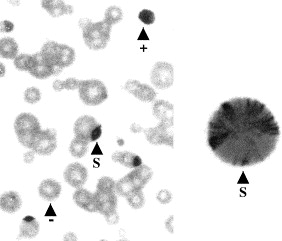
Some bacteria vary the antigens they show on
their surface in order to evade the immune system.
In this bacterium, the dark areas are stained for
one particular surface antigen. You can see that
some of the cells have none of that protein, some
have only that surface protein, and some have
discrete areas where that protein is expressed.

The defense is a good offense, so some bacteria attack. Pseudomonas strains kill the phagocytic cells that would try to eat them by releasing chemicals called aggressins. Staphylococcus aureus just confuses the phagocytes, producing toxins that stop their movement or make them move erratically.

These are but a few of the many bacterial defenses against our immune system. But they have evolved defenses against other threats as well, like our attempts to kill them with antibiotics.

We talked earlier about multidrug efflux pumps in bacteria that pump out the antibiotics with which we try to kill them. This is related to the stories in the media about antibiotic resistance in bacterial pathogens, but classic antibiotic resistance genes are often plasmid based defenses,as we have discussed. Recently, an additional defense against antibiotics has been recognized.

It seems most bacteria produce hydrogen sulfide (H2S, smells like rotten eggs), which was previously thought to be only a metabolic byproduct. A late 2011 studyshows that H2S is part of an integrated defense system used by almost all bacteria. The gas works to prevent oxidative damage. This is not unheard of since a few bacteria produce nitric oxide to do the same thing, but it is being recognized now that oxidative stress induction is a big part of how many antibiotics work. When the H2S system was turned off in several pathogens, they became much more sensitive to antibiotics. Maybe this a lesson we can exploit in the future.

In addition to our attempts to kill them, the universe itself is a tough place to survive if you are a bacterium. They may end up in bright sunlight for long periods of time, or hurtling through space on a rocket or meteor. Bacteria have ways to protect themselves here as well. Ultraviolet radiation from the sun is a mutagen (causes mutations in DNA), but it also can break down cellular molecules to release oxygen radicals, like hydrogen peroxide or superoxide.

It has been known since the 1950’s that pyruvate and catalase, as well as the newly discovered H2S discussed above, do some work in protecting the cell against oxidative damage, but a 2009 study described a whole new mechanism. It seems that E. coli has two proteins that seek out, identify, and repair oxygen radical-mediated damage to sulphur-containing cysteine amino acids within proteins.

Cysteine is the most reactive of the 20 common amino acids, which means that it are often located in the functional site of enzymes (where the enzyme reacts with the substrate). However, this reactivity also makes cysteine vulnerable to reaction with radicals, especially oxygen radicals, after which it becomes modified and non-functional.

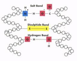
Disulphide bonds are formed between adjacent cysteines on the 
same peptide, far apart cysteines on the same peptides, 
or between cysteines on different peptides. When 
you (not me) get a permanent wave for your hair, the 
disulphide bonds are broken or rearranged by a reducing agent. 
To prevent this radical-mediated damage, cysteines often occur in pairs, where links between the sulphurs of the two cysteines help to prevent oxidation (called disulfide bonds, they also serve to link peptides together and give proteins their proper form). A 2008 study showed that this mechanism provides unusual oxidative stability to a cysteine-containing enzyme of the bacterium, Desulfovibrio africanus.

But there are exceptions; lone cysteines do occur, and these are the cysteines most vulnerable to oxidative damage. The DsbG and DsbC proteins of E. coli patrol the cytoplasm looking for oxidized cysteines to fix.

Here is how ingenious the system is – oxidizing a cysteine may or may not unfold the protein, so DSbG is charged and can interact with the still-folded proteins to correct the cysteine problem, but DsbC is uncharged, so it works better with proteins that have been unfolded. Amazing - and bacteria developed it all on their own – well, with the help of the evolutionary pressure of things trying to kill them.

I mentioned that radiation is also a DNA mutagen. The mutagenic properties of radiation affect bacteria just like they affect us; it is just that some bacteria can protect themselves better once their DNA is damaged. Follow me closely here - by using protein repair and protection systems, bacteria like E. coli, with its DsbG and C enzymes, can keep protein functions going when other organisms would break down and die. Some of these protein functions include DNA repair after mutagenesis. So - some bacteria don’t survive radiation because they protect their DNA better, they survive because they repair the damage better.
This is an overlap of different types of images of a
radiodurans bacterium. The circles of blue green and
pink show high concentrations of manganese, while
red is iron. The manganese is clustered around the
DNA and works to repair it after radiation damage.

Other bacteria have a different mechanism to maintain protein function. According to a 2010 study, a shield of manganese metal atoms and phosphates was found in D. radiodurans. It had been long known that manganese was present in very high levels in bacteria that are most resistant to radiation, but its function was unknown.

The recent study shows that these manganese complexes work together to protect proteins from radiation damage, but not DNA. The key for this system is to keep proteins functioning, which can then repair any radiation damage to the DNA. This mechanism allows D. radiodurans to withstand prolonged radiation that is 1000x stronger than that which would kill a human.

So, bacteria have defenses against immune and environmental attacks. Does anything else attack bacteria? How about other bacteria - it’s dog eat dog out there, competition for resources is brutal. Many bacteria have poisons (bacteriocins) that inhibit or kill bacteria that are distantly related (because related types of bacteria are likely to be in the same places looking for the same food). 

One type of bacteriocin are the lantibiotics. These protein toxins contain a nonstandard amino acid, called lanthionine. We mentioned above that cysteines are very reactive; lanthionine is a modified circular (polycylic) cysteine that gives the toxin its reactivity. And because it is cyclic, it is much less vulnerable to oxidative damage itself – funny how bacteria seem to cover their bases so well.

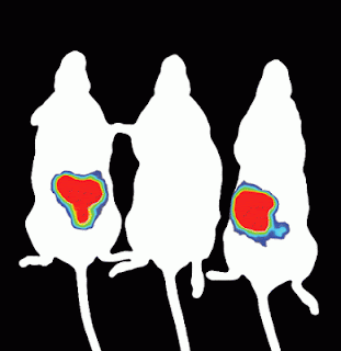
This is so cool. Bacteria that are engineered to produce
light were injected into rats. The rat in the middle was
also given a bacterium producing a bacteriocin to the
light producing bacteria. The whole rat bodies were
imaged while they were still alive to see if the bacteria
were alive and reproducing. Live animal imaging is a
great tool that is becoming more popular. Image by S.
C. Corr and P. G. Casey.
Lantibioticscome in two types, they either form pores in Gram+ bacterial cell walls or inhibit the cell wall formation. Because they attack only specific types of bacteria, lantibiotics are useful in cheese-making; they allow some bacteria to grow and ripen the cheese, while killing those that would cause the cheese to spoil. One type B lantibiotic just came through its phase I clinical trial in July 2012 with flying colors (phase I trials are meant only to test safety, not effectiveness).

A recent discovery illustrates just how bacteriocins are delivered to the target organism. It seems that bacteria can build a spike and a spike launching system anywhere on their cell membrane. The spike is spring loaded in a tube just 80 atoms long, and is fired at the target cell. Then the bacteriocin is released at the end of the spike to do its damage.

The release of toxin was already known, called a type IV secretion system, but the CalTech study that identified the spring-loaded spike as the delivery system is very new. Once fired, the whole system is broken down, ready to be rebuilt somewhere else in the cell. Amazing. (click for video)

Of course, for every punch there is an evolutionary counterpunch, so there are bacteriocin resistance mechanisms as well. Nisin, a bacteriocin active against strains of listeria, is approved as a food preservative. But listeria can spontaneously develop resistance to nisin. It appears that some strains change their membrane chemistry in order to render nisin ineffective. Therefore resistance could be a problem if we pursue the use of bacteriocins as antibiotics; we might end up back in the same situation that we're in now.

Regardless of this possible downside, scientists have found a way to bring bacteriocins into the battle against antibiotic resistance. An E. coli has been engineered to contain the gene for pyocin, a bacteriocin that kills strains of Pseudomonas bacteria. E.coliand Pseudomonas are not closely related, so E. coli would not naturally possess this toxin, scientists added the gene to the E. coli.

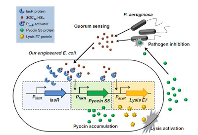
This is schematic of the engineered bacteria to kill Pseudomonas.
P. aeruginosa make chemicals when their numbers reach a
certain density. These trigger pyocin production in the E. coli,
but also triggers the production of the protein that lyses the
E. coli. When lysed, the pyocin attacks the P. aeruginosa.
When the engineered bacteria encounters Pseudomonas, it does two things; it produces the pyocin toxin to kill the target cell, and the engineered E. coli commits suicide. No release system has been engineered into the E. coli, so the only way they get the pyocin to the target is to have the E. coli produce a lysin that destroys its own cell membrane.

This suicide accomplishes two things, it releases the pyocin to kill the target, and it prevents the engineered E. coli from hanging around forever, possibly trading genes with other bacteria or causing havoc in some unforeseen way.

So it looks like bacteria have it made. They can resist immune system attacks, some can resist environmental onslaughts, they even have ways to protect themselves against competition and threats from other bacteria. No wonder they have always been the predominate life form on Earth. But bacteria do have foes of considerable power – veritable “Micro-Hercules” – we will meet them after Thanksgiving.

Let’s take a couple weeks to talk about the biology of turkeys and the so-called “tryptophan nap.”


Basler, M., Pilhofer, M., Henderson, G., Jensen, G., & Mekalanos, J. (2012). Type VI secretion requires a dynamic contractile phage tail-like structure Nature, 483 (7388), 182-186 DOI: 10.1038/nature10846

Saeidi, N., Wong, C., Lo, T., Nguyen, H., Ling, H., Leong, S., Poh, C., & Chang, M. (2011). Engineering microbes to sense and eradicate Pseudomonas aeruginosa, a human pathogen Molecular Systems Biology, 7 DOI: 10.1038/msb.2011.55

For more information or classroom activities, see:

Bacterial defenses–

Bacteriocins –
see Pubmed (http://www.ncbi.nlm.nih.gov/pubmed) for more information on these defenses.

A Meal More Powerful Than The NFL

$
0
0
Biology concepts – genetic code, neurotransmitters

A turkey dinner with all the fixins can lead to a
satisfying nap. But the meal usually takes a little
longer than this to have an effect. This fellow might
be more affected by last night’s activities than today’s
meal.
Turkeydinner at Thanksgiving brings the family together, celebrates the bountiful harvest, and puts you to sleep just as the NFL games are ready to start. Many people think that if you eat less turkey and fill up on the other goodies you can escape the post-Thanksgiving meal sleepiness. Other people look forward to eating seconds and thirds and then stretching out on the couch for a long nap, forcing Aunt Ethel to sit in the chair with the spring that surprises you every once in a while.

The culprit, or the hero, in this eat and sleep saga is said to be the tryptophan in the turkey. Other people think that it is simply how much you eat, not the turkey's tryptophan, but it isn’t quite that simple. What is tryptophan, and is it indeed responsible for the snoring that follows Thanksgiving dinner?  Some background will help.

Tryptophan is an amino acid, one of the twenty standard building blocks of proteins. Each amino acid has a similar basic structure, as shown in the picture below. The central carbon has an amino group (NH3) on one side and a carboxylic acid (COO-) moiety on the other; hence the name – amino acid. The third side group is a simple hydrogen (H), while the fourth side (R) refers to any of several different side groups and is what makes one amino acid different from one another.

Tryptophan is an aromatic amino acid, meaning that its side group contains a six-sided carbon ring structure (each corner represents a carbon). It also has a second ring group of four carbons and a nitrogen. As such, it is the largest and most massive of all the standard amino acids. However, tryptophan is the least abundant amino acid in plant and animal proteins; it accounts for only 1-1.5% of the total number of amino acids in proteins.

Amino acids are the building blocks of proteins. The NH3
is the amino part and the COO is the acid part. The R is
different for each amino acid. On the left, you see that
tryptophan’s R group is a big structure with two different
rings (each angle where two lines meet stands for a carbon,
they just don’t write in each “C”). Two lines means a double
bond. In producing the protein, the COO of the last amino acid
added gets connected to the NH3 of the next amino acid to be
connected. Which amino acid it is determine by the mRNA
and the genetic code.

Tryptophan’slarge structure and intricate rings make it costly to produce in terms of ATP invested. In fact, it takes so much energy to make that we have stopped making tryptophan all together. Tryptophan is abundant in a number of food sources commonly available to humans, so over evolutionary time we have turned it into an essential amino acid. True, it is essential for life, but here the word “essential” means that we MUST get it from our diet, we cannot produce it ourselves.

Of the 20 standard amino acids, 10 are essential in humans (9 that we must eat and 1 that we make from an essential amino acid), but bacteria make them all just fine - although the parents of newborns may wish it wasn’t so. Gut bacteria make tryptophan or use the tryptophan we eat. They transform it into molecules they need to survive, but the byproducts of these reactions are skatole and indole – these are the precious little molecules that give dirty diapers that wonderful smell!

Tryptophan is different from many other amino acids in another way as well; it gets no respect from the genetic code. Each amino acid is coded for by a group of three RNA bases, together called a codon. Since there are four different bases in mRNAs (A, C, G, and U – remember that T is used in DNA but not RNA), then there are 64 different codons (4 x 4 x 4). This is more than the 20 amino acids that the codons code for, so most amino acids have two or three codons that signals that they should be added to the growing peptide. But tryptophan is encoded by only one codon (UGG).

It may make sense that an amino acid that is not used often in proteins might rate only one codon, but the amino acid methionine is used much more often than tryptophan, and it's only coded for by one codon as well (AUG). You know nature must have a reason why tryptophan has a single codon, we just don't know it yet.

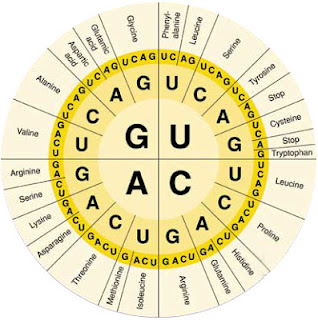
The genetic code is how mRNA codons (3 bases sequences)
get translated into a signal to build proteins from specific amino
acids. The first base of the codon is represented by the biggest
letters (ACGU), the middle base is the middle size letters, while
the third position (wobble position) is usually where you see an
amino acid coded for by more than one codon. For instance,
serine is coded for by UCU, UCC, UCA, or UCG. But tryptophan is
only coded for by UGG. Three codons signal the protein to stop
growing, called stop codons (UAG, UAA, and UGA).
Eventhough it is used sparingly in proteins, tryptophan is an essential amino acid - don’t eat enough of it and you die. This is because tryptophan’s most essential functions have nothing to do with protein synthesis or structure – tryptophan is important to your brain function. The crucial neurotransmitter, serotonin, is synthesized only from tryptophan.

It takes two enzymes to turn tryptophan into serotonin (also called 5-HT).  First is tryptophan hydroxylase; hydroxylase means it splits water, here it adds an OH to tryptophan. Next, the amino acid decarboxylase removes a carboxylic acid (COOH), producing serotonin.

Amongst the many functions of serotonin are a few that are not brain related. Serotonin is released by enterochromaffin cells that line your gut to tell your gut to move. The movement helps push the food along your digestive tract, but serves a protective function.

If you eat something toxic, the enterochromaffin cells produce more serotonin – your gut moves much faster, and you get diarrhea. If even more serotonin is made and released, it moves through the bloodstream to your stomach and esophagus and causes you to vomit.

But it is in the CNS that serotonin has its significant activities. As a neurotransmitter, it is responsible for controlling how electric messages are passed from one neuron to another. When serotonin is released in the synapse (the gap between the upstream and downstream neurons) and is taken up by adjacent neurons, it produces a sense of well-being.

Where one neuron ends and others begin there is
a gap called the synaptic cleft. Different types of
neurons use different neurotransmitters, of which
serotonin is one. It is released into the synapse, and
adjacent neurons with serotonin receptors can be
stimulated to conduct a nerve impulse. The serotonin
is broken down in the synapse by MAO’s and taken
back up to produce more serotonin.
It isn’t surprising that depressed individuals often have low blood levels of tryptophan, as well as reduced serotonin. Classic treatments for depression include increased tryptophan intake, monoamine oxidase (MAO) inhibitors, and serotonin reuptake inhibitors (SSRI). With more tryptophan, you make more serotonin – problem solved. On the other hand, MAO’s break down serotonin, so their inhibitors enhance the action of tryptophan. SSRI’s prevent the reuptake, this leaves serotonin in the synapse longer. Both types of drugs make tryptophan more likely to be taken up by downstream neurons.

Unfortunate, but interesting, is the study showing that the suicidal thoughts that sometimes accompany anti-depressant therapies (TESI – treatment enhances suicidal ideation) use may be related to polymorphisms in one form of the tryptophan hydroxylase enzyme that starts the serotonin production from tryptophan.

When non-suicidal patients were compared to those with TESI or those who were suicidal without treatment, a pattern emerged. Only those with TESI showed a polymorphism pattern in the tryptophan hydroxlyase 2 (TPH2) gene. This polymorphism had previously been associated with suicide victims and major depressive disorder. It seems that a slight alteration in function of TPH2 due to a single nucleotide change can contribute to the genetic background of treatment induced suicidal thoughts.

The feeling of general well being induced by serotonin also participates in the sleep/wake cycle. So is tryptophan – through serotonin – responsible for the post-Thanksgiving nap? Well… yes and no, it's an accomplice in a larger conspiracy.

Serotonin is use to produce the hormone melatonin, and melatonin promotes sleep, so you could say turkey dinner promotes sleep. But turkey doesn’t have that much tryptophan! Tofu has much more tryptophan than turkey, but you don’t get a post-Chinese takeout urge to sleep, so what gives?

Melatonin is made from serotonin in the pineal
gland. Sunlight stimulates the suprachiasmatic
nucleus (SCN) which inhibits the pineal from
making melatonin. As the sun goes down,
inhibition is reduced, more melatonin is made
and released from the pineal, and sleep is
promoted.
The melatonin effect has to do more with how much of everything else you eat at Thanksgiving dinner, especially carbohydrates. Here is how it works – eating lots of carbohydrates causes a release of insulin into the blood (to reduced blood glucose levels). Another function of insulin is to promote the uptake of some amino acids (but not tryptophan) into muscle cells. This leaves the blood higher in tryptophan as compared to other amino acids than it would normally be.

The brain takes in amino acids through a neutral amino acid transporter, which now finds more tryptophan than other neutral amino acids, so the brain level of tryptophan goes up. More tryptophan in the brain, more serotonin – more serotonin, more melatonin. More melatonin = nap time! So if you want to avoid the post-Thanksgiving nap, eat the turkey and skip the mashed potatoes.

You didn’t know how much tryptophan controlled your daily life, did you? Well, there’s more. Tryptophan is also important in synthesizing niacin, a.k.a. vitamin B3 or nicotinic acid. Niacin is important in production of NAD/NADH for energy metabolism, for production of steroid hormones and balance of lipid forms in the blood, and as an anti-convulsant.

The tryptophan-niacin connection is made stronger by recent evidence that high dietarytryptophan can prevent epileptic seizures in mice. In this study, a whey protein called alpha-lactoalbumin (ALAC) was found to have much tryptophan, much higher levels than in most proteins. Feeding epileptic mice ALAC resulted in reduced numbers of seizures.

So even if you don’t want to sleep or think happy thoughts, you still need to eat food that contain tryptophan or niacin. And many of those foods are plants, because plants use tryptophan to control their own activities. Tryptophan is easily converted to auxins, a type of plant hormone. Auxins are responsible for several different plant behaviors, namely the falling leaves in autumn and ripe fruits all year long.

Here is an interesting attempt to get kids to read
history. During the spring, captive warriors were
killed by cutting out their hearts, then their skin was
flayed off their body, and the priests would wear them
around for 20 days. This was meant to celebrate the
god who sacrificed himself to allow a new growing
season to begin. This time period corresponds
 to when they would have had the lowest amount of
 tryptophan in their daily die. No - I wouldn't want
to be an Aztec sacrifice!
Having dietary choices for tryptophan is good, and plants provide our major source. However, cooking grains and corn reduces usable tryptophan and niacin levels dramatically, so poorer environments where corn is the staple food need also to have additional dietary sources of tryptophan. A deficiency of this amino acid leads to some disturbing conditions. Low tryptophan leads to low serotonin levels and agitation, insomnia, and depression. A study in the Archivesof General Psychiatry stated that chronically low levels of tryptophan led to relapses of purging behaviors in bulimics.

More amazingly, studies in the 1970’s to 1990’s suggest that low tryptophan levels can lead to increases in religious fanaticism. Several studies from a single author correlate the Aztec human sacrificial ceremonies to the times of year when their diets depended more on foods that had less tryptophan. Think of all the lives that could have been saved by tofu!

But turkey is more than just tryptophan. You have to love an animal that has caruncles, a wattle, and a snood!


Musil, R., Zill, P., Seemüller, F., Bondy, B., Meyer, S., Spellmann, I., Bender, W., Adli, M., Heuser, I., Fisher, R., Gaebel, W., Maier, W., Rietschel, M., Rujescu, D., Schennach, R., Möller, H., & Riedel, M. (2012). Genetics of emergent suicidality during antidepressive treatment—Data from a naturalistic study on a large sample of inpatients with a major depressive episode European Neuropsychopharmacology DOI: 10.1016/j.euroneuro.2012.08.009


Russo, E., Scicchitano, F., Citraro, R., Aiello, R., Camastra, C., Mainardi, P., Chimirri, S., Perucca, E., Donato, G., & De Sarro, G. (2012). Protective activity of α-lactoalbumin (ALAC), a whey protein rich in tryptophan, in rodent models of epileptogenesis Neuroscience, 226, 282-288 DOI: 10.1016/j.neuroscience.2012.09.021

For more information or classroom activities, see:

Genetic code –


Neurotransmitters –
http://science.education.nih.gov/supplements/nih2/addiction/activities/activities_toc.htm


As A Bird - It's No Turkey

$
0
0
Mr. Carlson and Herb Tarlek had to deal with the
aftermath of bombing Cincinnati with live turkeys.
The line about turkeys being able to fly is one of the
most famous in TV history. But he should have at least
questioned whether they could fly, there are more than
50 species of flightless bird alive as we speak.

In a famous 1978 episode of the TV sitcom, WKRP In Cincinnati, station manager Arthur Carlson releases turkeys from a helicopter to a waiting crowd below as part of a holiday publicity stunt. The birds crashed to the ground (off camera), as intrepid reporter Less Nessman described the carnage. You can find the entire episode here; it’s as funny now as it ever was.

This comedy had a 1940’s parallel in real life, when the town of Yellville, Arkansas dropped Thanksgiving turkeys off the courthouse roof for several years in succession, and then from low flying planes. They didn’t seem to have any qualms about the flight problems of the domesticated turkey.

In contrast, the North and South American wild turkeys would have survived the stunts. In the genus Meleagris, there are several species of wild turkey, and they can and do fly short distances. In fact, they spend their nights perched in the low branches of trees from Maine to Peru.

The Aztecs introduced the Spanish to Southern Mexican turkeys (Meleagris gallopavo gallopavo), who took them back to Europe in the 1520’s. The Spanish trade capital at the time was Turkey, and from there turkeys spread all across the continent by the 1550’s. Therefore, the English called them turkeys, because they thought the birds originated in Turkey.

The Meleagris g. gallopavabirds brought back to Europe were domesticated and became the eating turkeys of today. They were bred for large breast muscles, and being raised in domestication caused them to lose much of their flying musculature; the domesticated turkey is flightless and mimics a bowling ball when released from a helicopter.

Here’s proof for you city folks that turkeys can fly.
Their perch is bird-like, drawing up one leg. This might
be to conserve heat, or to change their outline and
make them look like a plant in order to avoid predation.
Truly, that is one hypothesis… I mean it….really I do.
In animals, only birds have a vertical extension (keel) on their breastbone to allow for attachment of the large breast muscles required for flight. Birds are also the only animals to have fused collarbones, called a furcula. This bone attaches to the muscles important in the down stroke of wings, and also helps to pump air into the lungs - we know this structure as the wishbone. The furcula is more massive in the middle, and can flex and act like a spring during flight.

We use the furcula as a sign of good luck, but for domesticated turkeys it is just an unfortunate reminder that they used to have a fighting chance at avoiding a gravy bath. It's ironic that breeding to increase the size of their flight muscles is exactly why the domesticated turkey can’t fly.

Over many generations, the domesticated turkey’s muscles have become too big to allow it to fly and its legs have becomes shorter, so it has a hard time running. In fact, they are so large and cumbersome that they can’t even mate; they are inseminated artificially in order to breed them further. Many have been bred for white plumage, so that the small pin feathers left after plucking are harder to see.

But the noble turkey (Benjamin Franklin suggested the turkey as our national bird) can trace its line back to about 1100 CE, with the Spanish entering the picture about 400 years later. But new evidence suggests turkeys were raised in captivity much earlier than either of these estimates.

A recentstudy based on excavations of Mayan ruins shows that as early as 300 BCE there were male, female, and juvenile M. gallopava within the settlements, and some reduced flight morphology suggests that they had begun to be domesticated by that time. What is more, the native turkeys in southern Mexico were M. ocellata, not M. gallopava (from northern Mexico and America), suggesting that trade in the animals with the north had already commenced by this time period.

All this traveling suggests that by the time the pilgrims landed in Massachusetts they were already familiar with the turkey, and its inclusion in the first Thanksgiving feast was probably not a surprise to them. There is no evidence that turkey was the served at the first Thanksgiving, but it makes sense; both cultures were familiar with the bird. American Indians even had tribes named for turkeys and believed that their feathers had mystical powers; Central American Indians had turkey gods.

I find it a little odd that the turkey was revered as a god, considering its looks – that is truly a face only a mother could love. It has appendages and little growths everywhere. If a turkey spins its head around when startled, it could slap itself silly! But as nature proves again and again, everything has a purpose – or did.

You can see the differences between wild turkeys andthose bred 
for lots of meat. True, the domesticated version is puffed up in
a display, but he is much bigger weighing twice as much as 
the wild version on average (16-50 lb.s/7.25-22.5 kg for domestic).

The fleshy appendage around the head a throat of many bird species is called the wattle. In turkeys, the wattle hangs from under the beak and down the throat, but in pheasants it is located around the eye and cheek. Another name for this structure is the dewlap, and many animals have these. Even your grandmother might have a dewlap under her chin or upper arms!

The wattle is a mark of sexual dimorphism in many birds (di = two, and morph = shape). The males and females look different in species that are sexually dimorphic. It is hypothesized that birds’ wattles are a form of ornament for mate selection. A male with a larger wattle may be seen as more fit and a may have more reproductive success. The hypothesis states that a large ornament is energetically costly, so only the strongest, most disease resistant males will be able to survive the cost of a large ornament and still live to reproduce.

In terms of the female turkey, picking a male with a bigger wattle would be the same as picking a male with stronger genes. Indeed, a 2010 study inpheasants showed that there were different immune genotypes (MHC, major histocompatibility complex) associated with wattle size. The functional difference between the different MHC genotypes is not known, but they did show a significant difference in the genotypes of males with larger wattles, and those are the more highly preferred mates, so it may also represent stronger MHC types.

But domesticated turkeys don’t worry about selecting mates or appearing healthy, all decisions are made by the breeder, so why do they still have wattles? It may be because their breeding is anything but true natural selection, but it may also be that the wattle has another function. Being highly vascularized (having many blood vessels), the wattle can release body heat by placing a large amount of blood close to the surface, thereby acting as a physiologic control.

Use this picture to memorize the parts of the turkey’s
head.  Cousin Eddie asked Clark to save him the neck
in National Lampoon’s Christmas Vacation, but I doubt
that anyone ever specifically for the snood! There are
no snood recipes - believe me, I looked.
On top of a turkey’s head and down its wattle are smooth surfaced growths called caruncles. At the base of the wattle are larger growths called the major caruncles (not very imaginative). The exact function of the caruncles is not known, but they are significantly larger on males than on females, so sexual ornamentation might be one of their functions. Together, the wattle and caruncles are also a mood detector. When threatened or ready to mate, the wattle on a tom turkey will turn bright red.

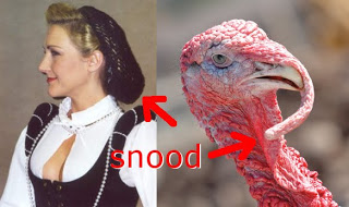
Thestrangest part of a turkey’s head is the snood. The English word “snood” was around long before the English were aware of turkeys. It referred to a decorative hair net or bag worn by women on the back of the head to confine their hair. The resulting mass of hair does look something like the snood that hangs over a turkey’s beak, and this might be where the name came from (see below).

While many animals have wattles, and several different kinds of fowl have caruncles, the turkey is the exception in that it is the only animal with a snood. Its functions may be similar to those of the wattle and caruncles, as they are much larger in males than in females. The snood length in males is linked to testosterone levels, and males are more likely to dominate or steal food from shorter snooded (just made up that word) males than long snooded (there it is again) ones.

I haven’t found any documentation that the turkey
snood is named after the hair snood, but it makes sense.
The snood as a garment makes a comeback every 100
years or so, now they are all the rage in McDonalds and
abattoirs (slaughterhouse) –turkeys with snoods are
processed by workers wearing snoods!

But turkeys are rarely served with the head intact, and eating them is what we are most interested in at Thanksgiving. Like chickens, turkeys have both dark and white meat. The difference in color is due to the makeup of the muscles and how they store and use energy.

The red meat of mammals and the dark meat of birds are similar in that they contain high amounts of myoglobin. The muscles that have myoglobin are for prolonged use; muscles used most of the time require lots of oxygen to make lots of ATP. Myoglobin is to muscles cells what hemoglobin is to red blood cells; it is a molecule that binds and holds oxygen. In the muscle cell, the myoglobin will release the oxygen as needed to allow the muscle to make more ATP and then use that ATP for contraction.

Myoglobinis highly pigmented, so the muscles look darker (redder). When denatured by temperature, the myoglobin turns a tan to dark brown color, giving the cooked meat its look.

Myoglobin is structurally similar to hemoglobin, in that it
looks like one of hemoglobin’s subunits. Each subunit in
hemoglobin can carry one oxygen molecule, but act in
cooperative behavior; the first one is hard to bind, the
second is easier and so on. Myoglobin stores oxygen within
the muscle cell. The more you exercise that muscle, the
more myoglobin it will produce.

White meat, on the other hand, has much less myoglobin. Why? The muscles with white meat (like flight muscles) need energy in short bursts, perhaps to evade predation. To do this, they need less oxygen most of the time, but need lots of glucose some of the time. Therefore, they have less myoglobin but more glycogen (a storage form of glucose) so they can react quickly, rather than waiting for the blood to bring more glucose. The glycogen makes the cooked meat look white and glossy.

So, we have a big bird (did you know that Big Bird’s costume is made of turkey feathers painted yellow?) providing us with a big meal on a big holiday. Next week, we look at a final implication of immune responses and when infections might be helpful - sick bacteria are good for us.


Baratti, M., Ammannati, M., Magnelli, C., Massolo, A., & Dessì-Fulgheri, F. (2010). Are large wattles related to particular MHC genotypes in the male pheasant? Genetica, 138 (6), 657-665 DOI: 10.1007/s10709-010-9440-5

Thornton, E., Emery, K., Steadman, D., Speller, C., Matheny, R., & Yang, D. (2012). Earliest Mexican Turkeys (Meleagris gallopavo) in the Maya Region: Implications for Pre-Hispanic Animal Trade and the Timing of Turkey Domestication PLoS ONE, 7 (8) DOI: 10.1371/journal.pone.0042630
For more information or classroom activities, see:


Sexual dimorphism –

Myoglobin –
http://www.getbodysmart.com/ap/respiratorysystem/physiology/gases/myoglobin/animation.html


Antibiotics Are Going Viral

$
0
0
Biology concepts – bacterial immunity, bacteriophage, antibiotics

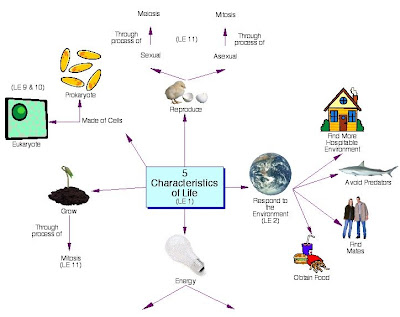
There are recognized characteristics that all living organisms 
share. However, they are not black and white; take a look at 
these characteristics and think about fire. Fire grows, 
consumes energy, gives off energy, metabolizes, reproduces. 
So if fire can be fit into some of these, maybe an argument
can be made for viruses?

Manyscientists do not consider viruses a form of life, but that doesn’t mean the idea is universal. Even virologists can’t agree. Viruses do blur the lines between life and non-life, and it gives us something to debate when parties get quiet. It makes for a great debate in Biology classes too, if you don't have a party to go to.

For many, it comes down to this - viruses don’t react to changes in their environment, grow, or metabolize, so they can’t be alive. They lack all these characteristics because these processes take energy, and viruses themselves don’t make or consume energy. This is a big sticking point for anyone trying to make an argument for including viruses as life.

But they seem to do O.K. at making their way in the world, and are becoming quite the model for immune stimulation. A 2011 study at the Emory Vaccine Center used virus-sized nanoparticles to try to induce life-long immunity as natural viruses do. It is hypothesized that virus particles bind to several different types of innate immune receptors (called Toll-like receptors, TLRs) and this diverse stimulation by one antigen is responsible for longer immunity.

The nanoparticles were composed of a synthetic polymer particle complexed with two stimulators. One is similar to a part of the bacterial cell wall, and the other mimics viral mRNA. The particles also stimulated several different TLRs in mice, and it is hoped they will do similar in humans.  Nice to see we can take advantage of viruses, since they take advantage of us so often. Important to our topic today, viruses can even take advantage of bacteria.

What a great image of a T2 bacteriophage. What
look like layers are …. layers. Each is a protein and
what is more, they self assemble! The head carries
the nucleic acid, the legs attach to the bacterium, and
the shaft creates the hole and injects the nucleic acid.

Since bacteria are prokaryotes, it would be right to assume that the viruses that infect them look and act differently than the viruses that infect eukaryotic cells. They even have a different name – bacteriophages (backtron = small rod, and phage = to feed on). Infection of a bacterium by a virus may seem a trivial event - we have our own problems to deal with. But there are several ways in which this infection affects animals.

Bacteriophages insert their nucleic acid into the bacteria from the outside; the virus doesn’t enter the cell. Similar to the bacteriocin delivery system recently discovered in bacteria, bacteriophage also use a spike system to punch a hole in the target cell. Scientists in Switzerland, Russia, and Indiana collaborated in 2011 to show that the bacteriophage spike has a single iron atom at the tip, and it punches, not drills, a hole in the target bacterium.

Once inside, the nucleic acid can have different fates. In many cases, the phage DNA is inserted into the bacterial chromosome and stays there for a while, not harming anyone, but also not making new virus particles. This is called lysogeny. Lysogens (cells infected with lysogenic phage) will then pass on the prophage (the phage nucleic acid that is integrated) on to their daughter cells.

Other bacteriophages don’t have the patience to just hang out in the bacterial genome; they take over the cell, make many copies of themselves and then destroy the bacterium by lysing it (breaking it open). These are the lytic bacteriophages.

You might recognize that lysogenic phage DNA, just sitting there in the chromosome, would die out with the cell (or daughter), so they must have another side to themselves. These phages can be lysogenic if the environment suits them, or lytic if they have the right signals, and they can switch from lysogenic to lytic if the environment changes, so they are called temperate bacteriophage. Do I have to point out that they can’t go the other direction (lytic to lysogenic); how could you insert yourself into the bacterial genome if you have already caused bacterial destruction?!

There are 19 recognized bacteriophage types (probably
more now). They have different kinds of head proteins, and
some are filamentous. Cystovirus (cytovirus) is the only
virus with RNA for a nucleic acid instead of DNA. Tectivirus
is the only phage that infects both archaea and bacteria.

There are currently 19 different classes of bacteriophage that infect bacteria and archaea. That’s a bunch of different ways that a bacterium would have to defend itself, but it can. Bacteria have several different ways to prevent bacteriophage infection. In some cases, the bacteria will produce cell wall molecules to prevent phage binding or nucleic acid injection.

In other cases, the bacteria will identify its own nucleic acid, usually by adding methyl groups to DNA. In some cases, the bacteria will methylate its own DNA, and then cut up (called restriction, this is where the restriction enzymes used in molecular biology come from) any DNA that isn't methylated. In other cases, the bacteria will methylate the incoming viral DNA and target all methylated DNA for restriction.   

Recent evidence show that bacteria even have a version of adaptive immunity. The CRISPR systemtakes spacer DNA (short repeats outside genes) from the bacteriophage and places them in specific CRISPR spots in its own chromosome. These serve as a memory in case that bacteriophage is encountered again. If it is, the appropriate spacer can be turned in to a piece of RNA that will target the phage DNA for destruction (called RNAi, the “i” stands for interfering, the process for another discussion).

Finally, bacteria can oppose phage by giving up. Like the apoptosis in our cells or the plant hypersensitive reactionwe have discussed, bacteria can kill themselves in order to prevent themselves from becoming virus factories. In the case of bacteriophage-infected bacteria, the process is called high frequency of lysogeny. This system prevents the bacterium from carrying the prophage and passing it on to daughter cells by having the cell die before it replicates.

So bacteria infected by phage can defend themselves, but in some cases, they don’t need to. In fact, it may help them out. Consider a lysogenic phage of one type and lytic phage of another type. Which would a bacterium consider living with – certainly not the lytic phage. But many viruses, including phage have mechanisms to prevent superinfection (infection with a second virus); phage of one type cannot survive in a bacterium infected with a phage of another type. If the lysogenic phage got there first, it could actually protect the bacterium from a death by a lytic phage.

Cholera toxin is carried by the CTX bacteriophage.
The phage needs TCP, a type IV pillus to infect the
V. cholerae. Once the bacterium is growing on the
intestinal surface, the phage is activated, reproduces,
infects other bacteria, and the cholera toxin is
produced. So to cause disease, the bacteria must
undergo horizontal gene transfer to gain the pillus,
and be infected by the CTX phage.

We maychuckle at the idea of bacteria getting infected – in many cases it serves them right – but it can also affect us. Certain bacteriophages possess DNA that can make an infected bacterium even better at causing humans distress. The cholera toxin of Vibrio cholerae is carried by the CTX bacteriophage, and the diphtheria toxin gene of Cornybacterium diphtheriae is also transferred from bacterium to bacterium by a phage.

But phage may also be turned from the dark side and used to help mankind. In the spirit of our recent discussions on when it is beneficial to be infected, how about letting your doctor infect you with bacteriophage to kill off your bacterial infection?

It is no secret that antibiotic resistance is becoming a large problem in medicine. If we know that viruses can infect bacteria, why don’t we use them as a type of antibiotic? This may very well be a good idea, but it isn’t a new one.

Before the advent of penicillin and other traditional antibiotics, bacteriophages were used to treat bacterial infections in the Soviet Union and Eastern Europe. However, the 1920-30's trials were not without their flaws, mostly because scientists didn’t have a good idea of how phages worked. For many years the West remained behind, because Soviet research was not widely distributed.

To kill bacteria, lytic phages would be the tools of choice. But there is a downside, we use bacteria to stay alive. You wouldn’t want to kill of your gut flora, you need them to digest food and absorb vitamins. So, bacteriophage must be delivered to the site of the infection only, replicate there but not travel, and kill only the target bacteria. This is a tall order, but trials are in progress for bacteriophage as antibiotics against drug resistant Staphylococcus and others. Bacteriophages are even being tested in bacterially-infected plants.

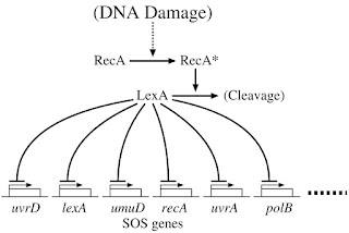
The SOS system is one way a bacterium can repair
DNA damage. The damage stimulates RecA protein
function. This is an important protein. It works in
many forms of DNA repair, as well as being responsible
for homologous recombination. The SOS repair genes
are controlled by RecA degrading the protein that
represses their production. They go on to fix the DNA
problem.

On another front, researchat MIT and Boston University from 2010 suggests that it may be possible to inhibit bacterial antibiotic resistance mechanisms, and once again making the resistant bacteria susceptible to conventional antibiotics. In this case, bacteriophage were engineered to target the bacterial DNA repair system in the target cells. The SOS system (see picture to right) is induced when bacteria are treated with antibiotics, but the bacteriophage-treated cells were more susceptible to the antibiotic. This could prevent resistance from developing, but may also be useful in strains that have developed some other antibiotic resistance mechanism.

Another potential bacteriophage aid to humanity has nothing to do with disease. May 2012work from the University of California has made use of the mechanical energy of the bacteriophage inside bacteria, turning it into electrical energy (piezoelectricity, piezo = to press or squeeze). While this is a very small amount of power per cell, it is hoped that this may soon be harnessed to run your smart phone and iPad.

Next week we will start a three-part series of Christmas posts, the biology of gold, frankincense, and myrrh.


Browning, C., Shneider, M., Bowman, V., Schwarzer, D., & Leiman, P. (2012). Phage Pierces the Host Cell Membrane with the Iron-Loaded Spike Structure, 20 (2), 326-339 DOI: 10.1016/j.str.2011.12.009

Lu, T., & Collins, J. (2009). Engineered bacteriophage targeting gene networks as adjuvants for antibiotic therapy Proceedings of the National Academy of Sciences, 106 (12), 4629-4634 DOI: 10.1073/pnas.0800442106

 
For more information or classroom activities, see:

Bacteriophage –

phage therapy –
http://www.popsci.com/science/article/2011-04/bleaching-threatens-coral-phage-therapy-could-prevent-ghost-coral

A Gift Worth Its Weight In Gold

$
0
0

Biology concepts – toxicity, trace elements

Say hello to the Holterman nugget of New South
Wales, Australia, supposedly the largest gold
nugget ever found. Strictly speaking, it isn’t a nugget,
but rather a huge vein of gold in a piece of quartz.
And Bernhardt didn’t find by himself, but he was a
shameless media hound and built himself a legend.
In 2ndcentury Rome, practitioners of Mithraism, a popular pagan religion of the time, had a feast on December 25 to celebrate the god Mithras, the “Invincible Sun.” This also coincided with other feasts for Saturn and the winter solstice. People gave gifts to one another during these holiday celebrations. This practice of gift giving was adopted by Christians when the pagan and Christian traditions were merged, as they often were.

Today, Christmas presents are most often associated with the gifts of the three kings who came to see the baby wrapped in swaddling clothes (although by the time they arrived, Jesus was already a toddler – camels will never be confused with jet planes). Their gifts were gold – a gift for a king, frankincense – a gift for a priest, and myrrh – a gift for one who was to die (used in burial rights).

These three gifts are not exempt from our search for biologic exceptions and amazement. In terms of biology, they are indeed gifts.  This week we will talk about gold, with the others to follow on successive posts, like the ghosts of Christmas past, present, and future.

At December 2012 prices, a 150 lb (69 kg) person has about 37 cents worth of gold in his/her body, excluding any dental work. Hardly worth trying to harvest, but nice to know you’re worth more than you thought. How did the gold get there and is it doing anything?

Living organisms rely on small amounts of some metals and other elements in order to carry out their metabolic reactions. As such, these elements that are needed in small amounts are called trace elements. Examples of important trace elements include selenium, iron, copper, iodine, and zinc. Zinc is probably the king of the trace elements, as it is used in over 200 different reactions in mammalian physiology.

Copper is used in many biochemical pathways,
and it is showing promise as an anti-inflammatory
agent. But NO! people - you can’t get the anti-
inflammatory effects by wearing the copper
bracelets or copper-impregnated compression
wear! On the other hand, copper impregnated
clothes are antimicrobial.
Zincworks to control what genes are activated to make proteins (zinc finger transcription factors), as well as DNA and RNA production and destruction. It is stored in the brain to control just how active some neurons become when stimulated, and plays an important role in neural plasticity – the reordering of neuron connections after experiences and sleep, you may know it as learning and memory. Make sure you take your zinc before studying for that big test!

Because you need only a “trace” of these substances to maintain growth, development and health, they are also called micronutrients. Their functions can be quite diverse. Iron is the oxygen carrier in hemoglobin, but you only need a trace in your diet because you are so good at preserving what you already have. Selenium is contained in a non-traditional amino acid called selenocysteine, which is important for antioxidant proteins (selenium replaces sulphur in the traditional cysteine).

The biologic rule is that gold is not a trace element! Supposedly, no living organism uses gold in its physiology, but you know there has to be an exception. In 2002, Russian scientists investigating a membrane bound enzyme of the aurophilic (au = gold, and philic = loving) bacteria, Micrococcus luteus, showed that the enzyme contained gold in its active site (the area that binds the molecule to be chemical reacted). The gold was important for converting methane to methanol, giving the bacteria a way to produce energy when traditional food sources were scarce.

But we, and presumably ever other organism don’t have this system, so why is there gold in our body? It turns out that we have many things in our body that we don’t use, they just accumulate, things like lead, mercury, cobalt, arsenic. Some are toxic at low levels and some are useful unless we get too much of them. We have discussed the problems associated with having too much iron, and copper excess can be toxic as well. We said zinc is important for many reactions, but too much zinc can hinder copper absorption and you can end up with a copper deficiency. This is just as dangerous as copper excess.

The liver is the site of much detoxification in the body.
Two systems are at work, one to break down or modify
fat soluble toxins, and the other to prepare them and
water soluble toxins for elimination in the bile or urine.
Toxic heavy metals often just get stored in the liver and
cause damage later.
If the element itself or amount of the element is toxic, then we have to get rid of some; this is the job of the liver. In some cases even gold can be toxic; as we accumulate more and more gold in the liver and kidney, it can disrupt their functions. Poor liver and/or kidney function – you die. The classic form of toxic gold found in nature is called gold chloride or “liquid gold,” which causes organ damage in humans and severe toxic effects in other organisms, but there is an exception.

A microbiologist and a professor of electronic art at Michigan State Universityhave worked with a bacterium that can withstand gold chloride levels that would kill every other known organism. They found that Cupriavidus metallidurans was hundreds of time more resistant to gold chloride than any other organism.

C. metalliduranstakes in the gold chloride and processes it to pure 24 karat gold, and then deposits it in a thin layer as part of the community of proteins and insoluble products that the bacteria builds around its colony. These organized layer of proteins, lipids and carbohydrates are called biofilms, and are being recognized as very important in bacteria ecology and pathology. In the case of C. metallidurans, the biofilm is intrinsically valuable to Wall Street.

Other organisms accumulate gold as well – bacteria, fungi, algae, fish, etc., but as does everything else in biology, it starts with the bacteria. It turns out that some bacteria excrete high levels of acidic amino acids – aspartate and glutamate (atemeans acid). Yes, amino acids that are used to build proteins are organic acids, hence the name.

To dissolve gold out of powdered and broken rock,
many mines like this one in Brazil spray the ore with
sodium cyanide in water. They collect the runoff and
precipitate out the purer gold. I guess they don’t worry
about all the living things contaminated with the
cyanide.
Thebacterium Chromobacteriumviolaceumactually makes and excretes cyanide. Cyanide binds stably to gold and silver, so it is used in gold mining to bind and concentrate very fine gold particles in rocks. Then the gold can be collected and precipitated. These examples show that if gold is in the immediate environment of these various organisms, it can be dissolved by the organic molecules and taken up by the bacteria when they feed.

Once gold is consumed and stored by the bacteria, it enters the food chain; millions of organisms feed on bacteria, and millions of organisms feed on the feeders, and so on. Eventually, we end up eating a little gold as well. This is similar to how fish that ingest food and swim in water contaminated by mercury runoff can end up increasing the human levels of mercury – the difference is that mercury is much more toxic than gold.

The accumulation of gold in sedentary organisms may provide someone with a gold rush. A 2010 study showed that the saprobicfungi (those that feed on decaying material) around an existing gold mine contain much higher levels of gold than ectomycorrhizalfungi (those that are parasitic)in the same area.  The accumulation of gold in soil bacteria and fungi may be able to provide scientifically astute miner with clues as to where they should dig the next mine!

The blue toadstool  (Entoloma hochstetteri) is an
example of a saprobic fungus. It gains all the
nutrients it needs from the soil and from decaying
organic material. Therefore, it picks up and
accumulates other things in the soil – like gold maybe.

 Bacteria may prove even more important to miners. December 2012 evidence indicates bacteria that dissolve and ingest gold in the rocks and soil purify it to some degree. When they form biofilms, the gold becomes insoluble again, and nuggets or flakes are formed. Veins of gold may be due to bacterial byproducts and corpses flowing into cracks in the rocks. Makes you look at your gold ring differently, doesn’t it.

We might be able to thank gold-loving bacteria for more than our jewelry – gold is finding its way into medical treatments and tests these days.  Because gold was rare, pure, inert, and costly, early physicians thought it just had to be good for you. Many remedies had gold incorporated into them, including a popular cure for alcoholism called the Keely cure.

The cure was so widely accepted (and patented by Dr. Keely) that even Theodore Roosevelt himself sent his brother, Elliott, to Dr Keely’s clinic in Dwight, IL to be cured of his addiction. It didn’t work, Elliott drank to the point of depression, and died from injuries that resulted from his jumping from a window.

More recent uses of gold include as an anti-inflammatory agent rheumatoid arthritis, including a compound called aurothiomalate. Just how this works remains a mystery, but a 2010 study in chondrocytes (the cells that make cartilage and are present in joints) showed that this drug down-regulates a signaling enzyme (MAP kinase phosphatase 1) that is important for expression of several inflammatory proteins, including cyclooxygenase, p38 MAP kinase, matrix metalloproteinase-3 and interleukin-6.

The aquatic or semi-aquatic plant Bacopa caroliniania can
be loaded with gold nanoparticles and made to give off
light.  Depending on the energy of the UV light shone on
it, it can glow from green to gold to red. Someday, maybe
our tree lined streets will have natural streetlights.
Gold isfinding a home as a treatment in other conditions as well including in cancer, viral infections, and parasitic diseases. But gold is being used most often as a carrier. Because gold is practically inert, nanoparticles of gold can be used to carry drugs to specific targets or to be used as imaging agents to illuminate very small structures.

Most spectacularly, gold may help light a dark world. Plants can now be grown with gold nanoparticles that are small enough to be taken up into the leaf cells. When exposed to UV light, the gold releases energy at a wavelength that stimulates chlorophyll to bioluminesce. The plants actually give off light like natural street lights.

That’s a lot of biology for a hunk of metal used for wedding rings and retirement watches – a way cool gift for any biologist. Next week, the biology of frankincense.

 Nieminen, R., Korhonen, R., Moilanen, T., Clark, A., & Moilanen, E. (2010). Aurothiomalate inhibits cyclooxygenase 2, matrix metalloproteinase 3, and interleukin-6 expression in chondrocytes by increasing MAPK phosphatase 1 expression and decreasing p38 phosphorylation: MAPK phosphatase 1 as a novel target for antirheumatic drugs Arthritis & Rheumatism, 62 (6), 1650-1659 DOI: 10.1002/art.27409

 Levchenko, L., Sadkov, A., Lariontseva, N., Koldasheva, E., Shilova, A., & Shilov, A. (2002). Gold helps bacteria to oxidize methane Journal of Inorganic Biochemistry, 88 (3-4), 251-253 DOI: 10.1016/S0162-0134(01)00385-3
 

The Resin For The Season

$
0
0

Biology concepts – sap, resin, latex, mucilage

Frankincense is a solid material than starts out as a 
liquid that oozes from a tree. In the presence of air, 
the resin turns hard. When burned, many 
fragrant and brain altering compounds are released.
We saw last week that gold doesn't just look good, it has a significant place in biology. This week we take a look at frankincense, a natural tree product prized for its use in sacred rituals. The Catholic Church is the number one purchaser of frankincense, but that may be about to change, especially for medicine. The wise men must have done some heavy thinking before they made their gift choices for Jesus – gift cards are so impersonal.

A 2008 study may have defined just why frankincense is used in religious rituals. Burning the resin releases incensole acetate (IA), one of the resin’s key components, which activates transient receptor potential vanilloid (TRPV3) ion channels in the skin and brain. This ion channel is responsible for mediating a warm feeling in the skin, but TRPV3 channels also mediate brain activity.

The researchers in Israel found that IA activates the cFos transcription factor in the brain, leading to anxiolytic (anxio = anxiety, and lytic = destroying) and anti-depressive feelings. Mice without TRPV3 channels did not show cFos activation or behavior changes when exposed to IA. It appears that burning frankincense makes one feel happier and more in tune with whatever activity is going on at the time, including religious rituals.

The fact that there is a psychoactive agent in frankincense is amazing enough, but there’s more biology to this second gift. Recent evidence indicates that the oils and other compounds in frankincense may save lives– if the trees that produce frankincense don’t disappear in the next 50 years. Unfortunately, their extinction is a distinct possibility – we must save this precious sap, or resin, or whatever it is.

Trees can produce various oozings and liquids. Pancake syrup most often comes from the sap of a maple tree, while your stick of Wrigley’s spearmint uses the latex that exude from many different kinds of plants. Gum drippings may also be used in chewing gum (Chiclets used chicle gum), but gums are now more commonly found in paints and erasers. The aloe vera you use on burns is a type of mucilage, rich in glycoproteins. But many plants, especially coniferous trees, exude resins when they are under attack or are damaged.

Amber is fossilized resin. Scientists learn much from organisms caught 
in it and thus preserved. Recent evidence also shows that amber can 
help us track bug attacks on plants from the days of dinosaurs. 
Gum is semi-solid, and rubbery. The gum shown is chicle, used 
for many years in Chiclets gum. Mucilage is produced by 
many pants, including as a treat and trap for insects in carnivorous
plants like this sundew. Maple sap is clear and dilute when tapped from
a tree. It must be boiled for hours to reduce it to syrup. Latex rubber is 
naturally white. The first car and bicycle tires were all white, not
just white-walled.
Gums can also be used for defense, but are made directly from disintegrating internal plant material. They harden to a certain degree after being exuded from the plant tissue, but are more known for their ability to increase the viscosity of a liquid, due to their long polysaccharide molecules. Bacterial agar plates use a gum from seaweed to grow microorganisms.

Sap is the sugary fluid that travels up and down in the xylem of vascular plants, providing the different structures with carbohydrate to produce ATP at the cellular level. Therefore, sap is a nutritive liquid and all trees produce it – but not all taste good.

Mucilage is similar to sap. It also contains glycoproteins and other carbohydrate-containing molecules, and is important for food and water storage in almost all plants, especially cacti. However, mucilage can be used for other purposes, like luring insects into carnivorous plant traps, such as the flypaper plant.

People used to lick mucilage everyday, but technology has reduced its role in our lives. When mixed with water, mucilage is an adhesive, like on the backs of stamps. You don’t have to lick your computer screen to send an e-mail, so mucilage is less important to us in these modern times.

Resins become definite solids when exposed to air. They are not nutritive, and contain primarily the byproducts and secondary metabolites of other cellular processes. While gums and saps are soluble (will dissolve) in water or fat, resins are stable in water but will dissolve in alcohol.

The reason for resin production is not fully understood. They may play a role in defense or tissue injury, but may instead serve to rid the plant of unneeded or unwanted waste products. Indeed, when trees are cut to harvest frankincense, the first resin produced is discarded, because it contains many toxins and foul smelling chemicals.

The Boswellia sacra tree grows in a harsh
environment. The roots can grip onto stones and
they grow out of the ground as buttresses to keep
the tree stable on the cliff sides.
Resinsare produced mostly by coniferous trees (like pine trees). This makes frankincense an exception, since it comes from the Boswellia sacratree, a deciduous tree (trees that lose their leaves in the winter). Frankincense is different from other resins in another aspect as well, it is technically a gum resin, since it has many compounds that are of the gum variety within its resin. The gum-like essential oils in frankincense are one of the reasons it is sought after as an incense.

B. sacra grows only in the middle eastern countries of Yemen and Oman, and possibly in Somalia. The tree is only 2-7 meters (6-23 ft.) when fully grown, and starts producing resin at a fairly young age of 8-10 years. Its small stature may be due in part to the arid climate that it lives in; there is so little water to be had that B. sacra survives only on the moisture it absorbs from fog.

However uninviting its environment might seem, B. sacra is well adapted to this area and is very finicky in growing anywhere else. In fact, a recent study indicates that they are more finicky than even previously believed. Though living in two different areas (Oman/Yemen vs. Somalia), it had been accepted that these plants were the same species. But based on chemical evaluation of the essential oils of the resins from trees in these two regions, the Oman/Yemen trees of B. sacra are truly different than the B. carterii trees of Somalia.

Initial gas chromatography-mass spectrum analysis did not show significant differences in the kinds of volatile molecules present, but there were large differences in the amounts of the individual compounds in the resin from each species of tree. Later experiments also showed chemical differences in the same compounds from each species.

Yemen and Oman are side by side and Somalia is
just across the Gulf of Aden. But recent studies show
that the frankincense trees that grow in Yemen and
Oman are distinctly different from those in Somalia.
This speciation difference shows that B. sacra REALLY likes to stay close to home. There’s nothing wrong with that, except that the small area that it grows in happens to be one of the most unstable parts of the world. The trees have been over harvested for resin, and this affects the rate at which the trees reproduce. Heavily tapped trees have seeds that germinate only 8-16% of the time, while trees that have not been tapped for resin germinate seeds at a rate of over 80%.

Add goats grazing on the existing trees, global warming, fires, and low genetic diversity in individual stands of trees to the low rate of propagation and this spells trouble for the B. sacra species. Estimates are as dire as a 50% decrease in frankincense production in the next 15 years, to a 90% loss of trees in the next 50 years – but there is hope.

A recent DNA study shows that trees from different parts of the Dhofar region are genetically distinct, and that there is a low level of heterozygosity in the trees of a single area. This low level of genetic diversity results in trees less able to survive changes in environment or biology (genetic diversity is key to natural selection). But some stands show more genetic diversity and arguments are now being made to initiate conservation efforts for the diverse stands, while increasing cross-pollination of the least genetically diverse trees. It is hoped that these efforts, as well as attempts to grow B. sacra in the Sonora Desert of North America, could stave off extinction of B. sacra.
 
The hippocampus is important in your sense of well-
being. Studies have shown that in people with
depression, the hippocampus is smaller, perhaps from
poor neurogenesisor from increased cell death. Why
the seahorse? In Greek, hippocampus means, “horse sea
monster.” I can see the resemblance.
Whyis it important that we save the frankincense trees? Because it is becoming evident that the resinous compounds in frankincense could have great medical benefits to humans – and unhappy mice.

We mentioned above that IA (incensole acetate) of frankincense acts on the brain to increase feelings of well-being. Mice bred to be submissive and to give up (quit) earlier in a test of depressive activity show a much stronger will to live and more positive behaviors when given IA. Recent research in Israel shows that IA influences brain molecular biology, especially in the hippocampus, altering depressive behaviors as much as other chemical interventions. It is hoped that IA may be a viable anti-depressant drug in the future.

This same group showed in 2008 that IA was a significant anti-inflammatory agent, through its inhibitor action on an important transcription factor (called NF-kB) that stimulates expression of inflammatory proteins. In mice with traumatic brain injuries, IA administration resulted in reduced inflammation and pressure on the brain, reduced neuron degeneration, and prevented loss of cognitive function. Their more recent study also indicates that IA is protective in stroke and in the damage that can come after strokes by reintroducing oxygen into the damage part of the brain (when blood flow resumes).

Boswellic acid is also of use in myeloid leukemia, a type
of cancer of the white blood cells. It seems that BA can
induce the cancer cells to commit suicide, and die after a
period of time like most cells do. BA trigger apoptosis by
stimulating the release of important compounds from the
mitochondria, suggesting to the cell that its energy making
organelles are irreparably damaged.
Anothercompound in frankincense is showing promise as an anti-cancer drug. An essential oil molecule called Boswellic Acid (BA) has been shown to slow the rate of cancer cell growth. A recent study has delineated at least part of the mechanism of BA-mediated inhibition of colorectal tumor growth.

Cancer is the result of mutations in genes that code for the production of proteins that keep cells living, growing, and dividing forever. BA stops the synthesis of some of these proteins. It turns out that BA stimulates production of a micro RNA (miRNA, a short RNA molecule of about 22 nucleotides) that can bind to the messages transcribed from DNA that would be translated into pro-cancer proteins and stop the proteins from being made. Do you think the three kings had any idea that they were giving a gift that can stop inflammation, depression, and cancer – or they did they just think it smelled nice?

Next week – the third Christmas gift, myrrh.

Takahashi, M., Sung, B., Shen, Y., Hur, K., Link, A., Boland, C., Aggarwal, B., & Goel, A. (2012). Boswellic acid exerts antitumor effects in colorectal cancer cells by modulating expression of the let-7 and miR-200 microRNA family Carcinogenesis, 33 (12), 2441-2449 DOI: 10.1093/carcin/bgs286

Moussaieff, A., Gross, M., Nesher, E., Tikhonov, T., Yadid, G., & Pinhasov, A. (2012). Incensole acetate reduces depressive-like behavior and modulates hippocampal BDNF and CRF expression of submissive animals Journal of Psychopharmacology, 26 (12), 1584-1593 DOI: 10.1177/0269881112458729

Coppi, A., Cecchi, L., Selvi, F., & Raffaelli, M. (2010). The Frankincense tree (Boswellia sacra, Burseraceae) from Oman: ITS and ISSR analyses of genetic diversity and implications for conservation Genetic Resources and Crop Evolution, 57 (7), 1041-1052 DOI: 10.1007/s10722-010-9546-8
 
For more information, see:

Resin –

Sap –

Gum –

Latex –

Mucilage –

Boswellia sacra –
http://www.iucnredlist.org/details/34533/0
 

One Myrrh-aculous Christmas Gift

$
0
0

Biology concepts – synergism, multidrug resistant cancers

The Commiphora myrrha is the classic source for myrrh
resin. It is a short tree that grows in low moisture and
poor soil areas. Its branches are very thorny; some
propose that the crown of thorns Jesus is said to have
worn was made of myrrh twigs.
The three original Christmas gifts are usually listed as gold, frankincense, and myrrh, but why that order? Some say it is because you give gold to a king, frankincense was used by priests, and myrrh was used to anoint the newly dead.

The order goes along with representations of how he was born (as a king), how he lived (as a preacher), and how he died. But I think that sells myrrh short. True, it was used in consecrating and embalming dead bodies, but it is so much more. As with gold and frankincense, there is “myrrh” here than meets the eye.

Likefrankincense, myrrh is a resin from a tree that grows in the Middle East, in this case Yemen, Somalia, Eritrea, and Ethiopia. Frankincense and myrrh trees even come from the same family, the Bursceraceae. Being deciduous trees, both frankincense and myrrh are exceptions to the rule that coniferous trees are more likely to be resin producers.

Myrrh resin is an oleo-gum-resin, since it is has essential oils (oleo) and long polysaccharides (gums), as well as resins. It is more complex than frankincense, containing over 300 individual secondary metabolites and other compounds. Being a more complex substance, it might follow that myrrh would have more uses than frankincense, both in ancient times and now. And here is an instance in biology when the logical answer is the correct answer. In addition to being used as incense in rituals and perfumes, it had other mystical properties. It was so prized that it was often worth more than gold.

Greek soldiers always carried myrrh in their travel kits because it was a potent antibacterial and anti-inflammatory agent. Being soldiers, they were likely to be wounded, and those wounds would get infected and swell. If they died, it is good that they had myrrh, because it was also used as an embalming agent and to consecrate the dead bodies.

In Greek mythology, Myrrha was a young lady who
committed an awful no-no, and was chased across
the desert by her father. The gods took pity on her
and turned her into a tree so she wouldn’t have to
run anymore. The myrrh resin that drips from the
tree is said to be her tears. But I don’t get the part
where she gives birth to Adonis while she is still a
 tree – family trees aren’t supposed to be literal.
In fact, the Egyptians were some of the first to use myrrh in this way. Combined with natron, a form of salt from the desert, they would stuff the bodies of the dead to pull out the water. This was a big part of the mummification process. The myrrh was there to prevent rotting and to help with the smell.

Myrrh smells good, but tastes horrible. In fact, the name myrrh originally came from the Aramaic word for bitter. To this day, the bitter taste of myrrh oil or powdered myrrh has limited it use in medicines. A recent study fiddled with making emulsions of myrrh in water in order to cover the taste, or adding fat-soluble compounds and using it as a suppository (there is usually good uptake of drugs from the south end of the gastrointestinal tract).

But the ancients still consumed myrrh despite the taste. It is said that someone gave Jesus myrrh dissolved in wine as a painkiller while he was on the cross. Others mixed it with red raspberry leaves to soothe a sore throat. Pliny the Elder wrote of using myrrh to kill bugs in wine and wine bottles before bottling the drink for transport and sale.

Though myrrh has been used for centuries, we have just started to explain how myrrh functions in these capacities. For example, it is now known that compounds in myrrh called terpenes can interact with opioid receptors in the brain. This is how they act as painkillers.

Myrrh and frankincense components are also being tested in combination as antimicrobial agents. Oils of myrrh alone can kill or slow down some microorganisms; so can oils of frankincense. But adding them together has been shown to be a case of 1+1=3.

This is a demonstration of the concept of synergism. Let’s say that one antimicrobial drug can kill or stop X number of organisms when given at a certain dose. It is often the case that as you increase the dose, you will kill or stop more organisms – up to a point. Almost any drug becomes toxic when you ingest a lot of it. The lowest amount you can give to do the job is the miminal effective dose, and the most you can give is the maximum recommended safe dose.

To get a bigger bang for your buck, sometimes you can add a second drug to the regimen. Drug 1 inhibits or kills X number of organisms and drug 2 affects Y number of organisms. Often, giving drug 1 and 2 together will then inhibit or kill X+Y organisms. This is an additive effect. Drugs with additive effects often work on different targets; they are like eating a foot-long hotdog from both ends. The hotdog goes away twice as fast because the two mouths aren’t competing for the hotdog.

The white dots are paper soaked in two different antibiotics.
They are put on a plate of bacteria (the hazy diagonal lines).
As the drugs diffuse out, they kill the bacteria (darker, clear
areas, but their concentrations go down the farther they
travel. But look between them, the area where they both are
low concentrations is a bigger cleared area (between red
lines). This is synergistic action.
Everyonce in a while, using drug 1 and drug 2 together gives you a bigger effect, greater than X+Y; this shows synergy. Synergistic effects are the exception, they don’t come around often and a researcher is lucky to find them. Synergism in drug activity can mediated by different mechanisms, but it may be caused by a second drug turning off the fall-back pathway a cell may use when the primary pathway is affected by the first drug - there are many redundant pathways in cells.

Synergism and additive effects are examples of pharmacodynamic effects, basically how the drugs work on cells. We will later see how some drugs have pharmacokinetic effects on each other.

When a groupin South Africa tested two myrrh oils in combinations with three frankincense oils, they found that a combination of B. papyrifera and C. myrrha oils were synergistic in controlling both Cryptococcus neoformans, a fungus, and Pseudomonas aeruginosa, a gram negative bacterium.

The anti-inflammatory mechanisms of myrrh are just being worked out as well. Recent studies from South Korea indicate that myrrh stops the inflammatory process by inhibiting the production of molecules that promote inflammation. Their 2011 studyindicates that myrrh turns off the enzymes that produce nitric oxide, prostaglandins, and some inflammatory cytokines (messengers that have many effects) when inflammation was stimulated by LPS, a cell wall component of many bacteria called lipopolysaccarhide, also called endotoxin. LPS is responsible for things like septic shock and necrotizing enterocolitis.

Rheumatoid arthritis (arthus = joint, and itis =
inflammation of) is mediated by an autoimmune
process that brings much inflammation. Myrrh
has been used for hundreds of years as an anti-
inflammatory drug, but we are just now figuring
out why it works.
They added work in 2012 that shows that myrrh is very good at controlling inflammation after a rupture of the large bowel (usually cases peritonitis and is very dangerous). This is probably due to its ability to stop inflammation induced by the LPS of the gut bacteria and its ability to kill the organisms as well. Those wise men were really quite wise – they didn’t know why it worked, but they knew it worked, and that was enough.

But even they did not suspect the wonders of myrrh. It is cancer that one myrrh component is turning out to be a gift. There are several species of myrrh trees, and a couple, C. mukul and C. molmol, contain a compound called guggulsterone (I love saying that name out loud – go ahead, it’s fun). Guggulsterone is not necessarily toxic to cancer cells by itself, but it may solve a big problem in that currently affects many cancer treatments.

We talked a while ago about how bacteria have pumps to kick antibiotics out of their cell, and thereby prevent their action. Cancer cells also have a pump to do this with many cancer chemotherapeutic drugs. The most common of these is a membrane channel protein called P-glycoprotein (P-gp). This protein is present in some normal types of cells, working to pump out toxic compounds, like liver cells and skin cells. This means that cancer drugs on these types of cancers have a hard time staying on the cells.

P-gp pumps cancer drugs back out of cells with
the help of changing ATP to ADP. This can lead to
drug resistant cancers. We are looking for inhibitors
that might block the action of P-gp by taking away
its ATP or by competing with the drug for the pump,
so less drug is pumped out.
Other cells can up-regulate the production of P-gp once they start to receive the cancer drugs. Either way, it leads to multidrug resistant (MDR) cancers – a serious problem. Many attempts have been made to develop P-gp inhibitors, but most have been either ineffective or toxic.

Enter guggulsterone (let’s call it GGS for short) – new research shows that this compound can reverse MDR in several types of cancer. The mechanism is just now being uncovered, GGS can act as a competitive inhibitor of P-gp, meaning that it is pumped out just like the cancer drugs. But the more time P-gp spends pumping out GGS, the less time it is pumping out cancer drug, so it is more effective. It does not appear that GGS stops production of P-gp or other actors in this play, it just keeps them busy – but it does it without being toxic. This is a pharmacokinetic effect, one drug (GSS) has an effect on how another drug (cancer drug) is acted on by the cells, in this case by keep the drug in the cancer cell much longer.

In the cases of pancreatic cancer and gall bladder cancer, very new studies show that GGS in combination with the cancer drug gemcitabine, works much better than the drug alone. The combination causes higher levels of apoptosis in these cancers, perhaps through the action of keeping more drug in the cancer cells, but GGS may have other cytotoxic effects as well.

Osteoporosis leads to less dense bones, which can alter posture 
and lead to bone breaks. It looks like the guggulsterone in 
myrrh can prevent bone resorption after menopause. It may 
even increase density and be a treatment for bone breaks.
And this is the most amazing part, even though it may be inducing damage in some cells, a new use for GGS is to prevent damage to heart muscle cells (cardiomyocytes). The cancer drug doxorubicin (DOX) is a very good cancer killer, but its use is limited because it damages the cardiomyocytes. GGS has recently been found to protect cardiomyocytes from DOX damage by preventing the up-regulation of many pro-apoptotic proteins. But GGS helps kill cancer cells by promoting apoptosis – what gives? Become a biologist and find out, it can be your gift to the rest of us.

Next week – the biology of New Years’ resolutions!


Xu, H., Xu, L., Li, L., Fu, J., & Mao, X. (2012). Reversion of P-glycoprotein-mediated multidrug resistance by guggulsterone in multidrug-resistant human cancer cell lines European Journal of Pharmacology, 694 (1-3), 39-44 DOI: 10.1016/j.ejphar.2012.06.046

Wang, W., Uen, Y., Chang, M., Cheah, K., Li, J., Yu, W., Lee, K., Choy, C., & Hu, C. (2012). Protective effect of guggulsterone against cardiomyocyte injury induced by doxorubicin in vitro BMC Complementary and Alternative Medicine, 12 (1) DOI: 10.1186/1472-6882-12-138

de Rapper, S., Van Vuuren, S., Kamatou, G., Viljoen, A., & Dagne, E. (2012). The additive and synergistic antimicrobial effects of select frankincense and myrrh oils - a combination from the pharaonic pharmacopoeia Letters in Applied Microbiology, 54 (4), 352-358 DOI: 10.1111/j.1472-765X.2012.03216.x


For more information or classroom activities, see:

Myrrh –

Additive and synergistic effects in pharmacology –

Multidrug resistance in cancer –
http://mayoresearch.mayo.edu/mayo/research/chang_lab/

It May Be A New Year, But It’s The Same Old Brain

$
0
0

Biology concepts – learning, habit, long term potentiation, neural plasticity

50% of Americans will make at least one New Year
resolution, but a quarter of them won’t even make it
one week before relapsing. However, those who write
down a resolution are much more likely to make
changes than those who don’t make a specific
demand of themselves.
I swear, this year I’m going to get these posts written a month in advance. Really, I mean it this time. I know I said the same thing last year, but this time I’ve got a plan in place –- yeah, sure. Biology is stacked against me here; making new good habits is definitely an exception. Our brains function to make it hard to change our behaviors – but it is possible.

First things first, I am not a neurologist. I don’t even play one on TV, but we’re going to delve into some neuroanatomy and neurochemistry here. I’ll try to keep it from making your brain hurt.

Before diving into the gooey mess inside our skulls, we need to know that keeping a resolution means creating a new habit, or breaking an old habit and replacing it with a new one. But, what is a habit anyway?

A habit (from old French meaning “to hold” or “customary practice”) is an extreme form of learning, ingrained to such an extent that we do not think consciously about performing the behavior. But we still have the ability to turn the behavior on or off consciously. This is what separates a habit from an addiction. A poor man’s definition – if you have to decide to do it, it’s not a habit, and if you can’t decide not to do it, it’s an addiction.

William James was trained as a physician, but was
the first professor to start offering psychology classes
at the college level. His brother was novelist Henry
James, who wrote about the social corruption of
England versus the brash selfishness of America. His
father was a theologian who worried about the moral
evil have thinking about oneself, and Sigmund Fred was
a family friend. No wonder William went into psychology.
The philosopher and psychologist William James said, “99% of our behavior is purely automatic ….. all of our life is nothing but a mass of habits.” This is mostly true, we need to save our thinking for things that are important and undetermined, not for everyday things for which we can easily predict the outcomes and do not threaten our existence. You don’t think about putting one foot in front of the other when you walk, you look for the bus that may stop you dead in your tracks.

Habits are important, they keep us safe and alive for the most part. Good habits aren’t easy to make, while bad habits seem so easy. Bad habits are rewarded at more primitive levels of the brain, and the rewards are more tangible and shorter term. Good choices may be their own reward, but in terms of our brains, they aren’t as strong as a big ice cream sundae.

Rewards reinforce our habits and learning in a chemical sense as well. The reward centers of the brain release a neurotransmitter called dopamine, and we will see below that dopaminergic neurons are very important in learning, memory and making habits.

We need to know how our brains make habits if we want to increase our chances of keeping our resolutions. First comes intent and motivation, then comes learning, then comes making the learned behavior an unconscious act. As it turns out, there are brain centers for all of these, and they are all tangled together.

Dopaminergic neurons release and may respond to dopamine. They are involved in reward, learning, and in reinforcing learning to make habits. Dopaminergic neurons are located in many parts of the brain and a new study shows just how important they are in forming habits.

To help uncover the mechanisms of habit making, a mouse model has been developed that can’t form strong habits. A certain receptor was eliminated from dopaminergic neurons, and then the mice were taught new conditioned behaviors, like stepping on a lever to give them food. They could learn that the lever motion provided food, but they stopped after a while. Normal mice will learn the habit, and just keep stepping on the lever to get more and more food.

NMDA receptors contribute to LTP by allowing calcium
into the cell. This stimulates a retrograde signal that
causes the presynaptic neuron to release even more
glutamate. This stimulates more NMDA action and even
more calcium influx. This loop can literally remain
turned on for months!
Thereceptors in question work with dopaminergic neurons are there to reinforce signals and strengthen nerve firing. They are called NMDA receptors, and they respond to glutamate, an amino acid and important neurotransmitter. In the synapses (gaps, Greek; syn = together, and haptein = junction) between neurons, NMDA receptors bind glutamate and then allow sodium and calcium into the downstream neuron. These work in different ways to make the firing of the neuron stronger. Calcium in particular can keep the upstream neuron firing and keep stimulating the down-stream neuron. This leads to long-term potentiation (LTP).

LTP results in repeated firing of those neurons, from minutes to months in duration. Every time they fire, that individual pathway gets strengthened. This is the key to learning, called neural plasticity. When neural pathways are repeatedly used, they become strengthened and a behavior is learned or remembered. If they are not used, the connections fade away. Dopaminergic neurons are especially important because they can generate LTP through NMDA receptors but can use additional mechanisms as well.

Many parts of the brain are involved in habit formation, like those that link intent with action. Peter Hall at University of Waterloo near Toronto has been looking at intent and brain function, specifically, a portion of the brain called the superior prefrontal cortex (SPFC), located just behind that place on our forehead where you smack yourself when you do something stupid.

Some people have better SPFC function than others, and they find it easier to act on intentions and make behavior match intention. But good habits can increase SPFC function – see the end of the post.

Adolescent brains are maturing at an astonishing rate
during the teen years, but the maturation is uneven. This
means that they often revert to the more primitive,
emotional brain for decision making. The emotional brain
includes the reward center, so teens are more likely to make
habits based on short-term rewards. Good school work and
behavior habits are tough to develop in these befuddled brains.
Theprefrontal cortex is more than just the SPFC. A 2009 study showed that the ventromedial prefrontal cortex is important in self-control, while the dorsolateral prefrontal cortexis important in meeting goals. And we all know that we need some hefty self-control to keep resolutions.

The entire prefrontal cortex is a big player here, as this is the seat of the executive function, those functions of the brain that control and manage other thinking; like planning, problem solving, resisting immediate reward, and mental flexibility. It boils down to this: the PFC is the chief weigher of risk vs. reward and is the boss decision maker – although he often listens to the primitive brain that, “wants what it wants, when it wants it.”

The signaling from the PFC communicates with other brain areas that are needed for habit formation. These include the nucleus accumbens and the ventral tegmental area that are deeper and older. These just happen to be those reward centers we talked about that reinforce actions based on the pleasure they bring.

Dopaminergic signaling in the nucleus accumbens has a lot to do with LTP and plasticity. A 2012 study shows that dopamine in the nucleus accumbens works to reinforce strong signals while inhibiting weak ones. So burgeoning habits get reinforced and become strong habits, while changing habits is difficult because the signals to do so are inhibited. Plasticity isn’t an easy thing to induce.

For every resolution you make, there is an unconscious
resolution not to change. One reason habits
(good or bad) are hard to break is because they have been
successful to this point; you aren’t dead yet. Changing a
habit means a journey into the unknown, and change is
evolutionarily dangerous; why change what has hasn’t hurt
you yet? This is why bad habits that take a long time to
manifest are so insidious – like a chain-smoking 2 yr. old.
Anotherreason habits are hard to break is the reinforcers; those things that trigger the behavior are a part of our everyday lives. You need to stay away from these reinforcers (temptations might be a better word) because your brain remembers those reinforcers for a long time. It stores the contexts in which the habits are triggered and can bring back the behavior of the context is encountered again. It takes time for plasticity to weaken these pathways.

It takes willpower to keep yourself out of those situations where bad habits are reinforced. It turns out that your willpower is a real thing, requiring energy to work and it can actually tire out. First proposed by RoyBaumeister in 1998, he showed that when people are asked to employ willpower to resist a temptation, it became harder for them to resist a later temptation. We all know this is true.

In addition, it seems that people with the best self-control use their willpower less often. A 2012 study of Wilhelm Hofmann from U. Chicago showed that people should set up their environments to minimize their temptations, so their willpower was energized for when it was really needed. If you want to stop gambling, don’t go to the track – duh!

Let’s put together all we have learned and get some tips from the experts (Peter Hall at University of Waterloo, B.J. Fogg at Stanford, and others) on how to keep your resolutions.

Exercise affects habit formation. A 2012 study from Brazil
shows that running rats on treadmills induced plasticity
in the habit formation portions of the brain. Proteins and
genes that control the formation and function of synapses
were affected in the striatum – which includes the
dopaminergic neurons of the ventral tegmental area.
1) Make your goal something concrete, you can’t resolve an abstraction.

2) Focus on tiny habits that can be implemented in small doses until you can build it up to something bigger. Don’t say you will learn to play the banjo – say you will learn to play one chord. Then do it over and over.

3) Don’t just say you have intent, make the implementation concrete as well. Where and when will you practice the chord on your banjo?

4) Place your new behavior directly after a good behavior that is already a habit – you will be less likely to avoid it.

5) Reward yourself – even just a nice thought about your ability to meet your goal for that day. It will help reinforce the pathways.

6) Limit your temptations, this will help degrade the pathways that lead to the behavior you wish to change and reinforce the new pathways.

7) Get some exercise– superior prefrontal cortex function in making habits and good executive function improves with physical exercise.

Next week we can start a whole new story. If you think that you are a product of your mother and father’s genes, you are mostly right, but boy are there a lot of exceptions!


Wang, L., Li, F., Wang, D., Xie, K., Wang, D., Shen, X., & Tsien, J. (2011). NMDA Receptors in Dopaminergic Neurons Are Crucial for Habit Learning Neuron, 72 (6), 1055-1066 DOI: 10.1016/j.neuron.2011.10.019

Wang, W., Dever, D., Lowe, J., Storey, G., Bhansali, A., Eck, E., Nitulescu, I., Weimer, J., & Bamford, N. (2012). Regulation of prefrontal excitatory neurotransmission by dopamine in the nucleus accumbens core The Journal of Physiology, 590 (16), 3743-3769 DOI: 10.1113/jphysiol.2012.235200


For more information, see:

NMDA receptors –

Long-term potentiation –

Neural plasticity -
http://www.google.com/url?sa=t&rct=j&q=&esrc=s&source=web&cd=10&ved=0CG8QFjAJ&url=http%3A%2F%2Fwww.acnp.org%2Fasset.axd%3Fid%3D852ca1c4-ece9-4f2b-988d-bd6b5222e5ac&ei=9Ty-UKeYM9S80QHLtYHgBQ&usg=AFQjCNER4QfEVPqNhq6jrFAXfcQE4DVN_A


Haploid, Diploid, And Those You Should Avoid

$
0
0
Biological concepts – ploidy, polyploidy, aneuploidy, cancer, therapy-induced senescence

Do your genes or your environment make you
who you are. The cop out answer would seem to
be that it takes both. But it is definitely true.
Certainly, how you are raised affects your outlook.
But we have known for a few years now that your
genes are affected by your environment as well.
What happens to you determines how your genes
are expressed – it’s called epigenetics (epi = beyond).

Wecan argue about whether your genes or your upbringing is more important for making you who you are (nature vs. nurture), but no one is going to argue that your chromosomes aren’t important in the process.

Mom and Dad both contributed to your chromosome number, you got a copy of each chromosome from each parent, so you ended up with 2 copies of each, a state of being diploid (di = two, ploos = fold, and –oid = like). So what if you weren’t diploid, is it a good thing or a bad thing? You know there must be exceptions.
           
Let’s start with how offspring get their DNA. In sexual reproduction, the rule is that the female and male each contributes half the genetic material to the offspring. Humans, for example, have 23 different pairs chromosomes, one from each pair comes from the egg and one from the sperm.

The egg and sperm then each have just half of the full complement of chromosomes. Therefore, the egg and sperm are termed haploid (hap = single). Ploidy in general refers to the number of copies of whole sets of chromosomes in the nucleus. The haploid sperm meets the haploid egg, they date for a while, and then voila, a diploid zygote that turns into a teenager one day.

Mitosis is the replication of cells in which each new cell gets 2 copies of each chromosome, while meiosis is the process where in cells split and give one chromosome from each pair to a developing sperm or egg. Simple, yet it doesn’t always work perfectly. Meiosis may foul up and make eggs or sperm with a diploid number of chromosomes. If a diploid sperm meets a haploid egg, then the resulting zygote will have three copies of each chromosome – triploidy. Since all cells develop from this single zygotic cell, all the following mitoses will produce triploid cells.

Polyploid chromosomes can be anything over
the normal complement of two copies of each
chromosome. Remember that additional
individual chromosomes does not make a cell
polyploidy; polyploidy refers to additional SETS
of all the chromosomes.
In a similar unfortunate incidence, a newly formed diploid zygote may replicate its DNA twice before it splits, leading to a tetraploid (4n) embryo. It is rare, but it does occur. Triploid, tetraploid, and even higher numbers of chromosome sets are possible (hexaploid = 6n, dodecaploid = 20n), they are all called polyploidy (many fold).

Fully 10% of spontaneous abortions in humans are due to the presence of polyploid fetuses, usually triploid or tetraploid. There are regulatory patterns in effect in mammals that just can’t deal with additional copies of chromosomes and the genes they hold.

For every gene whose product performs a function, our cells make a certain amount of that protein, not too much or to little - just enough to do its job.  What if we then add two more copies of that gene by being tetraploid? This is called dosage imbalance, and it may cause double the amount of that protein to be made, or even more. This could severely affect that biochemical pathway.

If there is not a regulatory mechanism to account for the additional protein, the polyploid problem can be big enough to cause spontaneous abortion. Now imagine that the genes that produce regulatory proteins to control whole biochemical pathways are there in higher numbers – it isn’t hard to understand that this could wreak havoc with fetal development.

Females usually have two copies of the X chromosome, but only one functions in any given cell. This X inactivation is one type of dosage compensation and we will talk about it later in this series of posts. With additional X chromosomes, X inactivation controls may not be strong enough to limit the effect of X-linked genes. The problem could also occur in males with extra Y chromosomes, since there isn’t a Y inactivation pathway. Sex chromosomes account for sexual development of the fetus; polyploidy can lead to problems in development that are incompatible with survival.

The karyotype (spread of chromosomes) on the left is from
a normal cell. It has 23 paired chromomsome copies,
including the sex chromosomes at the bottom right. The
karyotype on the right is from a cancer cell. It has two
copies of a few chromosomes, three or four copies of others,
and even 2 Y chromosomes. Normal cell is diploid, while
the cancer cell is aneuploid. The more aneuploid the tumor
cells are, the poorer the outlook for the patient.
But this isn’t the only problem that polyploidy can cause in mammals. Almost every cancer cell shows changes in ploidy. In many cases, there are too many copies of some chromosomes, two copies of others, one copy of yet others. All of these are referred to as states of aneuploidy(an = not, and eu = good).

Current hypotheses state that aneuploidy in most cancers starts out as tetraploidy; a 4n condition resulting from inappropriate replication without mitosis (called endomitosis, more on this next week), or from the merging of two cancer cell nuclei to form one 4n cell.

The formation of tetraploid cancer cells has many ramifications, including messing up the cell's system for dividing up the chromosomes between the daughter cells during mitosis. If they don’t get divided equally, you could end up with some having too many copies of individual chromosomes, and some with too few copies – aneuploidy. So what induces tetraploidy in the cancer cells? We don’t really know, but is the source of a current argument in the cancer field.

Cancer cells can enter senescence due to a number
of stressors, like cancer drugs and radiation.
Additional stimuli include other kinds of stress,
including oxygen stress, DNA damage, and mitotic
problems. This was said to be the end of the story
until it was discovered that some cancer cells can
escape senescence and come back with a vengeance.
When cancer cells are exposed to chemotherapeutic drugs or radiation (as in treatment for cancer), they sometimes just go into a holding pattern. They don’t die, but they don’t replicate or grow either; they enter a therapy-induced cellular senescence (TCS). Treat the cancer early enough and you could put cancer on hold; maybe even give your immune system time to kill the offending cells.

Sounds good doesn’t it? Some groups are looking to use TCS in cancer therapy, but other groups are warning that TCS may be a harbinger of bad things to come. Some cancer cells can escape TCS and become very nasty.

A group in Seattle has done significant work in this area, first showing that it is a cell cycle regulating protein called cdc2/cdk1 that allows the cells to enter senescence. Their 2011 paper showed that this also promotes expression of proteins that stop the cell from undergoing apoptosis (killing itself). If the cells escape from TCS, they are now primed to resist all treatment efforts to make them undergo apoptosis. They may be super-cancer cells.

This same group published in 2012 that TCS also promotes polyploidy developmentin the cancer cells. Their data indicates that polyploid development increases the chance that the cancer cells will escape senescence and begin to proliferate again. Their longitudinal study also indicated that TCS induction led to poorer outcomes for a group of patients with a certain type of lung cancer. Maybe telling cancer cells to go to sleep isn’t such a good idea, they don’t wake up nicely.

So polyploidy in mammals is a big no-no! Cancer and abortion aren't harbingers a of a long-life. But there is an exception - I give you the red vizcacha rat (Tympnoctomys barrerae). A cute little rodent, T. barrerae lives exclusively in the desert region of west-central Argentina. He seems to survive just fine being tetraploid, having an amazing 4x = 2n = 102 chromosomes! He even has a cousin that is reputedly tetraploid as well.

The red vizcacha rat is also known as the plains
vizcacha rat. It is on the IUCN threatened list and
due to destruction of its habitat. It concentrates it
urine to an amazing degree, which allows it to save
its water – it lives in a desert for gosh sakes.

 The reigning hypothesis is that T. barrerae developed as a polyploid species because of a meiotic error in his close relative, the mountain vizcacha rat (Octomys mimax), who has a diploid number of chromosome set at 56. But we had better study he and his cousin quickly, as their habitats are being destroyed at an alarming rate. In only a few years, there may be no red or mountain vizcacha rats left in the wild. Wouldn’t be awful if we lost this exception and then found out that it could have helped us conquer cancer?


The math doesn’t suggest that T. barrerae resulted from a simple meiotic error in its cousin (56+56102), so a study was undertaken to investigate whether the large genome size of the red vizcacha rat could have developed purely from duplication of repeated sequences. Using techniques like self-genomic in situ hybridization and whole genome comparative genomic hybridization, T. barrerae (tetraploid) and O. mimax (diploid) were compared for similar sequences and repeats of the same sequences.

The results, published in 2012 in thejournal Genome, indicate that despite some repetitive sequences around the centromeres of the chromosomes, it does not appear that the large genome is the result of sequence duplications. Comparative anaylsis with O. mimax also shows differences that do not suggest a mere doubling of the genome. Therefore, best evidence now says that T. barrerae evolved as a result of some hybridization of the mountain vizcacha rat and another species, with or without subsequent loss of some chromosomes pairs.

Having twice as much DNA in a cell nucleus most
often results in larger cells, and larger cells results
in larger organisms. But T. barrerae’s skull is not
appreciably larger than its diploid cousin, O. mimax.
This still leaves the question as to how this species overcomes the problems in embryonic and fetal development attributed to tetraploidy.  The unbalanced gene function in tetraploid cells has some how been overcome in fetal vizcacha rats. A study in 2008 showed that X-inactivation does indeed silence all but one copy of the X chromosome in the T. barrerae.

But even more stunning, experiments to look at the amount of protein made from certain crucial genes in T. barrerae show that it has the same amount of gene function as its diploid cousins. The rat has found some way to silence the extra copies of many of its genes. Scientists better keep looking at this exceptional animal.

Good thing we don’t have to worry about tetraploidy; it just makes life difficult. Thankfully, we don’t have any cells that are polyploid --- do we? We specialize in exceptions here, so you bet we do. And what’s more, we can’t live without them.


Suárez-Villota, E., Vargas, R., Marchant, C., Torres, J., Köhler, N., Núñez, J., de la Fuente, R., Page, J., Gallardo, M., & Jenkins, G. (2012). Distribution of repetitive DNAs and the hybrid origin of the red vizcacha rat (Octodontidae) Genome, 55 (2), 105-117 DOI: 10.1139/G11-084

Wang, Q., Wu, P., Dong, D., Ivanova, I., Chu, E., Zeliadt, S., Vesselle, H., & Wu, D. (2012). Polyploidy road to therapy-induced cellular senescence and escape International Journal of Cancer DOI: 10.1002/ijc.27810


For more information or classroom activities, see:

Diploid/haploid -

Polyploidy –

Therapy induced cell senescence –


When Too Much Is Just Enough

$
0
0
Biological concepts – endoreplication, endocycling, endomitosis, decidualization, trophoblast, megakaryocyte

Last week we learned that polyploidy plays a role in cancer development and is the number one cause of spontaneous abortions in humans. Polyploidy is just no darn good.


There’s alot to fret about once you hit the
atmosphere. But take heart, you’ve already found
a way to make a cancer-like pathway work for you.
Don’t worry about the details, you’ll get it all in
Biology class.
But what if I told you that this same evil process is crucial for the birth of every baby that has ever come kicking and screaming into this cold, cruel world? Without some very specific polyploid cells, none of us would be here. Many of the signaling pathways that contribute to cancer polyploidy also function in normal development, although they are dysregulated in the former and tightly regulated in the latter.

For example, osteoclasts (osteo = bone, and clast = to break) form from the fusion of two or more precursor cells. Since each precursor cell has its own nucleus with a 2n set of chromosomes (n=23 for humans), the fused cell may have 4n, 6n, 8n, or more chromosomes, in one or more nuclei. New evidence shows that not only can they fuse, but they can also fission to form more osteoclasts when needed. This had not even been hinted at before.

Osteoclasts eat bone; you are forever tearing down bone and replacing it with new bone. If you lift weights and build bigger muscles, you need bigger bones onto which you can attach your now stupendous guns. About every ten years or so, you have an entirely new skeleton!

Polyploid cells can be formed when diploid cells fuse, but it is more interesting when they are formed by the processes of endoreplication (endo = within). Normally, most cells just hum along, growing (G1), then replicating their DNA (S), then growing some more (G2), and finally dividing into two daughter cells by mitosis(M). The two new cells then repeat the process. This is called the cell cycle, and is abbreviated as G1, S, G2, M.

The mitosis portion of the cell cycle itself has several parts that we all learned in biology class – shout them out with me - prophase, metaphase, anaphase, and telophase! At the end of telophase, the two daughter cells finally decide they can’t be roommates any longer, and they divide up their belongings.  

The phases of mitosis finish up by dividing the cytoplasm and 
nucleoplasm. You can see that the cytokinesis starts first, with 
the appearance of the cleavage furrow, but karyokinesis is 
completed before cytokinesis is done. Therefore, you can’t have
complete cytokinesis with defective or incomplete karyokinesis.
Thereplicated chromosomes (each having two sister chromatids) had already separated in anaphase, so now the rest of the nuclear contents split in half and a nuclear membrane forms around each new nucleus – this is termed karyokinesis (karyo = nuclear, and kinesis= in motion). The last thing they do is divide up their cytoplasmic contents and pinch off a new membrane between the two of them, becoming two separate cells – cytokinesis.

In endoreplication, one or both of these processes is turned off, so the two daughter cells continue to share a room, but now the room has twice as much DNA (4n instead of 2n). The cell skips at least a portion of M phase, and the cell cycle becomes G1, S, G2 ----G1, S, G2, etc.  It may occur just once, producing a tetraploid cell, or it may occur several times, forming huge cells with 32n or more chromosome sets.

If the cell skips mitosis all together, the process is called endocyling. In this case, the chromatids don’t separate in anaphase, and you end up with chromatids that remain stuck together at their centromeres. If they replicate again in the next S phase, you end up with an octopus-looking chromosome with several arms sticking out – called a polytene chromosome.
The left side of the cartoon shows endocycling.
Skipping mitosis altogether keeps the chromatids
connected and forms polytene chromosomes.
Endomitosis is on the right, where the cell goes
through part of mitosis, then skips the part where
the two cells separate, either by skipping
cytokinesis alone or karyokinesis and cytokinesis.

Polytene chromosomes occur naturally in some animals, like the huge (1 mm) chromosomes in the salivary glands of larval fruit flies (Drosophila melanogaster). They benefit the fruit fly larva in that the cells can produce more proteins from the many copies of the genes. This allows the fruit fly larva to make enough of the proteins that are important in forming the pupal case when it undergoes metamorphosis. All due to endocycling and polyploid formation.

On the other hand, if a cell starts through mitosis and separates its chromatids, AND THEN decides to not divide, this is called endomitosis. Cells that have undergone endomitosis have many sets of chromosomes. Endomitosis without cytokinesis results in large cells with multiple diploid nuclei because karyokinesis separated the nuclei. Endomitosis without karyokinesis and cytokinesis results in large cells with a single polyploid nucleus. You can see that polyploidy would need to be highly regulated to keep it from getting out of control.

So how is that polypoloidy is crucial for our survival? It turns out that that some specialized cells of the embryo undergo polyploidization as the embryo implants into the wall of the uterus.

The embryo has an outer layer of cells called the trophoblast; these cells become the placenta, attach the embryo to the uterine wall, and create the blood vessel connection between mama and junior. The trophoblast is the first set of cells to differentiate in the embryo and they become several different types of trophoblasts.

One type in particular, the extravillous cytotrophoblasts (ECTs), spread out from the developing placenta and burrow into the uterine wall. This creates the tight attachment between mom and embryo. The ECTs also send out hormones to rearrange the mother’s blood vessels, forming the umbilical cord and vessels. This is how the growing baby gets all its nourishment until delivery.

ECTs have been studied most in rodents; they weren’t recognized in humans until just recently. However, a late 2012 study has shown that ECTs are released from the placenta and can be studied by collecting them at the cervix. The cells were sufficient to determine the sex of the child after only 5 weeks of gestation, and were generally of 4n-8n ploidy. Interestingly, female fetuses tended to form ECTs at a rate almost 7x higher than male fetuses – you’re guess is as good as mine as to why that might be.

In panel A there is a bunch of abbreviations. VT is the villous
trophoblasts that make the connection to the decidua (DD).
In the black box which is enlarged in panel B, you can see the
extravillous cytotrophoblast (EVT here) cells invading the
decidua. Both the EVT and the decidua are polyploid.
 The most amazing thing about the polyploid trophoblast cells is that they also regulate polyploidization of some of the cells of mom’s uterus. When the endometrium of the uterus prepares to accept the fertilized egg, it undergoes several changes that are together referred to as decidualization. Differentiation of stromal cells into decidual cells and other cellular differentiations make the uterus able to support embryonic growth.

The reason that cells of the decidua must be polyploid is unknown, but the fact that polyploidization begins at the point of implantation and spreads to a greater part of the uterus tells you that they are necessary. A new study points to a few possiblereasons. Comparing polyploid decidua to non-polyploid decidua showed that many genes were up-regulated or down-regulated.

The up-regulated genes had to do with metabolism, especially the mitochondrial energy production. On the other hand, down-regulated genes had to do with apopotosis and immune function. These results suggest that polyploidization of the decidua is meant to increase cell functions for the benefit of the embryo, and this takes energy (so more mitochondrial function), while at the same time making sure the cells survive to support the fetus until delivery (reduced apoptosis gene function) and protection of the fetus from the mother’s immune system (the baby is a foreign body after all).

So baby has polyploid cells that mediate joining with the mother, and mom has polyploid cells that also work in the formation of the link between the two. Everyone has to bring polyploidy to the party, or ain’t nobody getting born!

However, polyploidy in fetal development is only part of the story. You don’t abandon polyploid cells altogether once you are born or give birth. All of us have polyploid cells in our bodies right now. Take megakaryocytes for instance.

When you cut yourself, or there is a leak in a blood vessel, platelets arrive to help close the hole and stop the bleeding. Platelets are of irregular shape and are sticky, so they tend to get stuck along the edges of broken blood vessels. Then other things stick to them, a few dozen enzymatic reactions take place with myriad proteins, you form a clot (called a thrombus in the medical world).

I have described how platelets are important for coagulation.
This cartoon shows a break in the cell on the bottom, and
ALL THE STUFF that has to happen to forma thrombus (clot).
Platelets are central, but they are certainly not all the story.
Platelets are not cells, they are actually just fragments of megakaryocytes that pinch off and travel around in the circulation looking for holes to plug. A healthy adult will produce 100 billion platelets each day! Megakaryocytes can afford to give away lots of cytoplasm and membrane because they are large. And they are large because they are polyploid, with lobulated nuclei due to incomplete karyokinesis and no cytokinesis.

Hepatocytes(liver cells), smooth muscle cells in blood vessels, heart muscle cells – these can all be polyploid. In hepatocytes, polyploidization occurs in cells that are done dividing and specializing (terminally differentiated) and are now just doing their job. Fetal and newborn liver cells are exclusively diploid, but 30-40% of adult hepatocytes are polyploid.

Polyploidy may be a way to increase liver metabolism and function without going through cell division. Or it may help to protect the cell from the effects of individual mutations. Since the liver is involved in breaking down toxins, it’s a good guess that some genes will mutate. Having extra copies around would prevent a mutation from inhibiting cell function. One mutated gene can be compensated for by an additional normal gene.

On the other hand, smooth muscle cells seem to undergo polyploidization as a prerequisite to senescence; they are aged and they just stop working. This interesting, since we said last week that cancer cells are more likely escape therapy induced senescence by becoming polyploid. Once again, biology can turn the ordinary on its head.

We have discussed the appearance of a polyploid mammal and crucial sets of polyploid cells in humans. These are the exceptions in higher vertebrates. But in other organisms, polyploidy is a key to evolution. Next time we’ll talk about the exceptional role of polyploidy in the development of plants. 


Biron-Shental, T., Fejgin, M., Sifakis, S., Liberman, M., Antsaklis, A., & Amiel, A. (2012). Endoreduplication in cervical trophoblast cells from normal pregnancies Journal of Maternal-Fetal and Neonatal Medicine, 25 (12), 2625-2628 DOI: 10.3109/14767058.2012.717999

Ma, X., Gao, F., Rusie, A., Hemingway, J., Ostmann, A., Sroga, J., Jegga, A., & Das, S. (2011). Decidual Cell Polyploidization Necessitates Mitochondrial Activity PLoS ONE, 6 (10) DOI: 10.1371/journal.pone.0026774

Jansen, I., Vermeer, J., Bloemen, V., Stap, J., & Everts, V. (2012). Osteoclast Fusion and Fission Calcified Tissue International, 90 (6), 515-522 DOI: 10.1007/s00223-012-9600-y


For more information or classroom activities, see:

Osteoclasts and bone remodeling –

Endoreplication –

Trophoblast and decidualization–

Megakaryocytes –

An Evolutionary Ploy Employing Polyploidy

$
0
0
Biology concepts – polyploidy, autopolyploidy, allopolyploidy, gigas effect, heterosis

The Sixth Day was a cheesy science fiction thriller
about cloning. But plants do have different versions
of themselves in many cases, produced not by
cloning, but by polyploidization. I have no idea
what cloning Arnold had to do with the funky
lights in the eyes; maybe it is a vitamin D thing.
Imaginethat there are three different versions of you, each with different strengths and weaknesses, living in different places and surviving in different ways. Sounds like a strange Arnold Schwarzenegger sci-fi movie; maybe one version of you has really big muscles and an accent.

For some organisms, this isn’t science fiction, it is science fact. In the last two weeks we discussed how one mammal manages to survive while being polyploidy in all its cells. We have also discussed how our bodies have discrete sets of polyploidy cell types. While these cells are crucial for human development, they are tightly regulated; indiscriminate polyploidy in humans is deadly- it's called cancer.

Now we can talk about whole groups of organisms that use polyploidy as a key to their evolution. Not only can they survive as polyploidy beings, they thrive on it.

A study from late 2012 highlights the importance of polyploidy in plants. It turns out that plants can tolerate being polyploid much better than most animals can. In fact, being polyploid is the reason for much of their success in colonizing different habitats.

The researchers in the 2012 study were looking at a plant called Atriplex canescens, a drought resistant shrub that lives in the Chihuahuan Desert of the American Southwest. A. canescens has three versions of itself, called cytotypes. One is diploid in all its cells (except the ovule and pollen sperm of course). Another is tetraploid (4n), and the third is hexaploid (6n). It turns out that each cytotype lives in a slightly different habitat in the desert, depending on how much water is available.

The hexaploid version lives in the clay, the type of soil that is most likely to be water-poor. The diploid cytotype lives in the sandy soil nearest the regular sources of water, and the 4n shrub lives in between. Therefore, it was hypothesized that the different ploidys result in different physiologic and structural characteristics. This turns out to be so.

When plants have more than two copies of each chromosome, it changes the structures of their leaves and stems. Polyploid plants tend to have larger, but less densely packed pores in their leaves. We talked about these pores, called stomata, in an earlier post. They are responsible for releasing water and oxygen to the outside world. This regulates the movement of water in the plant. As more water evaporates from the stomata, more is drawn up from the roots by negative pressure, called transpiration.

Embolisms are bad for plants in the xylem and for human in the 
arteries. Divers have to ascend slowly from deep dive so that 
the gases in their blood has time to adjust to the pressure 
change. If the rise too quickly, the gases come out of 
solution and forms bubbles in their vessels. This is called 
decompression sickness or the bends, and it can kill.
Polyploidplants also tend to have thicker epidermis layers on their leaves, and this, together with the lower density of stomata means that polyploid plants tend to lose less water than diploid version of the same species. That could be helpful in low water environments.

Polyploid plants also have changes in their xylem. The xylem is the vessel-like tissue that moves sugars and nutrients throughout the plant. In time of drought, low water levels can cause an air pocket to form in the xylem. This stops the xylem flow, much like an air or solid object embolus can stop the flow of blood when it gets stuck in a blood vessel. You wonder why the nurse takes such care to remove the air from the syringe when she gives you a shot? An air bubble getting stuck in an artery in your heart, lung, or brain could very well kill you.

Emboli formation is less likely in polyploid xylem, because the channels are bigger. This is good for safety and remaining alive in drought conditions, but it is not good for growing fast when more water is available. Therefore, the diploid versions of a species are more likely to live where there is more water, and the polyploid versions where there is less water.

This is exactly what the researchers found out. The hexaploid cytotype had the high measured water resistance, with the largest stomata, thickest leaves and widest xylem channels. The opposite was true for the diploid version, and the 4n cytotype was in the middle. Therefore, they show that water conservation and movement is different in the different ploidy plants and this accounts for their different habitats.

The gigas effect isn’t just seen in the watermelon fruit.
The leaves and flowers of the tetraploid version (on
the right) or bigger than those of the diploid watermelon
plant. More DNA means a bigger nucleus, and a bigger
nucleus needs a bigger cell. So all the structures get 
bigger as well.
One species being able to live in several habitats is quite the evolutionary advantage. They don’t compete with one another and they can colonize a larger portion of the land. Being polyploid is quite the boon for some plants.

Theadvantages all seem to come from size; bigger stomata, thicker epidermal cells, wider xylem. If a cell has more DNA to house, the cell is necessarily going to be bigger. This leads to the bigger plant structures, and their size leads to less water loss. If the conditions arise where water is not available in a certain area, these characteristics will be advantageous and selected for by evolution.

But larger cells are supposed to be one of the disadvantages of polyploidization. Called the gigas effect, larger cells leads to higher energy needs and altered surface area to volume ratios. These change can inhibit interactions between the plasma membrane proteins and cytoplasmic elements can be disadvantageous, even lethal. However, for some things in plants, like fruits, huge increases in DNA, up to 126n or more work just fine.

Do you like watermelon? More watermelon is better then, right? Melons grow large because of the gigas effect. Many watermelon species are triploid or higher. The strawberries that come coated in chocolate and are as big as your palm are very likely to be octaploid (8n).

Autopolyploids can arise from genome duplications, or from
hybridizations between a diploid gamete and a haploid
gamete, with later stabilization of the genome by
polyploidization. But all the genes come from one version of
the organism. Allopolyploidization (on the right) come from
hybridization of two different organisms, often with a sterile
first generation (F1), and polyploidization to return fertility.
This plant has several copies of different genes, so it has quite
the chance to become a new species.
Manycrops are polyploid, the results of hybridizations and crosses over many years. These crosses have been meant to increase yields reduce disease susceptibility and expand the environments in which the crops can be grown. For hybrids of two different species, this is called allopolyploidy(allo = different). Using this method, we have developed strong wheat (hexaploid), apple (tetraploid), cotton (tetraploid), and sugar cane (octaploid) crops.

Many crop hybrids are often sterile in first generation, especially if they come about from autopolyploidyhybridizations. “Auto” means same, so these are crosses between variants of the same species, and are often associated with endoreplication events (see When Too Much Is Just Enough) giving a diploid gamete mating with a haploid gamete to give a triploid organism. Triploids are often sterile. This is how you have things like seedless watermelons and you know those little black dots in your banana, those are the undeveloped seeds. You have to propagate these plants by cuttings (called vegetative reproduction), not by seeds.

When you induce polyploidy in the triploid hybrids, they become fertile again, and they (and allopolyploids) also display another feature, called heterosis, also known as hybridization vigor. This heterosis is another reason why most of the cash crops of the world are polyploid. While the crosses are meant to alter traits, the resulting polyploidization increases heartiness. Still think GM crops are a bad idea – you’ve been eating them your entire life.

When plants undergo polyploidization, they have more
copies of each gene, called redundancy. This represented
by the green circles, only showing two here for convenience.
The plant may get rid of some copies, as in the left panel, or
may compartmentalize some of the functions in each allele
(subfunctionalization, on the right). In other cases, some alleles
may drift genetically, until they have new functions –
neofunctionalization, as show in the middle panel. New functions
could lead to a new species, if environmental changes make
them advantageous.
But heterosis could also have unwanted results. In the late 1800’s, hybrids of different spartina bush species were carried out in England in hopes of breeding a species that would better prevent erosion of the tidal mud flats. It turned out that the offspring underwent allopolyploidization and became too strong a species. The new species, Spartina anglica, underwent significant and rapid genetic changes and became invasive in salt marshes. It can crowd out other species and can grow dense enough to prevent some animals from moving from land to water.

The new talents of S. anglica are related to its polyploidization. When plants become polyploid, they may have lots of DNA with the same functions; therefore they tend to try and reduce their genetic load. This can occur by getting rid of some gene copies, or letting mutations run wild in some alleles, as others will still be around to perform the needed function.

This can lead to subfunctionalization(altered functions) or neofunctionalization(new functions) in the changing genes. New functions + change in environment can lead to new species, ie, speciation. Speciation due to polyploidy is apparent in 15% of angiosperms and 31% of ferns. In fact, 40-100% of flowering plants have some polyploidy in their past.

The sweet corn in your low country boil is a direct
effect of polyploidization. The sucrose produced in
polyploids is higher than in diploid corn, so
naturally we can’t get enough of the polyploidy
versions of corn. Sweet corn with shrimp, sausage,
and potatoes – can’t beat it.
But not every polyploid development is so simple. Sometimes the new cytotypes cannot quite overcome the problems inherent in having many more copies of genes all working at once. In corn for instance, some polyploid numbers are better tolerated than others. In 1996, Guo, the same primary researcher involved in the Atriplex work cited above, was working on haploid, diploid, triploid, and tetraploid versions of maize. He found that some gene products (proteins) did increase with increasing gene copy number, but others didn’t.

For example, sucrose synthase levels were twice as high in the 4n version as in the 2n version of maize as expected, but mRNA levels were 3x higher in the haploid plants and 6x higher in the triploid versions! Obviously, some regulatory pathways were not controlled as well at some of the polyploidy levels. In these plants, fully 10% of the genes had an “odd-ploidy” effect. This leads to less than stable cytotypes and poor endurance in the environment.

Next time, we will see that fish are one of the exceptions of the animal world. They tolerate polyploidy well, and we have even used that fact to increase our harvests, but also our headaches.


Hao GY, Lucero ME, Sanderson SC, Zacharias EH, Holbrook NM. (2012). Polyploidy enhances the occupation of heterogeneous environments through hydraulic related trade-offs in Atriplex canescens (Chenopodiaceae). New Phytol.

SALMON, A., AINOUCHE, M., & WENDEL, J. (2005). Genetic and epigenetic consequences of recent hybridization and polyploidy in Spartina (Poaceae) Molecular Ecology, 14 (4), 1163-1175 DOI: 10.1111/j.1365-294X.2005.02488.x

Guo M, Davis D, Birchler JA. (1996). Dosage effects on gene expression in a maize ploidy series Trends in Genetics, 12 (8) DOI: 10.1016/0168-9525(96)81463-6


For more information or classroom activities, see:

Polyploidy in angiosperms –

Polyploidy in crop plants –

Autopolyploidy and allopolyploidy –

Carp Diem - Polyploid Fish Seize The Day

$
0
0
Biology concepts – polyploidy, invasive species

There are 60,000 different species of weevils, a type of
beetle. And almost all of them are polyploid! Polyploidy
benefits speciation, so maybe them being polyploid is
why there are 60,000 species. On the left is
Trachelophorus giraffa, named for obvious reasons, and
on the right is Rhigus nigrosparsus. You’ll have to go to
Brazil to see one in person.
We think that polyploid animals are the rare exceptions, and they certainly are in the case of mammals, but there are other groups of animals don’t think twice about being polyploid. Arthropods are notorious for developing polyploid lines, while amphibians and reptiles are probably the most well studied polyploids. But there are more - that cedar planked salmon you enjoyed the other night – it was probably triploid as well.

We pointed out last week that polyploidy in plants has done a lot to promote speciation events, and this seems to be the case in fish as well. While some families have a few polyploid members, like the loaches, or the carps and minnows, other families are completely polyploid, like the Salmonidae (salmon). Wat is more, the families with the greatest number of polyploid members also have the highest number of species overall. Of the 28,000 known species and >60 orders of fish, 63% fall into the 9 orders that include polyploidy – coincidence? I don’t think so.

Remember that polyploidy in plants is well behaved, not much genome restructuring goes on even though there can be subfunctionalizationand neofunctionalization leading to speciation at the molecular level. In contrast, fish polyploidy seems to induce a tolerance of change, and gene ordering and genome restructuring seem to run rampant. This seems to be at least one reason for high rates of new species development in fish that are polyploid.

The effects of polyploidy on fish are similar to those we have talked about previously. Polyploid fish tend to be larger, ie. the gigas effect, and they tend to live longer and grow faster, ie. heterosis. Inductions of triploidy or formation of auto- or allopolyploid species tend to have fewer diseases. For some reason, sexual maturation in fish is linked to higher infection rates – most likely due to stress. Finding a mate and having kids is stressful, ask any adult. Stress is directly related to infection rates, as one of the effects of the stress hormone cortisol is to turn down the immune system.

The sunshine bass on top is a diploid female which
is filled with eggs (gravid). In contrast, the female
on the bottom is triploid. She is bigger, even
compared to a gravid fish. The gigas effect in
polyploids is real, and effects sport fishing. Everyone
wants to catch a bigger fish.
Whetheror not ploidy level itself has an effect on immune fitness is up for argument. A 2012 opinion paperfrom three prominent researchers states that increased gene numbers could lead to expression of more immune proteins, and antibodies to more different parasites, so it could increase resistance. They also offer that the mere increase in genes could end up producing more immune cells in total, therefore conferring more resistance.

In plants, a recent study indicates that a disease resistance cluster of genes in soybeans indicates that production of new disease resistance genes could develop by polyploid development. In an autopolyploid soybean, the number of disease resistance genes doubled, but they didn’t produce twice as much protein. It seems that they have begun to evolve independently. This may in turn produce newly functional resistance genes, or on the other hand, may eliminate one of the clusters. It appears that specific immune function and polyploidy may be interpreted only on a case by case basis.

But there negative effects that are similar to plants as well.  Triploid species are often less reproductively active, due either to difficulties in gamete production or to aberrant sex steroid levels as a result of dosage imbalance.

In some cases though, sterility has been used to the advantage of humans - triploid salmon are less likely to return to spawning grounds, which means they stay in the ocean longer, growing fat and happy. For wild salmon fisheries, this means a greater number of bigger fish. For salmon hatcheries and commercial growers, it means less stress on the animals and a greater harvest. Triploidy can be induced in the salmon (and other species) by cold shocking the eggs near the time of fertilization or using chemicals to prevent chromatid separation during meiosis.

Triploid oysters on the west coast are raised so that they can be 
harvested year round. They are bigger and taste better 
than spawning diploids. They are also more disease 
resistant, and this might affect pearl formation, since most 
natural pearls are induced by parasites that bore through the shell.
For a reason completely different than organism size or stress, oyster farmers have also induced triploidy in their product organisms. It seems that spawning reduces the sweetness and size of the oysters. They taste “spawny.” Since triploid oysters do not spawn, they retain their size and sweetness throughout the summer, when diploid oysters would be less tasty and are not harvested.

Pacific oyster species use up to 80% of their body weight for production of sperm and eggs – not good for food harvesting. This can last for most of the late spring and summer, so the triploids allow for harvesting when people are accustomed to avoiding oysters – typically, the rule is don’t eat oysters in any month without an “r.”

But if we harvest fewer diploids, and introduce more triploids – could we end up with a glut of oysters? The diploids that would have been caught are free to reproduce and we end up eating the triploids that wouldn’t have been reproducing anyway. What ecological niches might be disturbed by too many oysters? You can discuss amongst yourselves whether this is a good idea in the long run – I render no opinion one way or the other.

You can argue both sides of the polyploid introduction argument; human efforts to enhance (alter) the zoological face of the planet have met with some disastrous failures, but remember that majority of foreign introductions have been ecologically moot. This point is often overlooked, but we have talked about itbefore.

Polyploidy in wild salmon is extremely common, so would there really be that great a change? The use of triploid induction is more common in commercial fisheries and in the shellfish industry because they believe it provides a hedge against escapement and breeding with wild populations. Triploid fish and shellfish are sterile, so even if they did escape into the wild, they would be unlikely to breed with the wild type populations.

But Mother Nature always finds a way, doesn’t she? There have several cases of reversion to diploidy in triploid oysters. These shellfish are then free to breed with wild species. And what is more, induction of triploidy is not 100% efficient in fish, so some organisms will remain diploid. The incomplete induction of triploidy has been illustrated brilliantly by the invasion of the asian carp.

What we call the asian carp is actually four different
species. But they all get big. O.K. usually not this big!
They are moving up the Mississippi River and
threaten to enter the Great Lakes. If they do, they could
destroy a multibillion dollar a year fishing industry.
What we refer to as asian carp is actually a mix of four species, the bighead carp, the black carp, the grass carp, and the silver carp. The grass carp was introduced into Geogria from China and the USSR in the 1960’s as a way to control overgrowth of grass and weeds in local ponds – that's what they eat. The interested parties did consider escapement and breeding, so they instituted a program of triploid induction. However, since some eggs escaped triploid development and some fish escaped the ponds, they became invasive. In the late 1980’s a program was introduced to assure that all released fish were triploid, but by that time the damage was done.

The bighead carp and silver carp were introduced into the US in sewage treatment plants and aquaculture ponds as a way to produce clearer water. These two species eat zooplankton and the waste of other animals, so naturally they were a good choice to improve water quality. But as with the grass carp, they ended up in the Mississippi and now have become a great problem. As far as I can tell, bighead carp and silver carp were not required to be tested as triploid before release until 2005 or later.

Nobody wants to bite into their striped bass fillet and find a 
yellow grub. Black carp were introduced to destroy the 
snails that serve as one life cycle stages of the grub. My Gosh! 
Did this guy actually circle the grub with his wedding ring?!
The black carp was also introduced to help aquaculture farms. In raising striped bass, the yellow grub had become a major problem. The grub arrives in the waste of wading birds and can then wreak havoc by multiplying in snails and then attacking the striped bass fry. They burrow into the fish and cause large cysts to form. You can’t sell a food fish that releases a worm when you cut into it.

Black carp love snails, so they were introduced into fish farm ponds in the 1990’s to interrupt the yellow grub life cycle. The plan worked, and worked well; too bad the work to induce sterility did not work as well - the black carp has ended up in the Mississippi as have the other species of asian carp.

The result of these escapes is that rivers in 23 states are choked with asian carp, to the point that many native fish die off. True - the fish are big, very big, so they could provide a source of food. But they haven’t caught on as a food fish, and some places (like Canada) won’t even allow them to be sold for food. Fishing them for sport isn’t going to work as well, as their diets don’t help. How would you bait a hook with a piece of grass or a zooplankton?

The numbers have grown so large in recent years that other problems have developed. The silver carp has a strange habit of leaping out of the water when a boat motor approaches; there have been hundreds of instances where people have been struck by the fish. Noses have been broken, boats have been damaged, and this is all on top of losing the native species in the rivers. Check out this videoof the silver carp problem and the birth of a new sport, aerial bowfishing.

Jeff Goldblum said it in Jurassic Park – life finds a
way. There is no way of predicting which turn life
will take, or the lengths evolution will go to help
life persist in the face of huge obstacles. Too often,
we are that obstacle.
There are no exceptions to two rules of nature: one - life will find a way to exist in every form you can imagine and using strategies that you can’t even imagine; and two – altering nature through anything other than natural selection is going to have unintended consequences. Thus, polyploidy is a strategy that fish have employed to diversify and fill niches, and polyploidy used by humans has been both a benefit and a bane.

But we haven’t even talked about one of the most interesting exceptions in nature that is related to polyploid development; the link between extra sets of chromosomes and the abandonment of sexual reproduction. To illuminate this exception, we will focus on the insect, lizard and amphibian polyploids next time.


Ashfield, T., Egan, A., Pfeil, B., Chen, N., Podicheti, R., Ratnaparkhe, M., Ameline-Torregrosa, C., Denny, R., Cannon, S., Doyle, J., Geffroy, V., Roe, B., Saghai Maroof, M., Young, N., & Innes, R. (2012). Evolution of a Complex Disease Resistance Gene Cluster in Diploid Phaseolus and Tetraploid Glycine PLANT PHYSIOLOGY, 159 (1), 336-354 DOI: 10.1104/pp.112.195040

King, K., Seppala, O., & Neiman, M. (2012). Is more better? Polyploidy and parasite resistance Biology Letters, 8 (4), 598-600 DOI: 10.1098/rsbl.2011.1152
For more information and classroom activities, see:


Polyploidy in aquaculture –

Asian carp –
http://www.ecy.wa.gov/programs/wq/plants/management/aqua024.html

Exceptions Give Birth To Exceptions

$
0
0
Biology concepts – parthenogenesis, polyploidy, geographic parthenogenesis

Komodo dragons are the largest lizards on Earth, reaching
more than 10 ft (3 m) in length and upwards of 300 lb.s
(136 kg). It was believed that they used the toxic bacteria in
their moths to infect the prey they bite, then wait for it to die.
But later research shows they have a toxin in their saliva as well.
In early 2006, a female Komodo dragon in the London Zoo laid a clutch of 22 eggs – no big deal right? Well, she hadn’t been housed with a male Komodo for more than 2.5 years! She had four offspring come to maturity from that clutch, all males.

Later that same year, a Komodo Dragon in the Chester Zoo in England also laid a clutch of eggs, but she had never been house with a male! What gives? In both cases, DNA tests showed that the offspring had only their mother’s DNA – they were virgin births, technically called parthenogensis (parthenos= virgin, and genesis = birth).

The first incident had been attributed to storage of sperm from a past mating (many animals can do that), but the genetic tests proved that both mothers had resorted to asexual reproduction when faced with a lack of males.

A similar event occurred in 2008 in the Virginia Aquarium. A female black tip shark gave birth to several baby sharks, and they all had her DNA only. This made everyone go, “Hmmmm,” and then they started checking some other reports of shark births to females that hadn’t been housed with males. Like the Komodos, this had been reported, but they assumed they were cases of stored sperm. Low and behold, a 2001 family of bonnethead sharks from the Omaha Zoo showed that all the offspring had just their mother’s DNA as well.

Hammerhead shark come in different flavors. The bonnethead
has a curved front appendage (cephalofoil), while the
hammerheads have scalloped or straight edges. In the front
appendage houses their eyes, as well as an electrical sensor
and it also helps them to turn quickly.
By 2011we knew of over 70 species of vertebrates could undergo asexual reproduction by some form of parthenogenesis (or related mechanism), including some captive birds, like turkeys and chickens. But there were still more surprises to be seen. Scientists surmised that this abandonment of sex was due to their environment, being held captive without males around – a last ditch effort to save the species as it were.

But a study published in December of 2012 showed that pit vipers, specifically cottonmouth and copperhead snakes, can revert to asexual reproduction and undergo parthenogenesis in the wild, even with males all around! There are many known one-sex species of fish, reptiles, and amphibians that only undergo parthenogenesis as a reproductive strategy; finding a sexual species that will randomly switch to asexual in the wild had not been seen before, especially not in a vertebrate. This was a daunting task, since following the snakes around and proving that they didn’t mate. And then proving that the offspring (if you can catch them) have the same DNA as the mom ain’t easy.

Pit vipers are a group of snakes that can sense prey and
predator by their heat signature. The pit organ is an
infrared heat sensor, controlled by a protein called
TRP1a, a protein that is usually a chemical sensor
in other animals.
So science is now becoming more aware that parthenogenesis is not a freak way of reproducing, it is more common and has more variants than we ever could have imagined. But how does this fit into our previous series on polyploid organisms? believe it or not, in many cases, the two exceptions are linked.

There are two links between polyploidy and parthenogenesis, and they themselves are linked together. First is the issue of meiosis. We have discussed before that polyploidy messes with meiosis. Homologous pairs of chromosomes are hard to align when they don’t come in pairs (odd ploidys) or when there are more than one pair of the same chromosome (tetraploidy and higher even ploidys). The pairing gets mixed up with some left out, or more than one segregating together in meiosis I. This doesn’t even take into account how high ploidys seem to alter the production of centrosomes (centrioles + spindles), the apparatus that pulls the chromosomes apart.

As a result, gametes are more likely to be defective, and dosage problems (how much protein is made due to increased copies of a gene) can render a polyploidy organism sexually immature. These difficulties make it less likely that the organism will successfully reproduce if it has to rely on sexual means of propagation.

Therefore, through genetic drift and natural selection of the sex genes that were being used less, parthenogenesis appeared. With this strategy, the problems of meiosis can be avoided by merely skipping that step and making diploid (or higher ploidy) eggs. Being diploid, the eggs don’t need the contribution of sperm DNA to be complete, they “just” need to be jump started to develop into an embryo. That’s a big “just”, and we will talk about it in a bit.

The second link between polyploidy and parthenogenesis has to do with geography, and is often called the “rule of geographical parthenogenesis.” As a model, let’s use Alaskan bachelors. Men that relocated to Alaska first find gold and later to find oil were moving to a harsh environment. They were spread out over large areas so that the population density was low. So what were the chances that they were going to meet a nice girl, settle down, and have a family out there in the wilds?

In this figure, the desert regions (yellow) and the ice
and snow regions (blue) correspond to where the most
polyploidy and parthenogenic animals are found. In
South America, the ice/snow region is located high in the
mountains – remember that elevation is similar to
movement to extreme latitudes.
It’s the same way with all other species. If they are located in cold or particular harsh climates, or if they find themselves in a geography that separates them from others of their species, then they will be less likely to find a mate. This means that to keep the species going, they have two choices (O.K., neither is really a choice, it's nature finding a way): parthenogenesis or hybridization by mating with a closely related species that they happen to come across.

Either way, parthenogenesis is going to become more common in these habitats. One - they switch to parthenogenesis because they can’t find a mate, or two - they switch to parthenogenesis because they have hybridized and are now quite likely to be polyploid. Our model fails here, at least I hope it does, because I don’t think the Alaskan bachelors did either; they didn’t have babies on their own and I really hope they didn’t hybridize with a local species!

So geography is linked to polyploidy and it is linked to parthenogenesis. Here’s a simpler way of phrasing this geography idea - there is very little parthenogenesis and very few parthenogenic species in the tropics, but as you travel further north or south you gain more of both. In the polar and sub-polar regions, both polyploidy and parthenogenesis are much more popular.

Another factor is elevation. Most people, other than ecologists, don’t think much about it, but going up in elevation tends to mimic moving further north or south of the equator. In fact, every 300 feet of elevation equals one degree of latitudeor 70 statute miles north. Elevation brings the same changes in climate and habitat as do changes in latitude. So as you go up a mountainside, you are likely to find more and more polyploid species and more parthenogenic species.

So there is little parthenogenesis where it is hot, and much less sex going on where and when it is cold. That is sort of the opposite of humans; you ever wonder why more babies in the US are born in July through September?

Platythyrea punctata is a ponerine ant of Central and
southern North America, as well as amny Caribbean
islands. It has a nasty sting; it belongs to the same
group as the very toxic bullet ant.
A recent study of a neotropical ant helps to illustrate the idea of geographical parthenogenesis. Platythyrea punctata is a stinging ant that lives in Florida, Texas and Central America, as well as on many Caribbean islands. The 2013 paper shows that those colonies found on islands are exclusively parthenogenic, producing only females from unfertilized eggs. However, the continentally located colonies reproduce almost exclusively by sexual means. Those on islands have lost the genes to have sex, and those on continents have never developed the genes to undergo parthenogenesis. O.K. not quite, some colonies in Central America can produce parthenogenic offspring; I wonder if they were transferred from an island back to the mainland and the traits just haven’t disappeared yet.

The point is that islands are geographically isolated, so finding mates that are genetically different will be difficult, and if an ant isn’t going to gain the advantage of genetic diversity by sex, why go to the cost and energy of having sex. Parthenogenesis allows them to populate much faster and easier.

In general, insects that are parthenogenic are almost exclusively polyploid. No study has been carried out to see if P. punctata on the islands is polyploid, but they do have an abnormally high number of chromosomes for ant (84). As with many species, polyploidy and parthenogenesis in insects seem to be linked; those in tough areas do both because they need to.  

Insect parthenogens and those in other taxa also tend to be less mobile. Parthenogenic insects, for instance, are often flightless. This makes moving around harder, and that means they are more likely to not find mates (one reason to be parthenogenic) or to hybridize with those they can find, and become polyploid (another reason for parthenogenesis).

The New Zealand mud snail, Potamopyrgus antipodarum,
is an invasive snail that can reach amazing densities in
temperate waters, even though each individual is very
small. It was first introduce to England in the 1850’s and
spread to North America and the rest of Europe from
there. Wasn’t identified in the USA until 1987, in Idaho. The
US dime is 18 mm across.
But that doesn’t mean that the relationship between ploidy, reproductive manner and geography is always that defined. Take Potamopyrgus antipodarum, a New Zealand freshwater snail. This single snail species exists in diploid, triploid, and higher ploidy cytotypes– and they all live in the same climate. The polyploid versions of the snail live next to the diploids, with some lakes being <10% male and others being nearly 50% male, so the rule of geographic isolation as a source of hybridization and polyploidy doesn’t seem to fit.


What is more, a 2010 paper that was a collaboration of Indiana University, University of Iowa, and the Swiss Federal Institute of Technology (studying a New Zealand snail!) showed that the P. antipodarum has sexual and asexual reproductive strategies. It is usually assumed that the diploids reproduce sexually and the triploids and higher reproduce by parthenogenesis, but that is not what the researchers found that many of the diploid, triploid and higher ploidy males are offspring of asexual females, while some higher ploidy individuals likely come from sexual reproduction. Leave it to nature to screw up a good pattern.

Now that we know that parthenogenesis is widespread occurs in many different kinds of animals (and plants), let’s dive in a bit deeper. Even though it is reproduction without sex, it is still a battle of the sexes.

Booth, W., Smith, C., Eskridge, P., Hoss, S., Mendelson, J., & Schuett, G. (2012). Facultative parthenogenesis discovered in wild vertebrates Biology Letters, 8 (6), 983-985 DOI: 10.1098/rsbl.2012.0666

Kellner, K., Seal, J., & Heinze, J. (2013). Sex at the margins: parthenogenesis vs. facultative and obligate sex in a Neotropical ant Journal of Evolutionary Biology, 26 (1), 108-117 DOI: 10.1111/jeb.12025

Neiman, M., Paczesniak, D., Soper, D., Baldwin, A., & Hehman, G. (2011). WIDE VARIATION IN PLOIDY LEVEL AND GENOME SIZE IN A NEW ZEALAND FRESHWATER SNAIL WITH COEXISTING SEXUAL AND ASEXUAL LINEAGES Evolution, 65 (11), 3202-3216 DOI: 10.1111/j.1558-5646.2011.01360.x

For more information or classroom activities, see:

Parthenogenesis –

Rule of geographical parthenogenesis –

Polyploidy and parthenogenesis -


Just Leave The “Father” Line Blank

$
0
0
Biology concepts – apomixis, automixis, genomic imprinting, haplodiploid, facultative and obligate parthenogenesis

Kids questions can be exasperating, exhilarating,
and problematic –all at the same time. Questions
about biology are especially difficult because you
never know how much information to give at what
age. My advice – give the simplest answer that will
stimulate additional questions. Too much detail can
be a turn off to a kid and can lead you into subjects
that you aren’t ready to tackle with them. If you don’t
know the answer – find it out. Your kids should see
you demonstrate looking for an answer and learning.
“Mommy, why is the sky blue? Daddy, if atoms are mostly empty space, then why are objects solid?” These are questions with which every parent must deal. Unless you are familiar with Raleigh scattering or the quantum structure of the atom, you’re going to have to make something up. Use big words – it will confuse them into moving on to something else, and you get to look like you know something.

Parthenogenesisis a subject that baffles a lot of people for a lot of reasons, mostly because we know little about it yet. What has science’s response been to this lack of knowledge – give everything a new name and bury people in mountains of terminology. Jargon is job security after all.

Last week we saw that parthenogenesis, while an exception, is not as rare as we once thought. Now let’s take some time to get down and dirty and look at the in and outs of abandoning sex. We’ll use examples to keep the vocabulary monster at bay.

The whole point of parthenogenesis is to make an unfertilized egg develop into a whole organism. How can an egg develop on it own? In general, haploid eggs are useless (exception alert!) so moms need to construct a diploid or polyploid egg in order for parthenogenesis to have a chance.

One way is for mom to forego meiosis and produce diploid eggs. The term for this is apomixis(apo = free from, and mixis = mixing). It basically means "with no mixing of chromosomes;" neither by homologous recombination nor by random assortment in meiosis. Therefore, offspring produced by apomictic parthenogenesis are clones of their mothers.

Automixis results in half clones. The mother’s eggs
go through meiosis, so the joining of different
products to regain diploidy will necessarily join
unlike chromosomes. Therefore, the offspring can’t
be a full clone of the mother. The mixing can come
from joining two eggs that went through different
random assortment stages, or through
recombination that mixes different parts of
homologous chromosomes in meiosis.
On theother hand, if the mom’s gametes do go through meiosis, then the haploid egg has to be manipulated so that it is once again diploid. This is called automixis (auto = self) and comes in a couple of flavors. Two eggs can fuse, each being haploid, to produce a diploid super egg. Another way is for the egg to start to develop, go through a few divisions to form what is called a blastomere, and then two blastomere cells will fuse. Finally, a haploid egg or blastomere cell can fuse with a polar body, one of the meiotic products that was cheated of some cytoplasmic factors and did not become a full fledged egg.

The result of any of these fusions is the same, a diploid egg that can develop into a whole organism without fertilization. BUT---- they are not equal to the apomictic egg described above. In meiosis, there is a division of chromosomes and possible recombination to form new sequences. Therefore, no two eggs will have exactly the same DNA, even if produced at the same time by the same mom.

If you fuse these two different eggs (or blastomeres), the sets of chromosomes ARE NOT the same, so even though the offspring will have only maternal DNA, they will not be exact clone of the mom. Automictic parthenogens are therefore called half clones; apomictic parthenogens are full clones.

The fly in the ointment here is sex determination. It is possible for a clonal offspring of a parthenogenic mom to be of the opposite sex – weird enough for you? It all depends on the system that the particular group of animals uses to determine sex.

In mammals, the sex determination system is XX/XY. Females don’t have a Y, so even if by some miracle a mammal could give birth parthenogenically (it doesn’t happen, see below for why), the offspring could be only female. In other animals, this is not so.

There are different sex determination systems in different
groups of animals. The difference between human and
insects is that in humans males have two different sex
chromosomes, while in insects, the male just gets one copy
of the only type of sex chromosome. In the komodo dragon,
the sex determination system is the same as in birds – that
makes sense, birds and reptiles have a common ancestry.
Goingback to last week’s example of the Komodo Dragon, their sex determination system is ZZ/WZ, with ZZ = male. Therefore, the automictic fusion of a Z egg with a W egg could produce a female, while two Z eggs fusing would produce a male. On the other hand, apomictic parthenogenesis could produce only males, a Z egg doubles to become a ZZ egg, but a WW egg is not viable.

So komodos would produce more males than females, and their wild populations bear this out. Communities of Komodos can be up to 75% male. It would seem that this is an evolutionary strategy to help the Komodos colonize new islands. Say a female carjacks a log and lands on a new island. She undergoes parthenogenesis because no males are around, and produces males and a few females. Since parthenogenesis is quick (no time wasted on mating and seasonal fertile times) they can build a presence on the island quickly.

Then sexual reproduction can take over, increasing the genetic diversity of the species (because some drift and mutations will have taken place in the offspring). Now the Komodos might be more likely to survive an environmental change that would put on pressure for adaptation.

Another sex determination system is the XX/XO system of many insects. Pea aphids use this system, where XX = female, but those with only one X are male. Parthenogenesis in aphids can also produce only females. And wouldn’t you know it, there are terms for each. If only females are produced, it is called thelytoky; if only males, arrhenotoky, and if both can be produced, deuterotoky.

Most hymenopterans (bees, wasps) are haplodiploid.
This means that the two sexes have different number of
chromosomes. All the males are the product of unfertilized
haploid egg development, but despite this they are sexually
mature. The females are the result of sexual mating and
fertilization of haploid eggs to make them diploid. Even so,
only the rare diploid egg gets the right environment to
become a new queen.
I said above that there is an exception to haploid eggs being worthless, and here it is. Bees are haplodiploidin their sex determination. Males develop from haploid, unfertilized eggs, while females develop from fertilized, diploid eggs. The queen will mate with one or more males to produce new eggs that will be female, while she will lay unfertilized eggs to produce males, In some cases, the female workers will produce unfertilized eggs to become males as well. This is an example of arrhenotoky.

O.K., we’re almost through the terminology, one more set still to get through. Some animals, like the komodos and the pit vipers we have talked about, reproduce through sexual means, but can also reproduce by parthenogenesis under special circumstances. This is called facultative parthenogenesis. On the other hand, some species have abandoned sex all together and ONLY reproduce by parthenogenesis. This is called obligate parthenogenesis and their populations consist of only females – can you imagine the amount of gossip that must go on.

The vast majority of species that have completely abandoned sex (obligate parthenogens) are polyploid. Whiptail lizards are a good example. Of the all the species of whiptails, parthenogenic and sexual, 15 species are obligate parthenogens. And of these, all are polyploid.

Polyploid whiptails have trouble segregating chromosomes because of the increased number of them, and their spindle apparatuses are usually screwed up. If meiosis is going to fail, why use it? And if you aren’t going to use meiosis, why mate with males to produce embryos, just do it yourself? In addition, these species do tend to be found in extreme climates, where males finding females would be more difficult. Parthenogenesis is a way to keep the species going.

In genomic imprinting, genes from mother and dad are
differently regulated. Only one will be active in the
embryo, so you need inputs from ma and pa. The
silencing of the genes in one sex often are the result of
adding methyl groups to the cytosine or adenine bases,
so that they cannot be transcribed into mRNA;
therefore no protein is made.
Facultativeparthenogens only resort to asexual reproduction under certain circumstances, usually when males are in short supply, or when increasing numbers quickly is in the species best interest. I say this is usually true, but the paper we talked about last time concerning pit vipers showed that they use parthenogenesis even when males are present. May be they are just fed up with men.

What is common to facultative parthenogens is a lack of genomic imprinting, ie. there are not specific genes provided ONLY by the mom and other genes provided ONLY by the dad. If genes of the different parent must interact to work properly, this is one type genomic imprinting. If the genes exist in both sperm and egg, but one or the other is always silenced, this another type of imprinting.

If there is no genomic imprinting, an individual can survive with just the genes from one parent. However, imprinting is an important regulatory mechanism in all mammals, so we won’t be adopting parthenogenesis any time soon.

Mammals are the only group of animals in which we find genomic imprinting. Of course there is an exception- the monotremes, the platypuses and echidnas. But they’re known for being difficult to put into any one box. They’re mammals, but they lay eggs for gosh sakes!  In fact, it’s their egg laying that negates their necessity for imprinting.

A 2013 review paper looks into the evolution and mechanisms of genomic imprinting in mammals. The imprinted genes are largely involved in transfer of nutrition from the mother to the embryo, ie. the placenta. All mammals have a placenta of one type or another, but monotreme placentas are very short lived, just until the yolk sac forms.

The only surviving monotremes are the platypus and
four species of echidnas. They both look like science
projects gone horribly wrong. They both are mammals,
but they lay eggs. The platypus male has poison spikes
on its hind feet, but its bill is not like a bird bill. The
mouth is on the underside. The echidna has spines and
a long, narrow snout that house both nose and mouth.
Both monotremes have electrosensors in their
bills to find prey.
On the other hand, marsupial mammals (kangaroos, etc.) give birth to very immature young, which then grow bigger and stronger in their pouches. But while in utero, they are still tethered to mama by a placental connection. This makes sense, since monotremes diverged from placental mammals long before the marsupials did.

Since the placenta is so short-lived in monotremes, many of the reasons for imprinting of genes (placental nutrition) are not required. This would leave them free to pursue parthenogenesis as a reproductive strategy, but I am not aware of any documented instances of this.

Genomic imprinting is much more involved than we have described here, and it is involved in more processes than just placental function, including the size of offspring and the competition between males for female eggs. It’s the reason that ligers are so much bigger than tigons! I encourage you to read more about it.

Next week we can look at some very interesting examples of facultative and obligate parthenogenesis, and then some exceptions as to how parthenogenesis works. Exceptions to an exception!


Renfree, M., Suzuki, S., & Kaneko-Ishino, T. (2012). The origin and evolution of genomic imprinting and viviparity in mammals Philosophical Transactions of the Royal Society B: Biological Sciences, 368 (1609), 20120151-20120151 DOI: 10.1098/rstb.2012.0151


For more information or classroom activities, see:


Genomic imprinting –

Males – Can’t Live Without Them?

$
0
0

Biology concepts – parthenogenesis, gynogenesis, kleptogenesis, sperm-dependent parthenogenesis, pseudogamy, Muller’s ratchet

Last week we introduced the idea that species can be (facultative) or must be (obligate) parthenogenic. Both facultative and obligate species are diverse, interesting, and full of exceptions – what a surprise.

The pea aphid is a wonder of biology. Here, you see a winged male 
with offspring nearby. It is hard to tell if these are clonal 
offspring, but they are likely to be found in the Fall, 
as winged males are produced from late summer eggs. 
This is so they can fly to new food if necessary, before 
mating and the females laying eggs that will overwinter.
Pea aphids are a wonderful example of facultative parthenogenesis. There are several different cues that trigger parthenogenesis in animals that can produce both sexually and asexually, including temperature, behavior and a lack of males. In the case of aphids, they are only sexually in the summer. The rest of the year they reproduce by parthenogenesis.

Overwintered eggs hatch in the spring and become wingless females. These individuals immediately begin to give birth to clones of themselves,apomictic, thelytokic, parthenogens. These are all females, due to the sex determination system that aphids use, the XX/XO system. When diploid develop, they double their haploid chromosomes, so all are XX females.

The parthenogenic females reproduce quickly, giving birth to dozens of females over a period of just days. These females immediately begin to give birth to more clonal females. The reason it can be so fast is that the females are born pregnant! The process is called telescoping generations, because there is less and less time between birth and birth. This is one form of paedogenesis (paedo = child), reproduction by sexually immature forms.

The life cycle of the pea aphid is complicated, having
both sexual and asexual components. In the spring to
summer, females will produce off spring by
parthenogenesis. In the late summer and Fall, the
parthenogenic females will mate with males and lay
eggs that will hatch in Fall and later eggs that will
hatch the next spring.
In the heat of the summer, the aphid females will undergo a change of their egg production. Adding an extra step to their meiosis reduces their XX to an XO and produce males. These males then mate with the females and they lay eggs that will overwinter to produce next year’s females. Many generations of parthenogenic offspring are interrupted by one generation of sexually produced offspring.

The result is that millions of offspring can be produced from a single female in the spring (although they live only about 10-40 days). A comparison is warranted. If all the offspring from a female lasted an entire summer and they were lined up in a single line, they could circle the Earth more than four times! Maybe there is something to this parthenogenesis.

Bees are also facultative parthenogens, but with a different twist. Bees are haplodiploid, meaning that all the males develop from unfertilized haploid eggs, while the females come from fertilized eggs. Even the sterile female workers are the result of fertilization. The twist comes when in some species, the queen dies without an heir. In this case, some of the sterile worker bees can start to lay eggs. It is a futile effort though, they produce only males because they are sterile and have not mated. The hive dies out anyway.

The exception to this unfortunate affair is one species of South African bee, Apis melifera capensis, who can repopulate by hiring a new queen. The female workers of this species will fight it out when a queen dies, and some will start to produce diploid eggs to produce a new queen by parthenogenesis. She will be a clone of a worker, but she will mate with a male and introduce more genetic diversity into the hive.

Some species of whiptail lizards are females only –
no males at all. But they need the stimulation of
feigned mating to start development of the
unfertilized eggs. So females who have just laid
eggs act as males and perform male behaviors.
Females that are acted on by these “male fakers” are
more likely to lay eggs and have the young survive.
In some cases, females need some help to stimulate egg development for parthenogenesis. In a few instances, this help insures that maximal reproductive success is met. In the whiptail lizard, this takes the form of feigned mating. “But wait,” you say, whiptails are obligate parthenogens – they’re all female! Yep, but after they give birth they have a short burst of male hormones, and start to mimic male behaviors, including mating. The funny thing is, females who are not “mated” by these other females do not produce as many offspring. Something in the behavior helps stimulate more egg development.

Other parthenogenic species need more help to jump-start the egg development. Many species require sperm in order to stimulate egg development. The sperm does not contribute any DNA to the embryo, but it contains a chemical, hormonal, or physical property that makes the egg develop into a whole animal.

If many obligate parthenogens are strictly female, where does the sperm come from? A male of a closely related species usually does the honor, but it doesn’t really matter, since the DNA is not incorporated into the egg. This process has many names, and they all mean pretty much the same thing - sperm-dependent parthenogenesis, kleptogenesis, pseudogamy, gynogenesis– more names than those two fellas on “Psych” (when are they going to bring that show back?).

The triploid Amazon molly fish (Poeciliaformosa) is a good example of a gynogenetic species. It is the result of a hybridization of the Mexican and Atlantic molly species, and now lives in harmony with those species in an overlapping habitat It is good for P. formosa that they all get along so well, since they would die off with out the males of the other species. It is the mating process with those males that stimulates the amazon molly eggs to develop and hatch.


The amazon molly doesn’t live in the Amazon River.
It was named for the Amazon warriors of Greek
mythology, an all female warrior society. The amazon
molly is an all female species that reproduces by
gynogenesis. They mate with a closely related male,
but do not incorporate his DNA into the developing
embryo. The sperm is needed to stimulate egg development.
A 2011 studyshowed that male mollies of a close relative species fertilized P. formosa eggs about 50% as often as the eggs of females of its own species. The authors suggested that male-male competition for females was responsible for fertilization of the P. formosaeggs. These were the losers of the contest for females of their own species, but it really doesn’t matter, since the losers are not contributing DNA to the amazon molly offspring. Therefore, they are not weakening the species. Apparently this arrangement is enough to make P. formosa reproductively successful.


Many times, parthenogenesis is an animal’s only choice, but there are definite advantages to this mode of asexual reproduction. One, the offspring are clones, produced under a certain set of environmental conditions. Since the conditions were good enough to let the mother survive and reproduce. That means that offspring exactly like her should thrive in those conditions too. Little effort – maximum effect.

Two, we talked last week how rapid reproduction by parthenogenesis can help komodoscolonize new territory quickly, much faster than they could by sexual reproduction alone. And three, parthenogenesis doesn’t waste community resources and energy on animals that don’t give birth – males. I don’t think I like this advantage.
           
But there are also definite disadvantages to parthenogenesis. One disadvantage is that the very clonality that helps them in steady state conditions is a hindrance if the environment changes. Genetic diversity is important for adaptation, but parthenogenesis offers no chance for genetic diversity.

Another potential disadvantage to parthenogenesis is the loss of traits that are needed for sex, like mating behaviors, mating calls, etc. An example is a facultatively parthenogenic fruit fly. In 1961 they were separated from males and raised separately. Ten years later they were reintroduced to males. Only some mated, but they still had the genes that controlled mating behaviors. I 1981 they were reintroduced again, and none of the females participated in the mating behaviors; they had been lost completely.

Muller’s ratchet has more to say than just that unused
genes will drift. In terms of becoming parthenogenic, it
does surmise that genes that have to do with sexual
reproduction will mutate at a higher rate. However, it
also states that there will be deleterious mutations in
asexual organisms, resulting in a drop off in births. As
such, the ratchet is a commonly held argument for
why sexual reproduction is so evolutionarily important.
This is evidence for something called Muller’s ratchet. Muller states that if positive evolutionary pressure is not kept on a trait, mutations will build in that trait until it is lost or non-functional. This seems to be what happened in the fruit flies.

One last disadvantage - parthenogenic species seem to last only about 100,000 years on average, probably due to the lack of genetic diversity. However, some salamanders have been gynogenic for 1 million years, suggests that they have had a few indiscriminate fertilizations along the way that have introduced new DNA, about 1 in a million births. Some orbatid mites (1 mm soil mites that help recycle dead material) have been parthenogenic for 100 million years!

Even though species have been parthenogenic for millions of years, it is only in the last few decades that we have really learned anything about these behaviors. Now that we have some knowledge, it seems time to put it to use.

For instance, human eggs can now be induced to develop in the absence of sperm. Before release, pre-eggs are frozen in time in metaphase II stage of meiosis. This means that they are still diploid, it isn’t until anaphase and telophase that the chromatids are pulled apart and the eggs become haploid.

In this stage, if you prick the eggs with a needle on their membrane, or treat them with some chemicals, or apply a mild electric shock, it seems to bring the same response that penetration of a sperm head does. This triggers the initial stages of development in the egg (blastocyst), regardless of the fact that it doesn’t have dad’s DNA.

Under these conditions in the lab, the eggs will develop to the 500-1000 cell stage, and then they will die out. Remember that they do not have the paternally imprinted genes available to them, so they can never become a full-fledged embryo.

Human stem cells are produced by teasing out the cells of a
blastocyst and growing them separately. Then you can treat
them with different growth hormones and make them into
different types of cells. One way to get the blastocyst cells is
from fertilized eggs. But to avoid those ethical headaches,
now scientists often stimulate the egg to develop
parthogenetically, and then harvest the stems cells.
But, they can be teased apart and used as stem cells. Using these human parthenogenic embryonic stem cells (hpESC’s) avoids the ethical issues of creating stem cells from fertilized eggs. In the past five years or so, many efforts have been made to get these pluripotent (can become any time of cell) stem cells to mature into different kinds of cell types so that they can be used for research and as medical treatments.

For instance, one 2012 study showed that hpESC’s could be used to generate mesenchymal stem cells, that had the ability to differentiate into several different type of cells, include bone making cells and fat making cells. They compared the hpESC’s to stem cells generated from embryos and found they expressed very similar marker proteins. Because they are homozygous for immune markers, it is hoped that hpESC’s will be good replacement cells in tissue therapies.

Next week – birds can undergo parthenogenesis, but it is usually not a happy ending, unless you like omelets.


Chen, Y., Ai, A., Tang, Z., Zhou, G., Liu, W., Cao, Y., & Zhang, W. (2012). Mesenchymal-Like Stem Cells Derived from Human Parthenogenetic Embryonic Stem Cells Stem Cells and Development, 21 (1), 143-151 DOI: 10.1089/scd.2010.0585

Alberici da Barbiano, L., Aspbury, A., Nice, C., & Gabor, C. (2011). The impact of social context on male mate preference in a unisexual-bisexual mating complex Journal of Fish Biology, 79 (1), 194-204 DOI: 10.1111/j.1095-8649.2011.03009.x


For more information or classroom activities, see:

Sperm-dependent parthenogenesis –

Mueller’s ratchet –

Human parthenogenic embryonic stem cells –


The Yolk’s On You

$
0
0
Biology concepts – parthenogenesis, avian reproductive system

Some people practice a form of vegetariansim called veganism. The definition of vegan can be different from person to person, but generally it means that one does not consume or use animal products in which an animal was harmed to obtain them.

Vegans range from those who won’t eat animals or
products that require animals to be harmed, to those
that will not use any animal product whatsoever.
Plants make up their entire diet. How shocked will
they be to learn that research has uncovered that
plants might well have feelings and sensations.
Butthere are gray areas. Take chicken eggs for instance. Most chicken eggs laid by working hens all over the world are not fertilized. We will talk much more about this in a couple of paragraphs.

Unfertilized eggs can’t develop into a chick. Because of this, some vegans will eat chicken and quail eggs that are certified unfertilized. Now for a potential problem – chickens, quails, turkeys, and other birds are known to undergo parthenogenesis! Unfertilized eggs might have a partially developed embryo inside them. What is a vegan to do?!

The thought that birds might be able to undergo parthenogenesis is not that strange. Reptiles are the ancestors of bird species, and reptiles are famous for their number of parthenogenic species, both obligate and facultative.

The difference between parthenogenesis in birds and in reptiles is that the bird form rarely, very rarely, ends with a chick hatching from the egg. Most die at some point in development - usually early.

The first species in which bird parthenogenesis was studied was the Beltsville Small White(BSW) Turkey. It was recognized in the 1950’s that this breed had some eggs that had started to develop even though they had not been mated to a male.

The researchers then embarked on a long breeding project in which they increased the number of parthenogenic embryos. By breeding females that had a higher tendency to lay parthenogenic eggs  to males from mothers who were more likely to develop parthenogenic eggs, the scientists developed the breed for parthenogens that developed longer and longer.

In some cases (still less than 1%) the parthenogenic eggs would hatch. And some of those turkeys matured fully and lived to a ripe old age! The breed still exists and is still studied, so parthenogenic tom turkeys are born to these females every once in a while.

Zebra finches are native to central Australia, but have
been introduced into North America, Brazil, and
Portugal. They are good for research because they can
breed all year round, usually after strong rains, they
have clutches of five-seven eggs, and they are fast maturing.
Rememberthat they must be toms because of the sex determination system of birds. Males are ZZ, while their mothers have ZW sex chromosomes. By producing a diploid ovum (egg cell), the moms would double their own DNA, giving either a ZZ or a WW, but WW’s are nonviable. Therefore, all parthenogens would have to be male.

After studies in turkeys were publicized, it was recognized that parthenogenic development was also occurring in chickens. There was a single report of parthenogenesis in a pigeon. Then in the late 2000’s, parthenogenesis in both Chinese Painted Quails and Zebra Finches was recognized. It is important to note that in these later named species, only one parthenogen was noted to survive; that being a chicken in the 1970’s. At that time, molecular methods of genome identification were not available, so we are not sure if this was a true parthenogen.

The other point to note is that these are all domesticated, captive birds. We don’t know if parthenogenesis takes place in birds in the wild. Similar to the cases in other animals, we first recognized parthenogenesis in sharks and komodo dragons in zoos, because that is where people could control if females were exposed to males. Many assumed that parthenogenesis was caused by a lack of males and that they would not give birth from unfertilized eggs in the wild. We now know that isn’t true for komodos, and we have the report showing that pit vipers will undergo parthenogenesis in the wild, even if males are present. Who knows if this is the case for birds.

To understand parthenogenesis in birds, it would help to look at how eggs are produced; we’ll use the chicken as a model. Some weird things can happen with chicken eggs and their process of production is responsible for most of these oddities.

This isn’t the most pleasant of pictures, but it shows the
reproductive tract well. The yellow orbs on the left are the
ova with developing yolk in the ovary. The bigger ones will
be released first. The magnum is larger and adds albumen;
the isthmus is narrower and adds the membranes. Guess
what the shell gland does. The finished eggs exit via the vent.
First off, think of the laying of an egg as the equivalent of a human female’s menstrual cycle. Each month, a woman of child-bearing years will release a mature egg from her ovary (maybe two). In humans, the ovaries trade off each month, but in chickens, only the left ovary and oviduct are functional; the right is there when born, but degenerates over time.

If the human egg is fertilized, the embryo will implant into the wall of the uterus and the placenta will develop. If not, the uterine environment will flush itself out each month and the cycle will begin again. This is different in chickens. Whether the egg is fertilized or not, the ovum (and attached yolk) will be sent on along the oviduct and an egg will be formed.

Chicks are born with 13,000-14,000 ova and they produce no more. Not all will be laid as eggs, but every 26 hours or so, a new ovum with developed yolk (fatty nutrients for the developing chick) will be released from the ovary. The timing of the release is actually controlled by the laying of the egg. When an egg is laid, a new ovum will be released about 30-60 minutes later.

Ovulation is also controlled by the amount of sunlight in the day. Summer day lengths stimulate ovulation, so egg producers manipulate the lights so the hens always think it is summer.

Of course, there is an exception to this. Chickens won’t ovulate after about 3:00 pm! They must where watches. And the entire process for laying an egg takes about 26 hours. This is longer than a day – duh - so each day the chicken will ovulate about 2 hours later. This keeps up until she would be due to ovulate after 3:00. In this case, she just won’t do it, and will wait until the next morning to ovulate. As a result, a chicken will not lay an egg once every six days or so.

Double yolks aren’t really that uncommon, occurring
in about 1in 1000 eggs. However, they are usually
caught in the production process and used for other
egg products instead of putting them in your Styrofoam
box. Quadruple yolks are much less common, but they
do occur, and some breeds are more likely to give
them than others.
When ovum and mature yolk exit the ovary, they enter the oviduct. The first portion is called the infundibulum, and is where the ovum would be fertilized, if a rooster has been in the hen house recently. Whether or not it has been fertilized, the egg then passes into the magnum, which is about 4 inches or so long. In the magnum, the albumen is added around the yolk and ovum. The albumen is clear and provides protection and nutrients to the embryo. It is 90% water and about 10% protein.

After the magnum is the isthmus. This is where the egg is surrounded by the inner and outer membranes. The next stop is the shell gland, and you can guess what is added here. The calcium carbonate shell takes about 20 hours to form around the egg, so this is where the egg spends the majority of its time. Then it is laid by being squeezed out with muscle movement.

Like I mentioned above, weird things can happen during this 25-27 inch trek through the chicken. Sometimes two ova may be released at once. These can both be surrounded by a single albumen and shell and come out as a double yolk egg. There are instances where one or both yolks may be fertilized, but the lack of space and nutrients usually leads to at least one of the chicks dying in the shell, and usually both. The record is nine yolks in a single egg!

On a different note, when the hen is young no ovum may be released, and a small piece of loose tissue could be mistaken for an ovum. In this case, it will be wrapped in albumen, membranes and shell, and a yolkless egg will be produced. I have a student who is seriously considering investigating a way to manipulate chickens to give yolkless eggs all the time – could be a million dollar idea.

There was a recent story that illustrated one more weird possibility. If the muscular movement shoots the egg backward instead of out, it can happen that the developed egg will go through another round of the process. It can also meet up with the next developing ovum. In this case, the developed egg could be surrounded with more albumen, membrane, and then be wrapped in another shell - an egg within an egg! Don’t believe me? Watch the video.

In parthenogenesis, the ovum + yolk will be diploid, the result of endomitosis or fusion of two ova. They will be sent along the path of egg production, and once laid, they look like regular eggs. The embryo will not develop beyond three days or so, so they are hard to tell from unfertilized eggs or those eggs that are fertilized and undergo spontaneous early embryonic death. You probably wouldn’t know if you were eating one.

The Beltsville Small White turkey was developed in the
1930’s and quickly became the most popular turkey
on American plates. The name comes from the USDA
research farm n Beltsville Maryland, where they were
developed. However, in the 1940’s the broad breast
turkey was bred and the BSW quickly faded, except for
in research – they have high rates of parthenogenesis!
That should keep them popular.
The stimulus for diploid egg production is not known; however, the increase in parthenogenesis in the BSW turkeys after breeding them indicates that there is a genetic component to the development of unfertilized eggs. What that component might be is up for grabs. Maybe the breeding selected for females that have an odd hormone profile, or are more apt to undergo endomitosis in their gametes, or ….. you find out and get rich.

In the BSW turkeys, breeding led to later development and finally some live hatchings. This is now being tried in quails as well. Dr. C.D. McDaniel at Mississippi State University is investigating the idea that parthenogenic development actually reduces the hen’s ability to hatch fertilized eggs.

After nine generations of cross breeding females and males to increase parthenogenic development, McDaniel reported in late 2012 that quail that have more parthenogenic events do indeed have fewer fertilized eggs that hatch and develop to mature quails. Late embryonic death decreased, but early death increased dramatically. This is a significant economic question, as it would seem that lower rates of parthenogenesis will lead to greater production of quails.

Next week, we will see that parthenogenesis is not always the “choice” of the female. Sometimes, parthenogenesis can be forced on an animal.

Parker, H., Kiess, A., Robertson, M., Wells, J., & McDaniel, C. (2012). The relationship of parthenogenesis in virgin Chinese Painted quail (Coturnix chinensis) hens with embryonic mortality and hatchability following mating1 Poultry Science, 91 (6), 1425-1531 DOI: 10.3382/ps.2011-01692


A Marriage Of Inconvenience

$
0
0
Biology concepts – parasitic parthenogenesis, parental sex ratio chromosome

Henry the VIII had not quite divorced Catherine by the time Anne 
Boleyn caught his eye, although she was said to have six 
fingers and a large mole or goiter on her neck, so just how 
she caught his eye is up for grabs. She had a girl, the future 
Queen Elizabeth I, and then a stillborn child. This sealed 
her fate with Henry, and she was the first to lose her 
head over the situation of a male heir for Henry.
Henry VIII went through six wives in an attempt to generate a male heir to the throne. Some died, some were killed, some were divorced and banished. Producing a boy was apparently a very big deal to him (and to the country).

Believe it or not, there are bacteria that wield more power than the King of England. Some intracellular bacteria are powerful enough to dictate the sex of their host’s children. It would have been so much easier on the women of Henry VIII’s life if they just could have been infected with Wolbachia, the best studied of these bacteria - oops, maybe not.

Wolbachia is a gram negative bacterium related to Rickettsia (one rickettsia is the cause of Rocky Mountain Spotted Fever – a lethal infection) is symbiotic with its host, but it could be considered commensal.  Remember that commensalism is a type of symbiosis where one organism benefits, but the other is unaffected. In a strange way, even though Wolbachia will bias the sex of the offspring, it is not exactly a parasite. It does not kill the host, so the species does live on. Also, the physiology of the individual is not affected beyond the behavior of its “sex ratio distorter” guest. On the other hand, Wolbachia does prevent males from being born, so I’m sure they would consider it a parasite.

To ensure its own survival, Wolbachiahas developed a system in which it is transmitted vertically; that is, from mother to offspring. By doing this, it avoids the cold cruel world outside its host, including all the potential predators and environmental conditions that could kill it off before it found a new host.

This is a Drosophila fruit fly egg. The yellow dots are
all Wolbachia bacteria. You can see that they need a
lot of cytoplasm, so it behooves them to force
production of females (more eggs). This vertical
transmission is not rare, and usually does not result
in harm to the host – why bite the hand that feeds you.
The various species of Wolbachialive in the cellular cytoplasm of arthropod (insects, wasps, mites, etc.) and some nematodes (roundworm). It requires room to reproduce and release its progeny, so there needs to be sufficient cytoplasm for its life cycle to proceed. Since it is transmitted vertically, that means that it should be found in the egg of the female and the sperm of the male … but it isn’t in the sperm.

When you compare a sperm and an ovum, there is relatively little cytoplasm in the sperm; too little to support dividing Wolbachia. Therefore, males can't contribute to the vertical transmission of the bacterium.  As a result, any host that produces sperm is a literal dead end for our cytoplasmic guest – males are nothing but a graveyard for the Wolbachia.

What do you think evolution has done to alleviate this problem for the bacterium? Wolbachiahas learned to turn all the host offspring into females! It travels from mom to daughter in every case, because every child is a daughter.

So how does Wolbachiago about assuring that only females are born, thereby guaranteeing its passage to the next generation? Different species of the bacteria have different hosts and completely different strategies for inducing a female sex bias.

There are four ways that Wolbachia can bias the sex of offspring
in different arthropods. Examples of the species in which
mechanism works is on top, and the symbols for how it works are
down low. Feminization turns male embryos into females.
Parthenogenesisstimulated diploid egg production and
development. Male killing just gets rid of any male embryo
produced. Cytoplasmic incompatibility means that infected males
won’t produce offspring with uninfected females, so infected
females are preferentially represented in the offspring.
In some hosts, the bacteria will induce feminization, turning males into females. In others, it will simply kill off the male progeny while they are still embryos. In still more hosts, they induce an incompatibility between the male cytoplasm and the female cytoplasm during fertilization, resulting in the elimination of the male characteristics. But for my money, the neatest way that Wolbachiacan assure daughters is by inducing parthenogenesis.

Even in the induction of parthenogenesis, Wolbachia can vary its strategies. In some species of wasps, the bacterium causes a fusion of the gametes after meiosis. Remember that wasps and other hymenopterans are haplodiploid in their sex determination. Diploid eggs are a result of fertilization and generate females (XX), while unfertilized eggs are haploid and produce males (XO).

By forcing the fusion of two haploid eggs, the result is a diploid egg that is then induced to develop and generates an XX embryo -- Voila – a daughter! This turns out to be a every efficient way for Wolbachia to increase its own survival, so much so that in certain haplodiploid genera, EVERY known species is obligately parthenogenic and infected with Wolbachia!

However, there is an exception to this haplodiploid mechanism of sex distortion – there's always an exception. In a series of experiments on mites, Weeksand his colleagues found that all the parthenogenic haplodiploid mite species they chose to study were infected with Wolbachia, but that the daughters were full clones of the mother, not automictic half-clones as would be produced by fusion of the gametes in the wasp example above.  In the mite species, Wolbachia induced an endomitosis, so the egg itself duplicated its chromosomes without meiosis and developed from that point, not from the fusion of two egg cells. Wolbachiais imaginative if nothing else.

Just to prove how good a survival strategy altering sex ratios in vertically transmitted symbiots is, several other organisms have evolved it independently. Spiroplasmais a bacterium that induces a sex distortion in pea aphids by killing off the males. This seems odd, since pea aphids have a cyclic reproduction cycle; most of the year the females undergo parthenogenesis and produce female clones. Only a small part of the year do they complete sexual reproduction and produce males and females. It is in this time period that the Spiroplasma kills the male embryos.

Spiroplasma is a bacterium that shows you how it
got its name, it looks a corkscrew and moves in a
spiral fashion. Some species infect arthropods, while
others infect plants, like corn or citrus trees. In
either case, they live intracellularly. In plants, they
cause diseases like “Citrus Stubborn Disease”
and “Corn Stunt Disease.”
Why would it go to the trouble of killing males in such a short time frame? This may ensure bacterial survival in female lines, but it also appears that the host aphid benefits from male killing in some way, since the reproductive stages where male killing occurs persist despite the loss of nearly 50% of the male offspring.

A study by Simon et al. on Spiroplasma induction ofmale killing in the pea aphid hypothesizes that in species of aphid that are less dispersive, ie. more generations spend time on the same plant, there is decreased fitness in sexually reproduced offspring. By inducing male killing, it reduces the chance of inbreeding depression and loss of fitness. Everybody wins.

Other examples of forcing production of females do exist. There is the bacterium, A. nasoniae, that is a male embryo killer in wasps. There is a trematode (fluke worm) called Leucochloridiomorpha constantiae that very likely induced the development of parthenogenesis in a freshwater snail. The trematode dines on sperm in the mated female, causing a sperm deficit. Parthenogenesis likely resulted as a survival strategy in the sperm starved ova.

By no means lastly, but the last one I will mention, is the microsporidian (a small type of bacterium) Nosema granulosis that lives inside in a small crustacean called an amphipod. N. granulosis turns male crustaceans into female crustaceans (feminization), but this isn't all bad for the the little shrimp. It turns out that this somehow confers a survival advantage to the amphipod. This was first reported by Dr. Thierry Rigaud's group in Dijon, France in 2007

I asked Dr. Rigaud how a feminizing parasite could increase survival and he said, "perhaps females have an innate advantage in survival compared to males (this would fit the life-history trait theory postulating that the female sex invests more in survival while males invest more in reproduction). However, in the case of our experiment, the survival was measured during the pre-reproductive period. Therefore, here, males did not invest in reproduction, so that an increased investment in reproduction cannot explain an increased death rate compared to females." So for now, the answer is.... we don't know. But female humans survive longer than males, so if the human race was mostly female, it would look like that just being female would be providing some survival advantage as well - maybe girls know something guys don't.

But guys, all is not lost. If you are reader of this blog, you know that for every right cross that evolution throws at a species, a block and a nasty uppercut are soon to follow. If they didn’t, we would be talking in the past tense – that species would be extinct. Evolution is an escalating war of offense and defense, and in the case of Wolbachia, a parasitic wasp has developed a good tool to keep the bacterium at bay.

The fly in the top left is a Trichogramma. It lays its eggs
inside the moth egg. The lower picture shows how the
parasites grow inside the moth eggs, they appear as
dark spots. Eventually, the infected eggs will turn all
black, as shown in the right image.
Trichogrammawasps are parasitic organisms that are used to control certain moths and their caterpillar larvae in orchards and on vegetable crops. They are very small (about 4-5 could fit on the head of a pin), and they lay their eggs inside the eggs of certain moths. When their eggs hatch, they eat the moth eggs and prevent the development of the caterpillar that does so much damage.

T. kaykai males carry an extra chromosome, called the PSR chromosome. PSR stands for “parental sex ratio,” and it functions to force production of males on the females. After the first mitosis in embryos that carry the PSR chromosome, all the male chromosomes (except PSR) are lost. This makes the fertilized embryo haploid again, and therefore male.

Modeling experiments showed that with significant sibling mating (brothers and sisters from the same queen), this forcing of male production could keep Wolbachiainfection in the population at about 10%. This way, some lines could clear the infection by producing uninfected males, and mating of them to sisters could produce uninfected moms. These moms would be free to have both male and female offspring.  Take that Wolbachia!!

So the PSR chromosome is also a sex ratio distorter. It assures its own passage to the next generation since it is only carried in the sperm father to son. No wonder Richard Dawkins entitled his classic book on genetics and evolution, “The Selfish Gene” - everybody is out for themselves. And that is the key to evolution, he who persists in the offspring wins, no matter what tricks he has to play to achieve it.

In haplodiploid sex determination, haploid embryos
are male. If the males contain the PSR chromosome,
the female embryos are degraded back to haploid and
all males are produced. The ratios go from 90:10
female to 100% male. Notice that even thought the
PSR chromosome is from the male, it escapes
destruction in the embryo.
A new study has shed light on just how the PSR chromosome induces male production in haplodiploids. Published in 2012, Swim and colleagues showed that in another wasp species that carries the PSR chromosome, the function of the PSR genes is to modify the paternal chromosomes in such a way that they do not condense during mitosis.

If the paternal chromosomes don’t condense, they can’t be transferred to the daughter cells, so they are lost forever. However, the PSR chromosome itself is located on the periphery of the complex that modifies the paternal chromosomes, so it escapes modification and can be passed to the daughter cells. Hence, it forces haploidy and male production, but maintains itself in the male offspring for passage to the next generation. Amazing.

Next week, we will start a new series tackling the differences between venom and toxin, and the weird organisms that make one when all their kin get along without it – a venomous plant? A poisonous bird?


Swim, M., Kaeding, K., & Ferree, P. (2012). Impact of a selfish B chromosome on chromatin dynamics and nuclear organization in Nasonia Journal of Cell Science, 125 (21), 5241-5249 DOI: 10.1242/jcs.113423

Simon, J., Boutin, S., Tsuchida, T., Koga, R., Le Gallic, J., Frantz, A., Outreman, Y., & Fukatsu, T. (2011). Facultative Symbiont Infections Affect Aphid Reproduction PLoS ONE, 6 (7) DOI: 10.1371/journal.pone.0021831


For more information, see:

Wolbachia –

PSR chromosome –


One Man’s Poison Is Another Man’s Cure

$
0
0
Biology concepts – toxin, poison, venom, LD50, ED50, therapeutic index

The skull and crossbones is the most recognized symbol
for poison. It originated at the entrances of Spanish
cemeteries, so it has always been associated with death.
With advent of pirate toys and play acting, the United
States proposed moving to the Mr. Yuk symbol, shown
above, a registered trademark of the Children’s
Hospital of Pittsburgh, so they are going to want to get
paid. The design was by a fourth grader from West
Virginia in 1971.
There is a dangerous chemical that is all too common in the developed and developing worlds. Colorless and odorless, this poison is responsible for thousands of deaths and millions of injuries each year. Inhaling even a small amount can be harmful and more is certainly lethal. Likewise, ingestion of too much can also be lethal. In a gaseous state, it can cause severe to lethal burns.

And yet, there is no pending legislation to eliminate this compound or restrict its use for safety’s sake. You can find out more about this deadly substance at the only research site dedicated to its control. The molecule in question goes by many complex names in order to hide its true nature; Dihydrogen Oxide, Hydrogen Hydroxide, or simply Hydric acid.

But we know it most commonly as water…. yep, water. Look again at the list of dangers associated with water above. Are any of them untrue? Too much of even a good thing can be bad for you, like in drinking too much water. But water in your respiratory track can go bad very quickly; it’s called drowning. So next time you want to same something is harmless, think twice – just how much of something is still harmless?

This is not a new concept; one scientist was contemplating the nature of poisons and medicines 500 years ago. He called himself Paracelsus (para = as good as or better than, and celsus = the great encyclopedist named Celsus). Celsus lived just before Julius Caesar came to power. He wrote one of the first comprehensive medical encyclopedias, including books on pharmacology, pathology, anatomy, and surgery. What is more, this which was just one part of his more extensive encyclopedia of all the world’s knowledge.

Paracelsus, on the other hand, was a German-Swiss natural philosopher who lived from 1493 to 1541. He really liked himself, although I am kind of glad he adopted the pseudonym…. his real name was Philippus Aureolus Theophrastus Bombastus von Hohenheim!

Paracelsus was his own greatest fan. He traveled the
world in an effort to learn everything – he missed out
humility. Here is a quote to illustrate, “Let me tell you
this: every little hair on my neck knows more than you
and all your scribes, and my shoe buckles are more
learned than your Galen and Avicenna, and my beard
has more experience than all your high colleges.”
Paracelsusis often called the “Father of Toxicology,” although he also worked in metallurgy, botany, and astrology. He pioneered the idea of medicines; substances that could be used to treat diseases rather than just trying to adjust the systems of the body, such as that great medical technique --- bleeding.

Paracelsus believed that every chemical or substance had a good side and a bad side. His most famous quote goes like this, “All things are poison, and nothing is without poison; only the dose permits something not to be poisonous." We usually shorten this to, “The dose makes the poison.”

You can see from the water example above, Paracelsus was right - moderation in all things. How much water kills you? Well it depends on what body system it interacts with and what organism we are talking about. For drinking water, the lethal dose is about 2x10⁵ mg/kg. This is called the LD50.

LD50 translates to the dose that will be lethal in 50% of the organisms tested at that dose. It wouldn’t be fair to test substances out on humans (although we all know folks we would volunteer for that), so most commonly the LD50’s of known poisons and toxins are given in relation to the mouse model.

This even applies to medicines, and since rats and mice differ from humans in many ways (some more than others), LD50 in humans is most often a guess, but usually a very good guess. This is why your medicines come with a dosage – take enough to help you, but not enough to kill you.

ED50(effective dose for 50% of patients tested) is the term used for medicines that helps determine the dosage. It is the least you can take to reasonably insure that the medicine will do what you want. The goal in pharmacological development is to minimize the ED50 and maximize the LD50, so you have a big range (called therapeutic index) in which the medicine is safe. Sometimes this is calculated as the safety margin, or LD1/ED99; the dose that kills 1% of animals divided by the dose that is effective in 99% of animals.

The ratio of the LD50 to the ED50 is a drug’s
therapeutic index. The log of the dose is used on
order to produce nice curves. Here, the therapeutic
index for digoxin, used to treat congestive heart
failure, is 1.5-2. By way of contrast, for penicillin it
is more than 100. Penicillin is a safer drug ----
unless you’re allergic.
So,your medicines are just poisons under control. Sometimes we even use things that you wouldn’t think of as medicines. Take botulinum toxin A (BoNT/A) for instance; it’s a deadly poison, but that doesn’t stop people from injecting it into their foreheads to remove wrinkles! BoNT/A has also been used to treat muscular spasms in the larynx (spasmodic dysphonia) and it may be useful for chronic pain.

A new study from Rome shows that morphine + BoNT/A works better for chronic pain than morphine alone. In addition, BoNT/A keeps mice from developing a tolerance to morphine over time. It seems that even if morphine has been used for a while, administration of BoNT/A can up-regulate the morphine opiod receptors, so that the drug regains its maximum potency in the animal.  Studies like this show us that we must be careful how we use the word ”poison.”

Do you know the difference between poison, toxin, and venom? Some definitions are in order, because they are currently being used all wrong. A poison is any substance that brings about a change in a living organism. It doesn’t say a good change or a bad change, just a change. This is why I can say that medicines are poisons, and why water can be considered a poison. You would be hard pressed to find something that isn’t a poison.

Prohibition in the United States made the production,
selling, and consumption of alcohol illegal. Many turned
to wood alcohol (methanol) for the same high. For a
good description of the practices and outcomes of this
bad idea, I recommend a book called, The Poisoner’s
Handbook: Murder and the Birth of Forensic Medicine
In Jazz Age New York, by Deborah Blum.
A toxin is a poison that is produced by a biologic process. So man-made hydrofluorosilicicacid from phosphate production is poison, but the secretions from a poison dart frog’s back contain several toxins. Here is where things are being used incorrectly. Toxic waste dumps usually contain man-made chemicals that seep into the ground water or soil. But they are not toxins, they are poisons. People commonly use toxic to refer to anything that can harm a living organism – wrong, but well accepted.

Sometimes the toxin isn’t actually the toxin. For instance, many people died from methanol toxicity during prohibition. Unscrupulous producers would concoct wood alcohol (methanol) combinations and alcoholics would consume them, because they gave the same high as ethanol; but they could also kill.  

But, the methanol wasn't directly toxic until it underwent a process called toxication. The human body metabolizes the methyl alcohol to formic acid, and this is what does damage to the cells. Formate can damage the optic nerve at very low doses and cause permanent blindness. It attacks the mitochondria to stop ATP synthesis – something not compatible with continued life.

There are more definition problems; people talk about poisonous snakes and spiders. But their poisons are made biologically, so they are better described as toxins, not poisons.

Snakes and spiders provide another level of complexity. A venom is a toxin that is delivered into the flesh (subcutaneously – below the skin) by some deliver method developed by the organism. Toxins are often absorbed through the skin or mucosal surface, but venoms often cannot be absorbed, they have to be physically placed into the tissues. Poisonous snakes and spiders are better described as venomous (we will talk about exceptions to this rule as well).  Interestingly enough, many ants inject formic acid as their venom, the same chemical formed by toxication of methanol.

The Inland Taipan snake (Oxyuranus microlepidotus) is
also called the Fierce Snake.  Despite the word’s
Chinese origin, the snake is native to Australia, as are
so many things that can kill you. They can change colors
with the season to maximize heat absorption in the
winter, and are usually very shy. The only bites on record
have been to snake handlers, and an anti-venom is
available, so no deaths have been recorded lately.
So whatare the most deadly organisms on the planet? The deadliest snake is the Inland Taipan snake. One bite contains enough venom to kill about 100 people; the LD50is about 0.03 mg/kg of body weight! LD50’s for spiders aren’t as readily available, but the funnel web spider of Australia is considered very toxic, with an LD50 of about 0.16 mg/kg.

However, these pale in comparison to the most toxic organisms – and wouldn’t you know it, they are the smallest as well. In a list posted by the University of New Mexico, the top three toxins come from bacteria, as do half of the top 16! Number one is botulinum toxin, made by an anaerobic (grows without oxygen) bacterium called Clostridium botulinum– our Beverly Hills forehead flattener. Its LD50 is 0.000001 mg/kg, so you can imagine how little the doctors must use - doesn’t stop them from charging a mint for it! The toxin of C. botulinum is especially nasty as a food contaminant, since you can sterilize the food, but the premade toxin will still be active.

A close second are the shiga toxin of Shigella dysenteriae and the tetanus toxin of Clostridium tetani, each with an LD50 of 0.000002 mg/kg. The list contains plant toxins as well as marine animal toxins and spider venoms, but once again, bacteria show us who’s in charge of this planet.

Here are the three arthropods commonly referred to as daddy
long legs. The crane fly is on the left. It is an insect with no
venom what so ever. The middle picture is a harvestman. It is
an arachnid, but it is not a spider, and it is not venomous either.
You can see that its legs are attached to its only body segment.
The cellar spider on the right is a true spider. You can see it has
a cephalothorax and a large abdomen, and the legs are attached
to the cephalothorax. Everybody got that?
One last item while we are here talking about relative strengths of toxins and venoms - the daddy long leg myth. The myth says they are the most venomous spiders in the world, but their fangs are too short to penetrate human skin. Well, the brown recluse has very short fangs, and they are deadly. And just what daddy long legs are you talking about anyway?

There are three bugs commonly called daddy long legs; the crane fly, the cellar spider, and the harvestmen. Only the cellar spider is venomous, and no one has ever assessed its LD50 (except for a short segment on Mythbusters). I think the myth started because they will catch venomous spiders in their webs, and eat them. They kill something dangerous, so they must be more dangerous -the worst kind of scientific thinking. O.K., is that settled once and for all?

Next time, mammals have defenses, but it is the rare mammal that resorts to toxins.


Vacca, V., Marinelli, S., Luvisetto, S., & Pavone, F. (2013). Botulinum toxin A increases analgesic effects of morphine, counters development of morphine tolerance and modulates glia activation and μ opioid receptor expression in neuropathic mice Brain, Behavior, and Immunity DOI: 10.1016/j.bbi.2013.01.088


For more information and classroom activities, see:

Poisons –

Safety margin and therapeutic index –


The Best Offense Is A Good Defense

$
0
0
Biology concepts – venom, mammalian defenses, poison, toxin, sequester, honest signals

Humans are much of a match for most large predators, we
have to rely on our wits to get us out of dangerous
situations. Or better yet, KEEP us out of dangerous situations.
Don’t worry, no humans were harmed in the making of the
photograph; it is a re-enactment for a Discovery
Channel program.
Quick- name the mammal with the poorest sense of smell. O.K., then name the mammal with the worst ability to hold their breath. How about the mammal who is the weakest for their body mass? How about one of the slowest?

The answer to all – Homo sapiens. We’re a mess when it comes to protecting ourselves physically. Most mammals have defensive behaviors and some have built in protective anatomy, but not us. If we had to survive by fighting off a tiger, our species would be nothing but a footnote in history. We need some camouflage - or maybe a superpower.

Some mammals have super sight. Most prey animals have their eyes on the sides of their heads to scan 180˚ or more of the environment, but ours are on the front of our faces in order to allow for binocular vision and good depth perception. Don’t get cocky; even though we may be considered a predator, think of all the animals you wouldn’t want to run into in a dark alley.

Sense of smell in some mammals is quite developed.
Here a tule elk raises it nose to catch chemicals in the
breeze – always on guard for bears, and for good
reason. Bears are the champion smellers. Their brain
is 1/3 the size of humans, but 5x as much volume
is devoted to smell.
Most mammals can instinctively identify predators by shape, size and color - if it registers as a predator, prepare for fight or flight. Even behavior can be visually sensed; a sleeping lion is much less of a worry than a stalking lion. If a prey animal responded every time they merely saw a predator, they would need to eat five times as much everyday just to match the energy output. Truly, visual systems are very important to keeping mammals alive for another day.

But most mammals use other senses as well. Noses can be even more important than eyes. Chemicals from skin lipids or urine can identify predators before they are seen. Changing concentrations from spot to spot can provide evidence of time and direction; merely sensing a predator's odor alone would cause too many false alarms and wasted energy.

Added to the wealth of information prey mammals may pick up is sound. Again, differences in timing and loudness can give clues as to direction and distance, as we discussed with owls. Hearing is especially important for nocturnal mammals, as sight is of less value in the dark… duh!

Many animals have a way of avoiding predators even if you come into their range. Things like camouflage can help; predators may not attack if they can’t see you, even if they know you are there somewhere. But if they do spot you – it becomes decision time. Fight or flight are the two basic choices, although there are variations on these themes.

Some animals will freeze, based on the idea that predators are looking for grazing or some other motion. When motionless, camouflage has a better chance of working. Others choose speed to survive. Rabbits, gazelles look to outrun the predator, and turning on a dime is maneuver that a chaser may not be able to follow.

Pronking (stotting) is a defensive behavior meant to
display fitness. I am not aware of a study that shows
that he who pronks highest is least apt to be attacked,
but that is the idea. See the video for a good example.
In the fight plan, some mammals will display intimidation behaviors. Gorillas will stand up straight and become as big as possible. They show their teeth and shake nearby limbs to make them appear even bigger. They beat their chests in a show of bravado. However, if that doesn’t work, they are fully prepared to rip your arm off and beat you to death with it.

Likewise, elephants and rhinos will charge you with loud noises and clouds of dust. Same with bulls. These are called honest displays. They are true representations of fitness and/or aggressiveness. Honest displays can be used defensively as well. Stotting (also called pronking or pronging) is used by many species of gazelles, springboks and deer ostensibly to impress predators with their fitness. They bounce as high as they can using all four legs at once.

If they can expend this kind of energy when a predator is near, it must mean that they are the most fit; predators shouldn’t waste time trying to catch them. Many studies have been carried out to see if this type of behavior is stable in a species. My explanation is – if it didn’t work, they would all be dead or wouldn’t do it anymore. But other researchers want to be a little more specific.

A late 2012 study used game theory to predict the stability of signaling in prey animals. Their model showed that variable intensity signals would be stable; those where greater energy is expended in signaling the nearer the predator is to the prey. They also predict that fake signals (dishonest signals) would not be as stable because of wasted energy, as would on/off signals where an intense response would be elicited no matter the relative danger.

Sometimes signaling is not enough; pragmatic defenses are needed. Gorillas are strong, elephants are huge, porcupines have quills. These are all brilliant adaptations that serve their purposes, but I stand in awe of the mammalian biochemists.

Here is an example of a dishonest signal. The squirrel
chews up old rattlesnake skin and spreads it over its
fur. Now he smells like a rattlesnake; predators are
much more likely to leave him alone. Dishonest ---
but smart.
Opposums use thanatosis (playing dead), but that isn’t all. They have glands near their anus which release chemicals that smell like a rotting corpse. This is enough to ward off most predators – it isn’t smart to eat something you didn’t kill (unless you are a vulture and have the toughest stomach acid known to nature).

Skunks are very confident in their chemical defense, as well they should be. However, they may be a little over confident. It is hypothesized that many skunks are hit by cars because they see oncoming cars as just another predator that will cringe and run when faced with their backside and raised tail – oops.

Some mammalian chemical engineers are true exceptions. Did you know that there are venomous mammals? For one, there is the solenodon (solen = slotted, and don= tooth), a mammal that we were sure was extinct. Two species are now known to still exist, the Cuban solenodon, and the Hispaniola solendon, but they are exceedingly rare and are usually spotted only years apart.

The solenodons are very old species and have retained their ancient traits, this makes them interesting as example of what mammals were like during the dinosaur age. They have poisonous saliva that they grind into their prey with their slotted teeth, but this does not save them from their own predators. They have a tendency to stop and hide their heads if attacked; this is a less than optimal defense. Therefore, it's a good thing that they are nocturnal.

There is also the male platypus. We met the platypus when we discussed genomic imprinting, but being egg layers with the potential for parthenogenesis are just some of their exceptions. On each hind leg they have a talon or spur that is connected by a duct to the crural gland in their thigh. The venom, which is actually a mixture of many toxins, seeps out onto the spur and is transferred into the wound when the platypus kicks at a target.

But only the males have the spurs and toxin in adulthood. Females are born with the spurs, but they soon fall off. This is related to the notion that the venom is not fatal, it just hurts very badly, and that the venom is made mostly in the mating season. Put these three clues together, and the answer says that the venom is used in mate selection. The platypus has few predators, and they don’t need to subdue the worms and tiny shrimp they eat. But they do have rivals for females. A 2009 studyspeculates that the non-lethal venom probably developed as a mating selection device. The male who isn’t cringing in pain wins the girl. This is the only instance known of a temporal cue for venom production.

Here is a slow loris that is more than willing to show
you his toxin glands. They are not covered with fur
and are located halfway between his armpits and his
elbows.  I’m not sure I would be handling him
without gloves.
Finally, there are the slow loris primates. They seem to be both poisonous and venomous. A brachial gland between their art pit and elbow exudes a toxin that they lick and then transfer to their fur and saliva. The toxin is only mildly disconcerting for predators, but when mixed with saliva, it is something predators will back away from.

The slow lorises (all 9 species, living in southern Asia) will lick their young before stashing them away to go find food, protecting them from potential predators in a poisonous kind of way. They will also bite strongly and hold on, passing the toxin into the wound, which makes it a kind of venom.

To add to our mammalian exceptions, we should spend a minute talking about the mammals that are strictly poisonous. Two examples are known, both being toxin sequesterers. They don’t make their toxin, they gather it from another natural source and then use it for their defense.

One example is the southern vole (Microtus levis). They eat grass, and sometimes the grass is infected by a poisonous fungus. For most voles, the fungus is lethal, helping the fungus protect its grass habitat, but the southern vole is immune to the poison. In fact, the fungus toxin protects the voles from their main predator, the least weasel. One studyhas investigated why, with mixed results.

On the left is the Acokanthera tree. It looks harmless but is deadly.
However, the fruit is said to he edible and is used as medicine –
not for me it isn’t. In the middle is the crested rat. He has adopted
black and white colors, much like the skunk. Bright colors don’t work
so well when you are a crepuscular animal. On the right is one of the
hollow flank hairs of the crested rat that holds the oubain toxin.
Southern voles don’t poison potential predators, and the weasel can’t distinguish urine of a fungus eater from a non-fungus eater. In fact, the voles seem to freeze more, not run away better when they have been eating the fungi. And this may be the key; they seem to freeze more often. This may inadvertently protect them by fooling the weasels into thinking they aren’t there. Sometimes you’re lucky to be poisoned.

Lastly, there is the African crested rat (Lophiomys imhausi). A recent studyhas shown that this small mammal likes to moon its potential predators. It spends a lot of its time gnawing on the bark of the Acokanthera tree, which contains oubain, a curare-like toxin. It spreads this chewed up mess on its flanks, which contain specialized, hollow hairs (see pictures above). The hairs soak up the toxin, and then when threatened, the rat turns its flanks to the predator. This, along with its coloring and thick fur and skin in that area, is enough to keep the crested rat alive until the predator learns its lesson – it dies from the toxin.

Next week, how about discussing more exceptional animals - venomous amphibians and lizards?

Broom, M., & Ruxton, G. (2012). Perceptual advertisement by the prey of stalking or ambushing predators Journal of Theoretical Biology, 315, 9-16 DOI: 10.1016/j.jtbi.2012.08.026

Kingdon, J., Agwanda, B., Kinnaird, M., O'Brien, T., Holland, C., Gheysens, T., Boulet-Audet, M., & Vollrath, F. (2011). A poisonous surprise under the coat of the African crested rat Proceedings of the Royal Society B: Biological Sciences, 279 (1729), 675-680 DOI: 10.1098/rspb.2011.1169

Whittington, C., Koh, J., Warren, W., Papenfuss, A., Torres, A., Kuchel, P., & Belov, K. (2009). Understanding and utilising mammalian venom via a platypus venom transcriptome Journal of Proteomics, 72 (2), 155-164 DOI: 10.1016/j.jprot.2008.12.004

For more information, see:

Mammalian defenses –

Signaling theory –

Hang A Question Mark On It

$
0
0
Biology concepts – toxicofera hypothesis, evolution, venom, toxin, reptile

Bertrand Russell lived in England in from 1872 to
1970. He was a pacificist who spent time in jail for
his views during WWI. Amazingly, his Nobel
Prize came in literature, not peace or the sciences.
His philosophy was based in suing mathematics
to ground logic, as well as in metaphysics. None
of this means much to mean, I just like his quote.
The philosopher Bertrand Russell said, “In all affairs it's a healthy thing now and then to hang a question mark on the things you have long taken for granted.” The study of biology is such a good example of this idea. We should always be questioning the ideas that we have come to accept, and science should welcome efforts to overturn longstanding concepts.

This is why I don’t mind efforts to disprove evolution - IF they are carried out in a scientific manner – it’s the only way science can truly work. Unfortunately, many efforts are not carried out scientifically. They're similar to that old cartoon, where one scientist is charting the mechanism of a process on the board, and one of his steps is, “and then a miracle happens.”

On the other hand, when a radical change comes to us in a scientific manner, then we should accept it is another possible explanation; now we have multiple explanations to try and disprove. We're never be done testing a hypothesis.

And that brings us to the reptiles; the testudinata (turtles and tortoises, from Latin = movable armored shed), the squamata (snakes and lizards, from Latin = having scales), and the crocodilians (from Greek = lizard). Of these, the turtles and tortoises seem to be the oldest, appearing first more than 220 million years ago. Lizards were next, and snakes were said to have developed from lizards, about 100-120 million years ago. This is harder to follow in the fossil record because snake skeletons are delicate and do not fossilize well.

The snakes get all the publicity; I think it's the big fangs and the fact that they can swallow things bigger than their heads! Children can list the most venomous snakes, boys especially like to taunt young girls with writhing snakes; they even make movies about snakes on airliners!

But did you know that only about 20-30% of all known snake species are known to be venomous? Of the non-venomous majority, only the boas and the anacondas seem to get any attention. Their immediate ancestors, the lizards seem to get even less.

Gila monsters are black with pink, orange, or yellow
patterns. As such, it is one of the few animals with
pink pigmentation. Their tails are plump, and can store
fat for times when food is rare and over the winter.
There is a type II diabetes drug made from a protein
isolated from gila monster saliva. It is sometimes
called “lizard spit.”
Until recently only two lizards were known to be venomous, both calling North America home. The gila monster (Helderma suspectum) has a bite that, though usually not fatal to humans, can cause paralysis, convulsions, and difficulty breathing. It doesn’t have fangs that inject the venom into the flesh, but its saliva contains the toxin and he will literally chew on you, driving the poison into the wound. Toxic slobber, that’s all we need.

His cousin, the Mexican bearded lizard (Heloderma horridum) lives a little south of the gila, and he is considerably bigger, reaching up to a meter in length. The bearded lizard’s venom delivery is similar to the gila’s, with modified salivary glands in the lower jaw producing venom that flows with the saliva.

I said that recent events have changed our ideas about venomous lizards. Brian Fry at the University of Melbourne has published a series of papers since the early 2000’s that have expanded our understanding of venom evolution. Dr. Fry reported in early 2006 that beyond the heloderma lizards, the monitors and the iguanas also use venom in defense or food acquisition. One of the monitor lizards is the Komodo dragon (Varanus komodoensis), the largest living lizard on Earth.

Well, that’s all anyone remembered - the Komodo is venomous?! This really caught the public’s attention. It had been assumed that it was the vile state of the Komodo’s bacteria-infested mouth that was toxic to its prey, the bite transferred horrible organisms to the wound, which replicated, let loose with some bizarre toxin, and eventually killed the prey. Whole documentaries were made about how patient the Komodo was, following the prey around for days until it succumbed to one infection or another.

This is a computer rendition of the MRI of a Komodo
head. The dragon has six venom glands on each side of
the lower jaw, shown here as alternating red and pink.
The yellow glands produce the mucus that
gives the Komodo its famous drool.
Entire research programs were set up to discern how the Komodo could live with such terrible microorganisms in its saliva. Why didn’t the Komodos die too? Teams searched for new antibiotics in the saliva and blood of the Komodo. Now it turns out that the Komodo had a different strategy all along! Its venom is strong enough on its own to kill the prey animals. Fry’s follow-up 2009 paper showed the venom was the principal killing weapon of the Komodo; it's an anticoagulant and induces shock, so the victim bleeds out and its organs fail.

Two items were lost from Fry’s 2006 paper. One, the iguanas have venomous members as well. Iguanas live in North America as well as in Madagascar, Fiji, and Tonga. They have many representative species, several of which are common household pets. It turns out that many of them have venom of a mild potency, but strong enough to hold some predators at bay. The monitors and the iguanas have something else in common, the way the produce and deliver venom.

In both groups, the venom glands are located in both the upper and lower jaws, and the ducts that deliver the venom to the saliva are located between the teeth and at several locations in the mouth. This is different from the snakes that have venom glands only in the upper jaw and deliver venom through a single duct, and the gila monster and bearded lizard that have venom glands only in the lower jaw.

The differences between gila/bearded lizard venom delivery and that of snakes had led to the idea that these two systems developed independently of one another. But following the trail backwards through evolution, Fry showed that venom production started in the lizards, about 200 million years ago. The snakes that evolved from some lizards already had the mechanisms on board to produce venom – it came from a single common ancestor that was around before even T. rex!

The pink iguana is native to the Galapagos Islands.
It isn’t known whether it is a venomous iguana.
They were only discovered a few years ago and
were thought to be a variant of the common land
iguana, but genetic tests showed it was a
novel species on its own.
The two-jawed venom glands of the monitors and iguanas are ancestors to the single-jaw venom glands of the heloderma and the snakes, some evolved to house it only in the upper (snakes) and some evolved to house the glands only in the lower (gila monster and bearded lizard).

This was a radical change for those who studied the evolution of the reptiles. Like I said, one should never consider a theory to be absolutely proven. The questions then is - if lizards started the reptile venom idea, why are so few venomous now? Well, maybe they are venomous. We only seem to care if humans are threatened by them, so we haven’t even looked for venom or venom glands in most lizards. Fry estimates that perhaps more than 100 lizard species are venomous, and that perhaps all of them still contain the genes to produce venom.

Two of Fry’s co-authors jumped on the idea of all lizards being venomous, and expanded it to the idea that perhaps all snakes might have been venomous at one time. As such, they suggested a new categorization of all these animals into one clade (evolutionary group with a single common ancestor), called the Toxicofera clade. This idea became know as the Toxicofera Hypothesis… makes sense, I guess.

This is a cladogram that shows the hypothetical Toxicofera, or
venom clade. The emergence of venom glands occurred
about 200 million years ago, so all descendents of this animal
have the potential to be venomous. This includes ALL the
snakes (serpentes), the gila and Mexican (heloderms), the
Komodo and monitors (Varanid), and the iguanas. The one
in the middle that looks like a snake is actually the group of
worm lizards, their ancestor diverged before the
emergence of venom glands.
The Toxicofera (“those who bear toxins”) hypothesis is universally accepted, but it is gaining influence and has started discussions about the relative ages of venomous animals and who evolved from whom. It has also sparked investigations into seemingly non-venomous snakes and lizards, looking for remnants of venom glands and venoms. It is completely possible that some snakes and lizards are completely non-venomous, but they may still harbor the genes for venoms or some vestigial (regressed and unused) venom glands.

But don’t misunderstand the hypothesis. It applies only to reptiles, the Toxicofera family would not include venomous mollusks, like the blue-ringed octopus, or biting ants, like the fire ant, or the stinging bees and wasps. There are certainly instances where venom and venom delivery systems evolved independently. The toxicofera hypothesis applies only to the reptiles.

How about the turtles and tortoises? They are reptiles too; could they be venomous? Well…. no, at least not yet. These reptiles diverged from a common ancestor more than 200 million years ago, before the computer programs estimate the first reptile venom appeared. However, there is one sort of exception. The three-toed box turtle of North America isn’t venomous, but it can be poisonous.

In the wild, three-toed box turtles eat just about anything – insects, small dead animals, vegetation, worms, and fungi; it's the fungi that make the difference. They have a taste for the Death Cap Mushroom (Amanita phalloides). This fungus is related to the toadstools that some people use for their hallucinogenic properties, but the Death Angel is aptly named.

On the left is the three-toed box turtle, native to North
America. It is an exceptional in that it is the only turtle
that can completely close itself in it’s shell. You can
see the hard flap under its chest that will close the opening
when it ducks its head inside. On the right is the
Amanita phalloides death cap. It is very common and
very deadly. Its toxin is alpha-amantin, an RNA
polymerase II inhibitor, that causes complete
liver breakdown.
If the box turtle has eaten some of these mushrooms, the toxin will be in its tissues for some time. If you happen to like turtle stew, you could end up meeting the Angel of Death. Grilling up a wild box turtle was supposed to have made several boys very sick in the 1950’s, but I haven’t found another instance in the literature of this occurring any more recently. The question is - why don’t the mushrooms kill the turtle?

This is something we can investigate next week, along with the difference between just having a poison in your tissues because of something you ate or actively storing it for your own use. Along the way, we will meet a salamander with a lethal rib cage. Yeah- you read correctly, ribs that can kill.


Fry, B., Vidal, N., Norman, J., Vonk, F., Scheib, H., Ramjan, S., Kuruppu, S., Fung, K., Blair Hedges, S., Richardson, M., Hodgson, W., Ignjatovic, V., Summerhayes, R., & Kochva, E. (2005). Early evolution of the venom system in lizards and snakes Nature, 439 (7076), 584-588 DOI: 10.1038/nature04328 

Fry, B., Wroe, S., Teeuwisse, W., van Osch, M., Moreno, K., Ingle, J., McHenry, C., Ferrara, T., Clausen, P., Scheib, H., Winter, K., Greisman, L., Roelants, K., van der Weerd, L., Clemente, C., Giannakis, E., Hodgson, W., Luz, S., Martelli, P., Krishnasamy, K., Kochva, E., Kwok, H., Scanlon, D., Karas, J., Citron, D., Goldstein, E., Mcnaughtan, J., & Norman, J. (2009). A central role for venom in predation by Varanus komodoensis (Komodo Dragon) and the extinct giant Varanus (Megalania) priscus Proceedings of the National Academy of Sciences, 106 (22), 8969-8974 DOI: 10.1073/pnas.0810883106 

Fry, B., Winter, K., Norman, J., Roelants, K., Nabuurs, R., van Osch, M., Teeuwisse, W., van der Weerd, L., Mcnaughtan, J., Kwok, H., Scheib, H., Greisman, L., Kochva, E., Miller, L., Gao, F., Karas, J., Scanlon, D., Lin, F., Kuruppu, S., Shaw, C., Wong, L., & Hodgson, W. (2010). Functional and Structural Diversification of the Anguimorpha Lizard Venom System Molecular & Cellular Proteomics, 9 (11), 2369-2390 DOI: 10.1074/mcp.M110.001370
For more information, see:

Venomous lizards –
http://animal-world.com/encyclo/reptiles/lizards_venomous/Venomous.php
http://scienceblogs.com/notrocketscience/2009/05/18/venomous-komodo-dragons-kill-prey-with-wound-and-poison-tact/
http://animals.about.com/b/2009/05/19/komodo-dragons-pack-a-venomous-bite.htm
http://www.abc.net.au/science/articles/2005/11/17/1506321.htm
http://www.mapoflife.org/topics/topic_388_Venom-and-venom-fangs-in-snakes-lizards-and-synapsids/
http://www.pbs.org/wnet/nature/lessons/righteous-reptiles/lesson-activities/4683/
http://www.reptilesalive.com/teachers/teacherlessons.html
www.reptilesalive.com/teachers/teacherlessons.html
http://www.animallaw.info/articles/biusreptile.htm

Toxicofera hypothesis –
http://www.sworch.com/modules.php?name=Reptiles-MM&page=Toxicofera.html
http://www.askabiologist.org.uk/answers/viewtopic.php?id=4981
http://www.pueblozoo.org/articles/Monitorlizard.htm
http://www.hermanaresist.com/category/relationships/
http://lib.bioinfo.pl/paper:22446061
http://www.sworch.com/modules.php?name=Reptiles-MM&page=Toxicofera.html

Three-toed box turtle –
http://exoticpets.about.com/od/boxturtles/p/threetoeboxt.htm
http://www.tortoisetrust.org/care/ctriungis.html
http://mdc.mo.gov/discover-nature/field-guide/three-toed-box-turtle
http://users.ccewb.net/lonerock/turtles/NaturalHistory.htm
http://www.bio.davidson.edu/people/midorcas/research/Contribute/box%20turtle/boxinfo.htm
http://nationalzoo.si.edu/animals/reptilesamphibians/facts/factsheets/easternboxturtle.cfm
Viewing all 288 articles
Browse latest View live