Quantcast
Channel: As Many Exceptions As Rules
Viewing all 288 articles
Browse latest View live

A Salamander Superhero?

$
0
0
Biology Concepts – primary and secondary toxin, passive and active defense, venom, poison, toxin salamander, newt

Wolverine is a comic book character, as well as a
recurring movie character played by Hugh Jackman.
It’s hard to believe that this is the same man who
played John Valjean in 2012’s Les Miserables. Convict
24601’s tribulations would have been easier if he had
had those claws!
Wolverine from the X-Men is a mammal, but let’s use him as a model for a couple of particular amphibians. Amphibians have developed many different kinds of defenses against predation, but perhaps the most exceptional are found in a certain salamander.

Wolverine had a skeleton that was reinforced with an indestructible metal, could protrude some nasty claws or spines from his hands, and could heal his wounds very quickly. Well, so can Pleurodeles waltl, the Spanish ribbed newt. What is more, our newt friend can go Wolverine one better; P. waltl also produces a toxin and secretes it through its skin!

For clarity sake, P. waltl is actually both a salamander and a newt. All newts are salamanders, but not all salamanders are newts. Salamanders are newts if they fall into one of five genera. In general, newts spend a little more of their lives in or near water, and it is easier to tell male and female newts apart – but there are exceptions in each of these categories.

Egon Heiss at the University of Vienna published a study in 2009 that looked at the unique defense mechanism of P. waltl. It is definitely a poisonous amphibian, secreting a harmful substance primarily from glands at the base of its neck, but from other points along its skin as well. This toxin is noxious and irritating when absorbed through human membranes, but is lethal when injected into mice. And a method of injection is just what P. waltl has evolved.

The ribs of this (and the crocodile newt) amphibian are specially designed and aligned so they can be used as weapons. The ribs themselves are very thick at the proximal end (the end where they attach to the spine), but they taper to points at the distal end. They also have gentle curves downward in the first half, but back up again in the distal half. Each rib is also curved slightly forward.

On X-ray, these look impressive, but they are inside the salamander’s body so they still pose no danger. Beware the secret weapon! When P. waltl is threatened, they go to work. First, the salamander will start to secrete its toxin onto its skin. Then it will assume a hunched posture. By arching its back and holding that position, its rib points actually pierce its own skin and stick out like spines!

Here are pictures from Dr. Heiss’ paper. On the left you
can see the hinge’s on P. waltl’s ribs that allow them to
move to become weapons. You can also see the points
on the ends versus the strong bases to keep them from
breaking. On the right is the posture that P. waltlassumes
to force the rib tips through its skin. The picture only
shows a moderate attempt. They can stick them out
much farther if needed.
The ribs have joints where they meet the spine, and the muscles attached to them pull them forward. The hunching then pulls the skin taut - and here come the sharp points, just like Wolverine.

There are orange spots along the sides of P. waltl that correspond to the points where the ribs protrude, and these themselves are interesting. For some reason, the way the ribs are covered with a connective tissue sheath, and something about the orange spotted skin makes it so the salamander can very quickly heal his self-inflicted wounds - just like Wolverine!

The poison on the skin can be transferred to the wounds created on the would-be predator by the sharp rib points (whether on the skin or in the mouth) allowing the poison to enter the tissues, and making it much more toxic. Does this make P. waltlvenomous as well as poisonous? You could argue that point. A venom usually isn’t absorbed through the skin or mucous membranes, but this salamander’s poison is absorbed, so maybe it isn’t a classic venom. But it is a toxin that uses a natural delivery system to enter the tissues of the victim like a venom, so the argument is on.

We can use P. waltlto discuss a larger issue in poisonous amphibians. As a salamander, not much is known about where its poison comes from. He may take the poison from the food he eats, whether it be plant or insect, or he might produce the toxins through his own biochemistry.

I talked to Dr. Heiss about this issue in P. waltl. He said that he has raised eight generations of the spine ribbed salamanders in the lab, feeding them very non-toxic diets. Yet, he has been stuck in the finger by a rib and this finger swelled and stung much like a bee sting. He takes this as anecdotal evidence that P. waltl makes its own toxin.

The blue-ringed octopus is one of the most deadly
animals in the seas. It lives in the shallow coastal
waters off Australia and Indonesia. Its venom is a
secondary toxin. In 1989 it was suggested that
several bacteria might be producing the toxin in the
octopus' salivary glands. This same toxin is taken
up by many very different animals, like octopuses,
pufferfish, newts, and snails, so the idea that is
produced by something else is logical.
There is a possibility that P. waltl still sequesters toxin despite his non-toxic environment. Many animals, like the blue-ringed octopus, use toxins that are produced by microbes on, in, or around their bodies. Even if fed a non-toxic diet, if P. waltl harbors a bacterium that makes toxin and P. waltl passes that bacterium vertically to its offspring, it could be a source of newttoxin even in the laboratory environment.

Much more is known about poisonous frogs. Poison dart and other poisonous frogs gather their toxins from the food they eat, mostly poisonous beetles, ants, and especially poisonous orbatid mites. They then sequester the toxins in glands on their backs or elsewhere on their skin. As these frogs don't make their own poisons, the toxins are called secondary toxins.

Most poisonous frogs sequester toxins from their prey, but not all. Frogs of the genus Pseudophryne(like the Corroboree Frog of Australia) make their own toxin in addition to sequestering toxins from their diet. Dr. Alan Savitzky at Utah State University told me that diet does play a role, as the pseudophryne frogs make toxin themselves only when their toxin-producing prey is not available. This saves ATP; making toxin is a waste of energy when it is readily available from the environment.

Other amphibians, like most poisonous toads, use primary toxins, meaning that they produce them by their own physiology. True toads (family Bufonidae) commonly make a toxin called bufadienolide inside skin glands; the starting molecule they use is cholesterol. Too much cholesterol can kill you in several ways!

Cane toads are not native to Australia in 1935
from Hawaii to try and control the sugar cane
beetle. That’s weird, most times, it is things
introduced TO Hawaii that cause problems, not
FROM Hawaii. It isn’t too strange to think a cane
toad cold kill a crocodile when you realize that
an adult cane toad can weigh 2kg (4.5 lb)!
Cane toads introduced into Australia are a classic example of bufotoxins at work. The toads have killed thousands of native animals that have eaten them, so scientists are putting out sausages made with a little bit of cane toad poison, in an effort to teach native animals not to eat the toads.

However, the exception to primary toxins in toads is in the genus Mealnophrynicus. These toads make toxin, but they also sequester toxin, much like the pseudophryne frogs. These sequestering and toxin producing exceptions are discussed in the wider context of sequestered toxins in Dr. Savitzky's great 2012 review paper in Chemoecology.

Some animals are smart to use the toxins of their prey - some poisonous frogs are even smarter; they add their own biochemistry to the toxins they steal. Take some dendrobatid frogs for example. They aren't satisfied with merely using the toxin they sequester, they make it more potent by changing it's chemistry.  They can hydroxylate a dietary toxin called pumiliotoxin to become allopumiliotoxin. The allo-version is about 5x more toxic than the version they eat!

So, is this modified toxin a secondary toxin, or has it crossed over into being a primary toxin? So much is grey area. Let’s pile on another exception. In some cases, toxins that an animal eats are spread throughout is body, either in a biologic effort to make its tissues toxic, or just because they have not been broken down by the body yet. This is called toxin retention, and is a separate mechanism from toxin sequestration in glands specifically designed to concentrate the consumed toxins.

Here you can see the toxin from R. guttatusbeing squirted
out of a gland on its back. This is a unique activity for
toads (as far as we know), but there is a salamander
that can do it too. I guess nothing in biology is 100% true,
and nothing is unique to a single organism. The picture is
from Dr. Carlos Jared’s 2011 paper.
Primary and secondary poisons secreted on the skin are usually considered passive defenses. They only go into action when the animal is bitten. Predators usually learn quickly to avoid that kind of animal – if they aren’t already dead. This is why poisonous frogs are often colorful, it helps the predators recognize them as something bad to eat. This kind of defense is the opposite of camouflage, and is called aposematism.


Poisonous toads usually have a passive defense, even if some of their toxins are lethal, but there is an exception - wouldn't you know it. The Amazonian toad Rhaebo guttatus can become more active in use of its primary toxin. R. guttatuscan voluntarily squirt its toxin from the glands on its back, and can aim it at a predator! This defense was described in 2011 by Carlos Jared and his team in Brazil, even though the toad was first discovered over 200 years ago! The toad inflates its lungs, creating pressure in the skin on its back, and expelling the toxin. The direction is based on subtle movements of the toad's skeleton and musculature.

This leads to a final point that reminds us how much communication plays a role in science, and how it is a group activity. When looking for exceptions in amphibian toxins, I kept coming across papers about “toad venom,” but when I read the papers, they were really talking about toad toxin; they were never delivered below the skin.

I starting asking scientists why this might be, and I got several answers. Some tried to make an argument that venom just means that it is sequestered in glands – I reject this argument. Dr. Savitzky said that many scientists are guilty of using the term incorrectly, but it persists because of “cultural inertia.” I buy this explanation – I think it explains other weird occurrences, like Justin Bieber’s popularity.

Here is a recent scientific paper that uses the phrase “toad venom.” I did 
a computer search in the best medical research database there is, 
and “toad venom was used 88 times, while “toad toxin” was used 
only seven. Even more illustrative, a search for both words toads + 
venom showed 4500 papers, but toad + toxin, only 350. Misuse of the 
of the phrase is rampant, even in the people who study them!
Finally, Dr. Carlos Jared pointed out that in the latin-based languages of Portuguese and Spanish, the words venom and toxin mean exactly the opposite of what they mean in English. It is easy to see then how a term like “toad venom” could become ingrained even in the scientific literature. Warning – be precise in your language, or you could be the source of an unfortunate mess.

Next week, talk about some weird examples of venom in snakes; or are they poisons - if you spit it, is it still a venom?


Savitzky, A., Mori, A., Hutchinson, D., Saporito, R., Burghardt, G., Lillywhite, H., & Meinwald, J. (2012). Sequestered defensive toxins in tetrapod vertebrates: principles, patterns, and prospects for future studies Chemoecology, 22 (3), 141-158 DOI: 10.1007/s00049-012-0112-z

Heiss, E., Natchev, N., Salaberger, D., Gumpenberger, M., Rabanser, A., & Weisgram, J. (2010). Hurt yourself to hurt your enemy: new insights on the function of the bizarre antipredator mechanism in the salamandrid Journal of Zoology, 280 (2), 156-162 DOI: 10.1111/j.1469-7998.2009.00631.x

Trefaut Rodrigues, M., Felipe Toledo, L., Kruth Verdade, V., Maria Antoniazzi, M., & Jared, C. (2011). The Amazonian toad Rhaebo guttatus is able to voluntarily squirt poison from the paratoid macroglands Amphibia-Reptilia, 32 (4), 546-549 DOI: 10.1163/156853811X603724


For more information, see:

Spiny-ribbed salamanders –

Cane toads –

Aposematism –
http://en.wikipedia.org/wiki/Batesian_mimicry

Sneaky Snakes: Biters, Boobytraps, and Spit

$
0
0
Biology concepts – venom, toxin, poison, fangs, evolution, toxicofera hypothesis

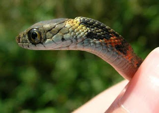
The California Red Sided Garter Snake is one beautiful
reptile.  It has round pupils, so it might be non-venomous,
but it has a highly patterned and bright colored body, so
it could be poisonous. It has a triangular head, but not to
broad, so it could be either. For many years we thought
it was non-venomous, but recent work says it has toxic
saliva. It’s probably not too harmful to humans, but it
goes to show that you can’t make assumptions.
If you know anything about this blog, you know that we look for exceptions in every rule, so I wouldn’t tell you that there are definitive ways to tell venomous from non-venomous snakes. There are just too many exceptions; any mistake in this area could be your last.

However, many people will tell you that the eyes of a snake will give them away –round pupils means non-venomous, while slit pupils (like cats) means venomous. Or that venomous snakes have patterned bodies while non-venomous snakes wear solid colors. Lastly, some people will tell you that venomous snakes have triangular heads, while non-venomous snakes have rounder heads.

Let’s tear down each myth. About 99% of snakes have triangular heads. So this is no help at all; although, if you stay away from all triangular headed snakes you probably won’t get in trouble. Venomous snakes do have a broader base to their triangular head, to account for the venom gland volume and associated muscles. However, are you going to take the time to determine just HOW BROAD is the head of the snake that’s about to bite you?

As for pattern versus solid color; this will also fail you. Ever hear of a black mamba (Dendroaspis polylepis)? Well, it’s the most venomous snake in Africa and it’s solid colored. Strangely enough, the black mamba isn’t black. Its name comes from the color of the inside of its mouth, but its body is silvery. The Inland Taipan viper (Fierce Snake, Oxyuranus microlepidotus) is the most venomous on land, but it has a solid dark tan body.

Maybe pupil shape matters. I am wondering why there would be an evolutionary link between the shape of the pupil and whether a snake has venom. Head shape – maybe, you have to account for venom glands. Body color – maybe, patterns would warn a predator to stay away (aposematism). But pupil shape? How would that be linked to venom or no venom?

The black mamba is one of the most aggressive of venomous
snakes. It’s as if it goes out looking for things to bite. And
they are fast, moving up to 20 ft./sec (that’s 14 mph, or 22.5
kph), although I think Kobe Bryant can still move faster than
that in short spurts. The mamba grows to over 10 ft in length
and will attack a lion, so it backs up the deadly look of its
open mouth – it ain’t just for show.
We can use the black mamba and inland taipan snake examples for debunking the pupil myth as well. The mambas and the fierce snake are round, as are the pupils of the coral snake (very dangerous). More to the point, who wants to get close enough to a snake’s pupils to see if they are slit shaped or round? Let’s err on the side of caution; if you see a snake in the wild – assume it’s venomous. Problem solved.

So let’s find some real exceptions in the realm of venomous snakes. Unfortunately, there are so many different combinations of fang type, venom type, and venom gland type that it is difficult to call any kind of venomous snake an exception – there aren’t many rules. There are true venom glands and false glands, based on whether they can store venom. Then there are rear fangs, front fangs, and front fangs that can fold up. And then there are systems that deliver venom to several upper teeth, and those that deliver venom only through the channels in front fangs or rear fangs.

There may be different ways to deliver and different venoms to deliver, but a 2008 study says it all venomous snakes derive from a common ancestor that lived about 60 million years ago. The study of Dr. Vonk looked at front and rear-fanged venomous snake embryos, and saw that the venom gland ducts ALWAYS start out attached to a rear tooth, but in the front-fanged snakes, the tooth and duct move forward during fetal development!

The rear-fanged snakes have back teeth that deliver venom,
but not by injecting it like a hypodermic. They are not hollow;
some have grooves for the venom to run down, while others
are just sharp, bigger teeth. Snakes with rear fangs have to
take a bigger bite, and hold on longer to grind the venom into
the wound. Doesn’t sound like the perfect system, but
evolution is striving for perfection, it just uses what is there.
What is more, it would seem that the tooth that developed into a fang became linked to the development of the venom gland rather than the other teeth, suggesting that they co-evolved in all types of venomous snakes, linking the type of venom with the type of delivery and type of fang. Perhaps they all started out the same, but they developed their own combinations independently.

It may be that this occurred with all snakes; those that aren’t venomous just lost the ability to produce or deliver venom. This is part of the toxicofera hypothesis of which we have spoken. We don’t even know what percentage of snakes are venomous. Scientists have focused on the highly venomous snakes for so long, that the so-called non-venomous snakes have been ignored.

Many snakes that were once thought to be non-venomous are now known to have venomous bites. Colubridaesnakes (garter snakes, hognose snakes and many more) were thought up to the 1950’s to be utterly non-venomous. But Dr. Fry showed that many of these snakes do indeed deliver venom, though most may be harmless to humans.

As recently as ten years ago it was said that only 10% of snakes were venomous; now that percentage is somewhere near 30%. Where might it end – could most snakes be venomous?

Even though scientists now know more about the evolution of venom, there are still mysteries. With millions of years to perfect a venom system, why is it that some snakes have venom that is WAY TOO POTENT for its purpose? With each bite the fierce snake delivers enough venom to kill 2000 mice or 50 humans – why so much? It must be advantageous in some way; or else it doesn’t cost any more energy to make the venom that potent.

This is a juvenile tiger keelback snake. The raised part on the
back explains the name. It is actually the nuchal gland that
stores cane toad toxin as a defense. How could a small
juvenile have toxin before it is big enough to start eating cane
toads? It can be passed on from mother to offspring, if she
ate a cane toad before egg format
Great diversity in venoms and fangs aside, there are exceptions in venomous snakes. Let’s talk about the keelback snakes. They may be venomous, but they are also poisonous.

Debra Hutchinson published on the keelback snakes in 2007. Tiger keelback snakes (Rhabdophis tigranus) live in Asia, and enjoy a diet of cane toads – poisonous cane toads! Cane toads kill most things that try to eat them, but for some reason the keelback snakes don’t seem to be bothered by the toxin.

In fact, they sequester the cane toad toxin to two nuchal glands, located on the back of their necks. Then, when the snake is threatened by a predator, they turn their back to the aggressor and dare them to bite down on the nuchal glands! Most predators have learned not to take the bait.

The nuchal glands are purely for storage. They don’t have ducts or deliver the poison to the skin or a fang. The sequestered toxin is purely defensive. But the keelbacks also have venom glands, of the false gland type, delivered to the base of several upper teeth. The keelback then bites, chews and grinds the venom into the wound.

This is the way it goes for many rear fanged snakes. Delivering venom by the front fangs is 100x more efficient than the rear fangs, so rear fang snakes must hang on longer to their target in order to envenomate them. This means that they are more vulnerable to being bitten when the target fights back. The keelback has made this less likely by storing another toxin in its neck. Pretty smart, huh?

When threatened, the tiger keelback assumes a familiar neck
arch position. This was described in Akira Mori’s 2012 paper.
If disturbed, it will also try to bump the nuchal glands into the
aggressor, They will spray toxic contents if bitten or pinched.
Now for one more venomous snake exception. Your parents always told you not to spit, but a few snakes are expectorating geniuses. The spitting cobras (genus Naja, and a couple others) have an additional modification to their front fangs that gives them the ability to spit their venom, in some cases, over twenty feet.

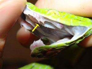
Injecting venom from front fangs is controlled by specific muscles around the venom gland. Spitting snakes combine this quick delivery under pressure with a targeting system. Instead delivering venom from the tips of their fangs, they have an aperture (hole) in the front face of their fang (see picture). Some cobras aim for the eyes of their targets, while others aim for mouth, nose or skin.

The aim is incredible in all, but it is even better in some species. I will use guns as a model. Most guns and cannons up to the time of the US Civil War were very inaccurate. By gouging curved grooves down the barrel, a spin was placed on the cannon ball, and the spin made it much more accurate. This “rifling” was invented in the 1500’s, but didn’t become common until the 1800’s.

The same is seen in the African (not so often in the Asian) spitting cobras. The fang and aperture have rifling grooves that make them even more accurate. I would say that humans stole the idea from nature (like we so often do), but I don’t think we knew about spitting cobras when guns barrels started being rifled.

But how does spitting (really squirting, no saliva is involved) venom at an aggressor help, other than grossing them out? We know that venom must be injected below the skin in order to be effective, but a spitting cobra’s toxin can be cytotoxic (lots of inflammation and tissue destruction) to the skin, and can blind if it hits the eyes. The black-necked cobra and the red Mozambique cobra have been shown to aim only for eyes.

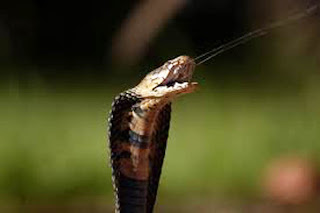
This is a Mozambique spitting cobra. Notice how the spray
comes straight out from the front of the fangs and is directed in a
narrow, pointed direction. This isn’t strafing fire, it’s sniper work.
In a 2005 study of spitting cobras, the red and black-necked spitters recognized faces and eyes, but would not spit at photographs of faces. That’s mighty evolved; you don’t want to waste the toxin, and you must be accurate to avoid waste as well. The black-necked was able to hit the eyes 80% of the time, and the red spitter never missed.

Most spitting cobras actually have a mix of toxins; some neurotoxic, some hemotoxic, some cardiotoxic, and some cytotoxic. Somewhere along the way, evolutionarily speaking, the spitting cobras concocted a toxin that has both the ability to harm by surface contact, and the ability to harm on contact. Evolution at its best.

Next week, is there a group of animals where every species is venomous? And how about a group where none of the species are venomous.


Vonk, F., Admiraal, J., Jackson, K., Reshef, R., de Bakker, M., Vanderschoot, K., van den Berge, I., van Atten, M., Burgerhout, E., Beck, A., Mirtschin, P., Kochva, E., Witte, F., Fry, B., Woods, A., & Richardson, M. (2008). Evolutionary origin and development of snake fangs Nature, 454 (7204), 630-633 DOI: 10.1038/nature07178

Mori, A., Burghardt, G., Savitzky, A., Roberts, K., Hutchinson, D., & Goris, R. (2011). Nuchal glands: a novel defensive system in snakes Chemoecology, 22 (3), 187-198 DOI: 10.1007/s00049-011-0086-2
 
Hutchinson, D., Mori, A., Savitzky, A., Burghardt, G., Wu, X., Meinwald, J., & Schroeder, F. (2007). From the Cover: Dietary sequestration of defensive steroids in nuchal glands of the Asian snake Rhabdophis tigrinus Proceedings of the National Academy of Sciences, 104 (7), 2265-2270 DOI: 10.1073/pnas.0610785104


For more information and classroom activities, see:

Snakes –

keelback snakes and nuchal glands –

spitting cobras –

It’s An All Or None Proposition

$
0
0
Biology concepts – toxin, venom, cnidarians, kleptocnidae

In our discussions of venoms and toxins we have looked at many groups (phylums) of animals. In each phylum we have identified at least one venomous animal. We have talked venomous amphibians (frogs, salamanders), venomous reptiles (lizards, snakes), venomous arthropods (insects and spiders), and even venomous mammals. Even though we haven’t talked about them in this series, there are also venomous sponges, and sponges are the most primitive animals on Earth.

Sinornithosaurus was a raptor dinosaur with feathers;
a very early proto-bird. It was still a reptile, but with
features that would come to be typical of birds. One
thing that wasn’t typical was its teeth. A 2009 study
indicated that one of its long fangs had a groove down
the side – channel for venom! So, while no modern birds
are/were venomous, maybe an ancient ancestor was.
If even the most primitive animal phylum has members that are venomous, then all phylums probably do – right? Well, no – birds are the exception. We know of no venomous bird; NO bird makes or sequesters a toxin that is then delivered by bite or talon scratch or other natural mechanism.

Birds are the evolutionary descendents of the reptiles; they diverged from the reptiles about 240 million years ago. The toxicofera hypothesis says that all reptiles were at one time venomous, so why aren’t the birds? It may have something to do with the timing of the divergence. The oldest venom genes and delivery systems are associated with the lizards, about 200 million years ago, AFTER the divergence of birds. Mystery solved. Of course, mammals and arthropods had diverged hundreds of million years earlier, and they have some venomous species. If they could do it on their own, why couldn’t birds?

Well, a few birds have found a way to use the toxins produced by other organisms. Members of the pitohui family of birds in Papua New Guinea tend to feed on toxin producing choresine melyrid beetles.  The toxins remain in the bird’s tissues and feathers for some time before they are broken down or excreted.

Just rubbing against the feathers can induce numbing, and consuming a bird would be lethal for many animals, including humans. The Hooded Pitohui seems to know this and is very social and loud. It suspects that predators know it is a bad meal and will leave it alone. An added benefit - lice that usually live in the feathers are also affected by the toxin, so these birds are relatively parasite-free.

This is the spur winged goose. It lives in the wetlands
of Saharan Africa. It also feeds on toxic insects, so it
comes by its toxins in the same way that the pitohuis
do in New Guinea. Not all the geese are toxic, since
they have a large range. Only some live near the
blister beetles of Gambia that are poisonous and
render the birds toxic.
This is odd, since the toxins involved are from the batrachotoxin family, just about the most potent neurotoxins on the planet. They work by binding to the ion channels in neurons and holding them open. This prevents the neuron from recovering after it sends an electrical signal. Therefore, the neuron can’t fire anymore, and whatever it is connected to can't be stimulated. If it is connected to a touch receptor, you will feel numb there. If it is connected to a muscle, the muscle will be paralyzed. Batrachotoxins seem to work in every animal that has this type of neural system, so why isn’t the beetle the bird’s last meal?

Birds are certainly an exception to the rule that at least some organisms in each phylum developed venom. How about the other end of the spectrum? It would also be an exception if we had a phylum of animals that were ALL venomous. Well, we do.

The cnidarians are the phylum of animals that include the anthozoans (corals and sea anemones), and the medusozoa (jellyfish, box jellyfish, and the hydras). It turns out that EVERY species of cnidarian is venomous, though some might not be venomous enough to harm humans.

Cnidarians all have cnidocytes (cnida = nettle, like plant nettles that stick you and cyte = cell); cnidocytes are the secret handshake required for membership in the cnidarian club. There are three main flavors of cnidocytes; nematocysts, spirocysts, and ptychocysts. It is the nematocysts that make cnidarians venomous.

Nematocytes are the cells that house the actual stinging apparatus, called nematocysts. They have a barbed shaft that together looks like a harpoon end. This is housed in a cavity filled with venom and covered by a trap door (operculum). There are about 35-40 different shapes and lengths for nematocysts, but they all work basically the same way.

The left image is a cartoon of a typical cnidarian nematocyte. You
can see the operculum, the trap door on top, as well as how the
barb is packaged inside the cnida. The entire volume is filled with
venom that is sprayed out under pressure when it fires. The right
image is a photomicrograph of a fired nematocyst. The scale
shows how small they really are ( a micron - µm - is a millionth
of a meter).
When triggered by mechanical pressure on a hair cell sticking out, and sometimes when accompanied by a chemical signal that a prey organism is near (they “taste” the water), the pressure inside is increased, reaching more than 2000 pounds per square inch, and the cell bursts. 


The operculum opens and the shaft is everted at the prey in just 700 nanoseconds (about 700 billionths of a second), with an acceleration of more than 5,400,000 x gravity. It isn’t a surprise that the prey’s skin is pierced by the shaft! The pressurized venom is then injected into the wound through the hollow shaft and/or hollow tubule.

For some cnidarians, like the sea wasp (Chironex) or the Portuguese man-of-war (Physalia), the venom is important because their prey is strong, including large fish. Most cnidarians, especially jellyfish, are fragile animals; they don’t have a strong internal skeletal and can be ripped apart easily. Therefore, it is important for them to immobilize their prey quickly. The venom does the job. It works very well, for some jellyfish it works well enough to severely harm (Irukandji jellyfish) or kill (sea wasp) humans.
Inside the vial is an Irukandji jellyfish. It is small, but it
packs a wallop. This is one of the few jellyfish that
possess nematocysts on its bell (the round part at top) as
well as on its tentacles. The lower image shows that
while the body s about 5 mm long, the tentacles of the
Irukandji jellyfish can be up to a meter long! One, you can
hardly see it, and two, it can nail you from long distance.

Spriocysts and ptychocysts are the other types of cnidocytes. I was worried about making the statement that all cnidarians are venomous, on the off chance that some cnidarians possess only spirocysts and/or ptychocysts. I contacted several researchers that study cnidarians, and they all stated that as far as they know, all cnidarians possess nematocysts, while only some have spirocysts and/or ptychocysts.

Many cnidarians rely primarily on spirocysts. These cnidocytes are very similar to nematocysts, except that they don’t have an associated venom. Spirocysts are used primarily by cnidarians that prey on less vigorous animals, animals that aren’t as able to pull them apart at the seams. Most corals, for example, prey on small invertebrates, so they rely less on venom and more on entanglement. This is the function of spirocysts, they substitute adhesive for venom.

The bubble tip anemone (Entacmaea quadricolor), for example, relies on a combination of nematocysts and spirocysts to bring in its prey and for defense. But it doesn’t have to hunt much to gather food. It has symbiotic relationships with other animals that help out. The bubble tip is often green colored, because it has intracellular photosynthetic dinoflagellate organisms that provide it with carbohydrates.

The bubble tip also has a relationship with the clownfish (think Finding Nemo). The fish clean away parasites and devour any dead tentacles, while they also scare off predators and provide the anemone with scraps from its meals. Though the bubble tip does have nematocysts, it seems that the clownfish is immune to the toxin, so living amongst the tentacles provides the clownfish with protection from its predators.

Entacmaea quadricolor is the scientific name for the bubble
tip anemone. It comes in four different colored varieties,
pink, red, orange, and green – hence the name “quadricolor.”
And the “Nemo” fish it protects is actually called a
cinnamon anemonefish. I’m wondering who decided
it tasted like cinnamon. Sometimes it is called a fire
clownfish – so does it taste like fire too?
The third class of cnidocytes are the ptychocysts. These are restricted to a group of sea anemones called tube anemones. They are used to build the tubes that these animals live inside. The threads of the ptychocysts are mixed with mucus and debris and become fibrous houses for the animals inside. As such, they are mainly for defense, not for catching food. Yet they do have nematocysts for defense as well.

Some people think that there are some cnidarians that have lost the ability to sting using nematocysts. The TV show, Survivor, went to Palau for a season, including an episode where the winners of some challenge were rewarded with a chance to swim in the lakes with the jellyfish. These golden jellyfish and moon jellyfish are related to the species that live in the nearby ocean, have been separated geographically for thousands of years.

This separation has led to the misconception that they have lost their nematocysts due to a lack of predators. But it is not so, moon jellyfish stings in the lake will be noticed, just not as much as those fro the ocean. Perhaps a genetic drift is taking place, but swimmers do report numbness around their mouths and fingers, so the jellyfish in the lake do still have venom.

Venom from cnidarians protects cnidarians, but it also protects others. Nudibranches are a type of sea slug, related to snails and other molluscs. They eat cnidarians, but not only do they eat them, they use them as well. The use of cnidarian nematocysts by nudibranches is discussed in a 2009 review by Paul Greenwood. 

Berghia coerulescenslikes to eat sea anemones; it may or may not be susceptible to the venom. But that doesn’t really matter since the nudibranch eats the nematocytes whole, perhaps without triggering them to fire.

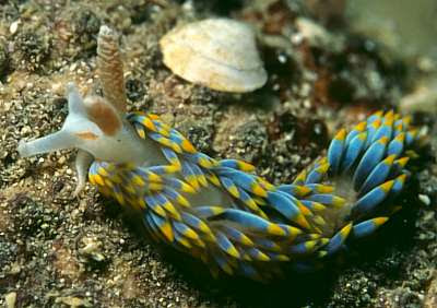
Nudibranch sea slugs are some of the most colorful
animals in the world. You can see the cerata on its back,
like so many dreadlocks. These are where the
nematocysts are housed for defense. They should make
a Disney movie about a sea slug – Where’s Nudi?
There are two hypotheses as to how B. coerulescens can consume the nematocytes and then place them into its cerata on its back. One hypothesis is that they coat them with mucus and that keeps them from firing as they are eaten and moved through the digestive system. The other hypothesis states that they mature nematocytes do discharge, but the immature nematocytes cannot; they are then sequestered and mature while being it held in the cerata.

Either way it occurs, when the nudibranch is threatened, it stiffens its cerata and the musculature moves the nematocytes to a pore. When they contact the seawater, they fire. This is supposed to keep the predators at bay. The review of Greenwood discusses whether this defense is effective – maybe, maybe not. It needs more study.

We just scraped the surface of the weirdness that is the cnidarians, so we will talk more about them in the future. However, next week we will finish the stories on venoms and toxins by looking at the poisonous plants. Did I say, poisonous? Well at least one is venomous.


Gong, E., Martin, L., Burnham, D., & Falk, A. (2009). From the Cover: The birdlike raptor Sinornithosaurus was venomous Proceedings of the National Academy of Sciences, 107 (2), 766-768 DOI: 10.1073/pnas.0912360107

Greenwood, P. (2009). Acquisition and use of nematocysts by cnidarian predators Toxicon, 54 (8), 1065-1070 DOI: 10.1016/j.toxicon.2009.02.029

For more information or classroom activities, see:

Cnidarians –

Nematocysts –

Sinornithosaurus –

Toxic birds –

Kleptocnidae -


A Death Apple A Day Keeps…..

$
0
0
Biology concepts – toxin, poison, urushiol, oleander, hapten, allergic contact dermatitis

A cloudburst threatens to ruin your summer hike. You dart under a tree for protection from the rain and break out a granola bar. You decide to wait it out, but after a few minutes, your skin starts to itch and your eyes sting. After a few more minutes, you notice a rash on your arms and your throat feels like it's closing. Is it bad granola? Is it acid rain? Are you going to die?

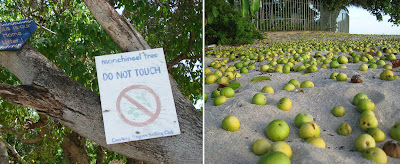
The manchineel tree is toxic enough that just touching it can
do you serious harm. The sign should say, “Don’t even get
near me!” because dripping sap can be just as bad. On the
right you see the apples of the manchineel. They look good,
they are sweet, and the price is right – and then you die.
No, no, and maybe. You just picked the wrong tree to use as an umbrella. This is the manchineel tree (Hippomane manciella), native to several countries in the around the Americas and the Caribbean.

The latex that oozes from this tree contains the toxins hippomanin A and B. Both toxins are present in the latex, leaves, bark, wood, roots, fruit, flowers, and nectar of the manchineel. Eat it or rub against it, and you get sick. Cut it up and the sawdust makes you sick; get it in your eyes (even the smoke from burning it) and you can go blind!

Most deaths have occurred from eating the apple of the manchineel; hence the common name for the tree – the Death Apple. Your mucosal surfaces blister, your larynx swells shut, your GI system rebels loudly and explosively. Massive hemorrhage can follow the closing of your throat, so you drown in your own blood.

In the 1500’s, South American Indians threw death apples down their own wells to poison the invading Spanish conquistadors. It worked because this toxic plant is an exception; it is sweet. Most often, plant toxins taste bitter and that's how we know to avoid them. The death apple’s taste prevents us from making a judgment that could save our life. Definitely, this is a plant to be respected and feared.

Now that I have your attention, let’s talk about a seemingly more common plant toxin. Urushiol is the name for the offending group of molecules in poison ivy, poison oak, poison sumac, even more exotic plants like mango, lacquer trees and cashew nuts.

Mangoes (left) and cashews (right) are in the same family
of plants as poison ivy and poison oak. They contain
urushiol and can cause significant damage to those who have
a heightened allergic response. Urushiol is present in the
skin of the mango fruit and in the shell of the cashew nut.
This is why you can’t buy cashews in the shell, the shell
must be removed to make them safe. Cutting a mango will
give you a small exposure, but most people tolerate this well.
The urushiol (Japanese for lacquer) is exuded from the leaves and stems of the offending plants, and is found in the cashew nut shell. Skin contact leads to blisters and a rash; these are seen earlier in patches that get a larger dose. Urushiol is only partly water soluble, so it can stay on the skin or other surfaces and be spread for quite a while. It can stay on clothes until they are washed; even if that may be years, as in the case with my teenagers.

Urushiol toxicity comes from the immune reaction it generates in about 60-80% of the population. However, urushiol doesn’t spark an immune response on its own. It turns your body against itself. Immune responses are aimed at antigens (not born of, so not self), but urushiol breakdown products are haptens (to fasten to); think of them as half antigens. Haptens must combine with something else to become full antigens. In the case of urushiol, they combine with proteins from our own cell membranes.

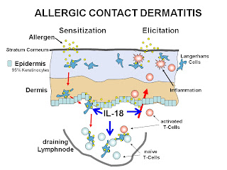
When portion of the urushiol combines with the integral protein, now the protein is seen as foreign and your immune system might start to attack, in a process called type IVdelayed hypersensitivity. This produces inflammation and tissue damage in a reaction termed allergiccontact dermatitis.

Allergic contact dermatitis is different from irritant contact
dermatitis in that irritants damage the skin directly, while
allergens invoke an immune response that causes the
damage. The hapten, in the case of urushiol, penetrates and
is modified by Langerhan cells. The lymphocytes are
exposed to the modified urushiol + membrane protein and
initiate a response. The activated T cells then circulate and
react the next time the urushiol is touched.
As with other allergy reactions, contact dermatitis requires a sensitizing dose, in which your body is exposed to the allergen and ramps up a small reaction. Subsequent episodes are worse because part of the allergic reaction in the immune system sticks around (immune memory).

Not everyone’s immune system recognizes or overreacts to the hapten + membrane protein, so not everyone gets a rash from poison ivy – lucky devils! Other permutations are possible as well. You can be resistant and then develop an allergy late in life, or you can have contact dermatitis when young and later on become resistant. We know a lot about allergic hypersensitivity, but there's also a lot we don’t know. Much research is underway on plant toxins and allergens.

And herein lies the rub - pun intended - with many toxic plants. They cause pain, damage and irritation, yet Paracelsus said, “only the dose makes the poison.” Does that mean that a lower dose has no effect? Well, most of our medicines – antibiotics, anti-cancer, anti-depressive - come from fungi and plants. It isn’t just that less may not be harmful; less might actually be helpful!

Take urushiol for instance. A 2011 study shows that urushiol can kill H. pylori, the bacterium that causes many stomach ulcers. Within 10 minutes, urushiol can strip the membrane off of the bacterium and cause it to lyse. Traditional treatments were found to eradicate the disease in 75% of cases, but adding urushiol brought a 100% cure rate. It even worked in a mouse model, but no one asked the mice if their stomachs itched. Even hippomanin A is an inhibitor of herpes simplex virus 2 replication. It seems that every toxic plant we talk about here has some medicinal use – nothing and nobody are completely evil.

There are several plants that could wrestle for the title of most toxic, but anyone’s top five contenders would have to include oleander (Nerium oleander). You can die just from eating honey collected from bees that landed on the plant and partook of the pollen or nectar.

Nerium oleander is a bush like plant that can have red or
white flowers. It is deadly, but is repairing its reputation
by being used as a medicine. Oleander is the official flower
of Hiroshima, Japan, as it was the first flower that grew
after the atomic bomb was dropped. In Texas, oleander is
used as a decorative plant in road medians – don’t mess
with Texas medians!
The principal toxin is oleandrin, a cardiac glycoside. This type of toxin messes with the electrical impulse generation in heart muscle cells. As a result of the toxin, cardiac activity is dysfunctional, often to the point of arrythmia and heart attack. In other cells, it interferes with calcium levels and can induce cell death. Despite these evil tendencies, oleandrin is proving to be a very useful medicine.

A 2012 study has shown that oleander distillate is therapeutic in diabetes. Rats with induced diabetes were treated with oleander extracts for 12 weeks. Those treated rats had better blood sugar levels, reduced insulin resistance and cholesterol, and improved insulin levels. Not only was the diabetes positively affected, but fat levels were also positively affected – all through treatment with a lethal poison.

But wait - there’s more! Oleandrin has been show to be effective in inhibiting cancer. In at least five different kinds of cancer, oleandrin can stop cancer cells from increasing in number (proliferating), and can even induce the cancer cells to kill themselves (apoptosis). That’s a good start, but it gets better.

A 2005 review discusses the idea of resistance to treatment that develops in many cancers over time. Wouldn’t it be great if we had something that could make the cancer cells sensitive to the drug treatments again? Well, this review discusses studies that show oleandrin can do just that. Oleandrin acts not only as a chemosensitizer, but makes cancer cells more sensitive to radiation therapy. Therefore, oleander is synergistic with other cancer therapies and makes them work better.

Can you stand any more wondrous uses for this poison? A more recent study indicates that oleandrin reduces infectivity of HIV. AZT, a traditional HIV drug, reduces replication but not infectivity, while oleandrin reduces infectivity but not replication, so they could work together.  Oleander can save you from infectious diseases, cancers, and metabolic diseases – but eat the berries on your next hike and you’ll die a horrible death.

Both the cinnabar caterpillar (left) and the moth (right) are
brightly colored. This is called aposematism, warning predators
that they are toxic. It usually works, but the common cuckoo is
apparently opposed to aposematism; it has learned to avoid the
most toxic portions of the larvae and adult.
So humans are animals that can’t just willy-nilly start munching on toxic plants. But other animals can. We have talked about animals that use the toxins they eat (2˚ toxin sequestering), usually from either insects or plants. But is there an exception – does any animal sequester a toxin that its prey sequestered from a plant? I looked for one.

There is a cuckoo that eats the cinnabar moth caterpillar that eats toxic ragwort. The plant has alkaloids. In the stomachs of most animals, they are quickly converted to toxins. But a 2012 study shows that the cinnabar moth caterpillar’s enzymes can convert the metabolic products back to their non-toxic alkaloid forms. Then they are ready to poison the unwitting animal that eats the caterpillar and hasn’t had the forethought to evolve a detoxification process!

However, the common cuckoo (Cuculus canorus) avoids the toxins in the cinnabar caterpillar by biting off the head of the larvae and discarding it, then shaking the carcass to expel the liquid toxin. This is like how some birds can eat monarch butterflies. Monarchs are toxic, having sequestered milkweed toxins they ate as caterpillars. Shining cuckoos in New Zealand and some North American birds know to get rid of the most toxic portions and just eat the rest. Therefore, these birds are not 3˚ toxin sequesterers. I couldn’t find an example – can you?
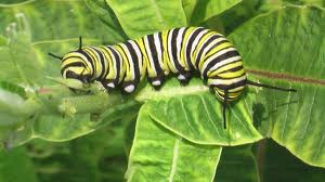
The monarch caterpillar eats only milkweed – ONLY milkweed.
This is where is picks up its toxins. The adult will drink nectar
of many flowers, but the toxin is maintained as the larvae
metamorphed to the adult.

Most birds just stay away from monarchs most of the time, but even this has weirdness associated with it. Monarchs lose toxicity as they age, and males usually have less toxin than females, yet somehow the birds can sense it. Research has shown that monarchs with higher levels of toxin are less likely to be attacked by a predator. How do the birds know?

We have just touched the surface of toxic plants; there are more than we can mention. In Australia alone there are said to be over 1000 toxic plants! Let’s next look to our exceptions; plants that aren’t just toxic, they’re venomous.


Bas, A., Demirci, S., Yazihan, N., Uney, K., & Ermis Kaya, E. (2012). Nerium oleander Distillate Improves Fat and Glucose Metabolism in High-Fat Diet-Fed Streptozotocin-Induced Diabetic Rats International Journal of Endocrinology, 2012, 1-10 DOI: 10.1155/2012/947187

Suk, K., Baik, S., Kim, H., Park, S., Paeng, K., Uh, Y., Jang, I., Cho, M., Choi, E., Kim, M., & Ham, Y. (2011). Antibacterial Effects of the Urushiol Component in the Sap of the Lacquer Tree (Rhus verniciflua Stokes) on Helicobacter pylori Helicobacter, 16 (6), 434-443 DOI: 10.1111/j.1523-5378.2011.00864.x

Garg, A., Buchholz, T., & Aggarwal, B. (2005). Chemosensitization and Radiosensitization of Tumors by Plant Polyphenols Antioxidants & Redox Signaling, 7 (11-12), 1630-1647 DOI: 10.1089/ars.2005.7.1630


For more information, see:

Plant toxins –


Venomous Plants – A Hairy Situation

$
0
0
Biology concepts – venom, toxin, poison, nettle, urticating hairs, trichomes, defense behavior

The cobalt blue tarantula is a beautiful old world
tarantula, but not very cleverly named. They are
popular as pets, even though they are fast,
aggressive and have a potent venom. Fortunately,
they don’t have urticating hairs.
A tarantula, a jellyfish, and an ongaonga tree walk into a bar – O.K., maybe not the best start. But these three organisms do have something in common, something that has been recognized since the time of their classification and naming. Follow along.

Tarantula spiders are a popular example of venomous arthropods, arachnids to be exact. “Tarantula” is a vague term as it is used in the general population. The name comes from Taranto, Italy and came to mean any unknown, hairy, long-legged spider. In scientific taxonomy, tarantulas belong to the family Theraphodsidae, a group containing at least a dozen subfamilies and more than 900 species.

Many tarantulas have impressive fangs that deliver potent toxins to their victims. The fringed ornamental tarantula (Poecilotheria ornate) has produced a coma in a human; however, no known tarantula possesses venom that is acutely lethal to people.

But biting isn’t the only way tarantulas can defend themselves. Besides giving you the heebie-jeebies, two subfamilies of tarantula spiders have defenses called urticating hairs. These hairs are easily lost from their hairy backs or legs when the spider is touched by a predator. These small hairs can lodge in the eyes or skin of predators and cause significant physical irritation, enough to ward off a predator.

There are at least four types of urticating hairs, each differing in size and in the type of predator against which they are most effective. The old world tarantulas have type II urticating hairs that are dislodged by touch, but some tarantulas from the Americas can go one step further. They can fire their urticating hairs from a distance (types I, III or IV).

Urticating hairs often cause urticaria (hives), but
sometimes the red bumps will coelesce and form a
rash. And if you allergic, as seen here, the rash will
become big, ugly, and painful. You can see blister
development at the bottom. Explain to me why he is
affected on his belly?!
Species like the chilean rose and the mexican red-knee tarantulas have urticating hairs that can be fired by kicking their back legs against the back of their abdomen. When threatened, the tarantulas turn and rise up on their legs – ready! They point their abdomen at the threat – aim! And then they rub their legs against their abdomen and release a cloud of hairs toward the target – fire! This leaves a bald patch on their back and a very annoyed predator.

Most urticating hairs are mildly irritating to humans, unless you hold the spider up to your face. This is what happened in 2013 to a three year old boy at his birthday party. He held a rose tarantula up to his face to get a good look, and got two eyes worth of uritcating hairs! He cried for days, as they are so small as to become completely buried in the cornea and cannot be removed. He has made several subsequent trips to the hospital for care.

Other tarantulas have more damaging urticating hairs.  The Goliath Birdeater has larger hairs that can cause very bad rashes, and feel like fiberglass shards embedded in the skin. Some people will become allergic to the hairs, and the rash and reaction will be even worse (see the picture above).
So what has tarantula hair got to do with jellyfish or the ongaonga tree? Urticais the Latin word for “nettle,” and the ongaonga tree is also known as the tree nettle or Urticaferox. And the Greek word for nettle is “cnida,” as in cnidarians – like the jellyfish and coral we talked about two weeks ago. All three of these types of organisms use stinging cells for defense or offense.
Cnidarians use nematocysts to envenomate their prey, shooting toxin filled harpoons at the target. Tarantulas (and some caterpillars) use urticating hairs, not to poison but to irritate their predators. And there are some plants, the nettles, which use urticating hairs as venom delivery systems – the best of both worlds.
The nettles (genus Urtica, approximately 80 species) have hollow uricating hairs that can deliver toxins when they are broken off and embedded in an unfortunate victim. The hairs are actually modified trichomes, epithelial structures found in many plants that are merely raised areas on the plant surface.
Trichomes evolved many variations, those termed “hairs” can be thick or thin, long or short, fuzzy or smooth. Some may be used for water absorption or evaporation, while others will physically impede the movement of insects along the plant, or act as sensors. Venus flytraps (Dionaea muscipula) have three different kinds of trichomes; two secrete digestive juices and one is the sensitive trip wire for closing the trap.
These are the trichomes (stingers) of the ongaonga
nettle. Most nettles have smaller hairs, but this makes
for a more ominous picture. Remember that it isn’t
just their sharp points, they contain venom too.

Typical toxins included in nettle tricomes are formic acid, like in many ant species, and neurotransmitters like serotonin, and histamine. The pain or itch goes away in a few hours. They raise red welts that itch, called hives. In scientific terms, all hive-producing reactions are called urticaria. Get the connection? Most nettle trichome envenomations, like those from Urtica dioica (common nettle) are irritating, but little else.

However, the ongaonga tree (Uritca ferox) is the exception. There has been at least one death associated with just brushing against it. The ongaonga has unusually large spines; the lightest touch brings pain for more than five days.  Its neurotoxins also include an acetylcholine (Ach)-like chemical, yet another neurotransmitter.

The late symptoms can include breathing problems, blindness and paralysis. A 21 year old student developed a paralysis after a brush with the ongaonga. The neurotoxin caused her motor nerves to malfunction, firing too slowly and without pattern. It took weeks for her to recover.

But the news isn’t all bad. Nettle toxins may be used to in medicine, including diabetes, infection and even liver damage. A 2013 study in India treated rats with common nettle oil before performing a partial liver removal. The oils helped promote liver regeneration and decreased cell death after surgery. They also reduced the amount of oxidative damage in the surviving cells. So if you plan on destroying your liver, go run through a nettle patch first. However, I couldn’t find any studies using ongaonga oils – it is just too toxic. So be sure of your nettle patch species prior to your liver-protecting frolic.
A strange picture to see here, but follow along. You can
have part of your liver removed if it is damaged and live
just fine. A partial removal is called a hepatectomy. Some
parts can even regenerate after you have them removed.
Hepatectomy is important, as it makes it possible to have
living liver donors – you give someone part of your liver,
and you grow it back. This is where the nettle medicine
could be useful.

Our king of venomous plants comes from a different genus of the same family of plants as the nettles. You would think a plant that could kill you by touch would have a tough name, but it turns out to be just another insult added to the injury. You have tell your best buddies that you are laid up for weeks by a plant; and when asked, you have to tell them it was the “gympie gympie!” I can hear the laughter now.

The gympie gympie (Dendrocnidae moroides) lives in Australia, the land of painful deaths. The Australian Geographic website says that being envenomated by the gympie gympie is like, “being burnt with hot acid and electrocuted at the same time.” It has killed people, horses, and dogs.
Minor stings can last for hours to days with increased heart rate and sweating. The gympie’s trichomes seem to be silica based, like glass. You can heat them with a flame until they glow red, but they will still hold their shape. Add being stabbed with glass shards to the description of the gympie's sting.

Severe encounters can bring pain for months, with symptoms waning and then brought back by hot or cold air, water, or rubbing. Some people have shot themselves to relieve the pain, while others have had to be strapped to the bed.

There isn’t much you can do to treat the pain, but you might be able to shorten the length of your torture. The best first aid is to immediately apply hair removal wax and yank out the trichomes. You go for a hike and end up with silky, smooth skin and a pain that won’t stop – oh, wait, that could be just be describing the waxing.
The gympie gympie has huge leaves, like it is trying to ruin
your day. You can’t even see the hairs here, they are too
small. But you know it if you touch it. Did this guy lose a bet? 
Just being this close is a very bad idea.

Usually the pain comes from rubbing against the leaves, stem, or twigs. But the gympie wants to reach out and touch you, even if you don’t reach out and touch it. It sheds its urticating hairs all the time, so if you hang around a tree long enough, you will get a nosebleed and start to sneeze painfully. And you can’t wax the inside of your nose ….. I hope.

Fortunately, few deaths have been associated with the gympie gympie. It grows in the rainforests of northeast Australia where the population is very low, about 5-10 people per 2.5 sq. mile. The aborigines live here, and they actually eat the berries of the gympie gympie. Since all its trichomes point one direction, the natives know how to move along the stems and leaves in the right direction to harvest dinner. Apparently the berries aren’t poisonous.

D. moroides toxins include those said to act as neurotransmitters Ach, serotonin,  and histamine, but their chemical structures are different. They also include moroidin, a short peptide toxin that was first isolated from the leaves and stalks of thegympie.

No, this isn’t a picture of some electrical spark experiment
gone wrong. The green spines are the mitotic spindle and
the red blobs are the chromatids being pulled apart during
mitosis. More mitoses, more cell divisions. More divisions,
more cells. Too many more cells = cancer. It would be nice
to stop the spindles in that case.
Moroidin is a mitotic inhibitor; it interrupts the polymerization of tubulin during the formation of the mitotic spindle. If no spindle forms, then there is no alignment or segregation of chromatids during mitosis, so no cell division. Moroidin is supposed to be the factor that makes the sting pain last a long time, but not enough research has been done in this area. No one can even tell me specific chemicals the gympie possesses or how it causes pain! How can we make use of it in medicine if we don’t know how it works? I would think that a mitosis inhibitor might work well against cancer – let’s get to work people!

School is winding down, so why don't we start our summer posts. Each week will be a separate question in biology, from misconceptions to things that make you wonder, to weirdness galore. Next week - how good are different species at going without oxygen, and who can hold their breath the longest?

Oguz, S., Kanter, M., Erboga, M., Toydemir, T., Sayhan, M., & Onur, H. (2013). Effects of Urtica dioica on oxidative stress, proliferation and apoptosis after partial hepatectomy in rats Toxicology and Industrial Health DOI: 10.1177/0748233713480211

Hammond-Tooke, G., Taylor, P., Punchihewa, S., & Beasley, M. (2007). Urtica ferox neuropathy Muscle & Nerve, 35 (6), 804-807 DOI: 10.1002/mus.20730


For more information, see:

Tarantula urticating hairs –

Nettles –

Gympie gympie –


It’s An Airtight Case

$
0
0
Biology concepts – respiration, aerobe, anaerobe, CAM plants, plastron respiration, cutaneous respiration

Question of the Day – what living thing can hold its breath the longest?

It may seem like an exaggeration, but people whose
tissues are low on oxygen (hypoxic) can have a bluish
hue (cyanosis). Blood that is oxygenated is redder
than blood that is deoxygenated. In animals with more
hemoglobin than humans, like whales, the blood can
actually turn almost purple. Blue Man Group will turn
you blue - from laughter, not disease.
The world’s record for holding one’s breath (voluntarily) was set in May, 2012 by Denmark’s Stig Severinsen. Even though it wasn’t technically cheating, he did hyperventilate with almost pure oxygen for 19 minutes before he then held his breath for an amazing 22 minutes and 0 seconds!

Hyperventilation works by reducing the CO2:O2ratio in your blood. CO2 concentration is measured by sensors in your large blood vessels. Signals are sent to the brain to increase the breathing rate if there is too much CO2 in the blood or to reduce the breathing rate if the pCO2 is too low. By breathing fast and breathing pure O2, there will be less CO2 in the blood and your brain will tell you to stop breathing until the pCO2 increases to normal levels.

Stig said he used two mental tricks to increase the time he can hold his breath. One is scientifically valid; biofeedback allows Stig to slow his own body activity. By concentrating on his heartbeat, Stig has learned to stimulate neural pathways to reduce his cardiac output and his overall metabolism rate -lower metabolism and heart rate - less need for oxygen. His second technique? He thinks about dolphins.

Twenty-two minutes seems like a long time; indeed, this feat required arduous training by Stig. In truth, 22 minutes isn’t very long at all. In fact, humans are weenies when it comes to going without oxygen. Lots of animals can hold their breath longer than we can.

Sperm whales and elephant seals are the banner carriers for the marine mammals in our contest. Sperm whales can submerge for more than two hours, while elephant seals may stay under water for an hour or more. Being mammals, they have physiology very similar to humans in some respects, but they do have modifications that allow for the long submersions.

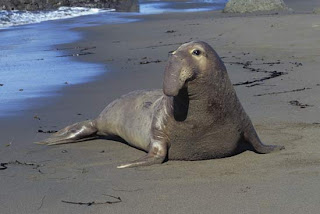
Elephant seals have a large proboscis; hence the
elephant name. There are two species, Northern and
Southern, with the Southern species being slightly
larger. Males can weigh over 5 tons and they fight for
females. The females are more like 1500-2000 pounds,
making this the largest relative difference in weights
of two sexes for any mammal.
Whales and seals have more blood than humans do – duh! Even compared pound for pound, they have 3x as much blood. More blood means more oxygen carrying capability. They also have increased oxygen carrying proteins in their blood and tissues. In addition, they can reduce their metabolism in all but the most necessary organs, and they can divert their blood to just these organs.

New research shows that marine mammals also have oxygen carrying proteins in their brains, called neuroglobin and cytoglobin. So, while blood levels of oxygen may plummet when diving, the brain remains oxygenated.

Sea turtles and crocodilians are examples of reptiles that can hold their breath for amazing lengths of time. Aligators and crocodiles can stay submerged for a couple of hours, while a Galapagos sea turtle can easily stay underwater for 4-7 hours, depending on its level of activity. Maybe they think about Stig in order to stay submerged longer.

Sea turtle hibernation is controversial, but some freshwater turtles do just that. They can stay submerged for weeks or perhaps months at a time! But many turtles cheat at our contest, they have a bimodal respiratory system, through their lungs and through their skin. Cutaneous gas exchange is apparent in all freshwater turtles to some degree, but it is much more efficient in some soft shell turtles, according to a 2001 study.

Let’s look at a different type of reptile. The Belcher’s sea snake (Hydrophis belcheri) is the most venomous snake on the face of the Earth, or under it, as the case may be. It is a sea snake that can remain submerged for 7-8 hours. Sea snakes like H. belcheri have a single lung that runs almost the entire length of their body, and their trachea can also transfer oxygen to the blood. This reduces the “dead space” in their respiratory system and allows them to absorb more of the oxygen they inhale.

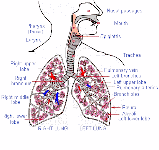
This diagram gives you an idea of how little respiratory
space humans use for gas exchange. The only places
that move oxygen in to the blood are the pinkish
alveoli at the ends of each airway. Sea snakes use
their available respiratory space to exchange gasses.
Humans, as a comparison, only absorb about 15% of the oxygen in each breath, partly because the gas exchange takes place only in the alveoli (terminal air sacs). Oxygen in the nose, pharynx, trachea, bronchi, and bronchioles is just exhaled without any chance to be used by the body.

However, sea snakes are cheaters as well. Their bodies have been streamlined to help them move through the water. One adaptation in this direction is the complete loss of scales. As a result, these snakes have evolved the ability to exchange some oxygen and carbon dioxide with the water through their skin. So they aren’t really holding their breath when submerged.

Almost all amphibians are cutaneous (skin surface) breathers as well. In air, most amphibians can survive exclusively by exchanging gasses through their skin, and in water, adult gills or rudimentary lungs are supplemented by exchange of gas from the water. Cold water and turbulent water contains more oxygen, so in these environments amphibians can survive indefinitely by garnering oxygen from water.

Indeed, the largest family of salamanders (the plethodontidae), don’t have any lungs at all. They exchange gasses only through their skin and the mucosa surfaces of their mouths. And many of these salamanders are primarily aquatic, they don’t take a breath in their entire lives – but they aren’t holding their breath either.

But none of these animals are the champion breath holders. There are organisms that laugh at holding their breath for a couple of hours. But let’s limit our discussion to those organisms that require oxygen. It’s no fun watching an anaerobic bacterium hold its breath; it doesn’t need oxygen! In many cases, air kills them!

Cockroaches, ticks, and ants do last a long time underwater, just try flushing one. But they can’t win our contest either. They seem to trap a bubble of air as they submerge. They have long hairs on their abdomens that trap air via the surface the surface tension and cohesion of water.
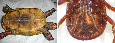
A plastron is the bottom portion of the turtle or tortoise shell,
made up of flat pieces. On the right is the plastron of a tick.
In some ticks there can be gas exchange from water to bug
through the plastron. This is called plastron respiration.

Surrounded by the bubble, they can oygenate their tissues via the breathing holes on the sides of their bodies (spiracles). This allows them to be underwater for nearly an hour and still be breathing. New research in ticks shows that the plastron (flat portion under the abdomen) is capable of some gas exchange itself via the air trapped by the hydrophobic hairs on the abdomen.

We make a big deal about how plants take in carbon dioxide and give off oxygen, and they do during photosynthesis in their chloroplasts. But that’s only half the story. They also have mitochondria that produce ATP from photosynthesis products via oxidative phosphorylation, just like we do.

For plants that grow in hot, dry environments, loss of water is a serious threat. To minimize water loss, some can close the pores in their leaves (stomata), but this also prevents gas exchange, including taking up carbon dioxide and oxygen. The stomata will open only at night, when the temperatures are cooler and water loss would be lost. This is the only time they exchange CO2 and O2 with the environment as well.

CAM (crassulacean acid metabolism) plants can store the carbon dioxide they take in at night in the form of malate. They then can perform photosynthesis even though their stomata are closed. CAM physiology also reduces the amount of O2bound by RuBisCo enzyme instead of CO2. RuBisCo + O2leads to inefficient carbon fixation, so waiting until night time when CO2is relatively more abundant and more soluble will increase photosynthesis productivity. As a result, CAM plants hold their breath for 8-15 hours every day!

CAM plants close their stomata during the hot day, but
exchange gasses during the cooler night. They convert
carbon dioxide to malate as a temporary fixation, which
they store in the central vacuole. During the day, they
convert the malate to carbon dioxide and then to
carbohydrate in the chloroplast using normal
photosynthesis pathways. CAM plants include the
prickly pear, as shown on the extreme right and left.
But the winners of our lack of oxygen survival contest – bacteria, of course! Bacteria come in many flavors, including those that don’t need oxygen for respiration (chemosynthesizers and anaerobes), those that can take or leave oxygen (facultative bacteria), and those that must have oxygen in order to make ATP (obligate aerobes).

Mycobacterium tuberculosis is an example of an obligate aerobe. I talked to Martin Gengenbacher at the Max Planck Institute in Berlin about M. tuberculosis and its survival time without oxygen. He has recently published a great review of M.tuberculosis biology. In a series of experiments that resulted in the development of something called the Wayne model, M. tuberculosis was sealed in a vessel in which they consumed all the available oxygen over time.

However, even after the oxygen was gone, the organisms remained viable for 25 days! They do seem to go dormant, but this dormancy is not the same as ceasing activity completely. It seems that some metabolism and respiration is maintained in the complete absence of oxygen – even though we know that M. tuberculosis absolutely requires oxygen to survive.

These 25 days make M. tuberculosis better than any of our other example organisms at living without gas exchange, though there may be other obligate aerobes that can perform similar feats. But there’s more to the skills of the tuberculin bacterium. In tuberculosis, the body has a difficult time killing off the organism, so it does the next best thing – it walls off the bacteria and traps them in a prison cell of immune cells. These whirls of cells are called granulomas and are very complex structures.

On the left shows a tuberculin granuloma forming and breaking
down. You can see in the middle a formed granuloma, with
macrophages surrounded by a fibrous cuff and lymphocytes.
When immunosuppression sets in, the granuloma breaks down
and the organisms is released to cause disease. On the right is a
photomicrograph of granulomas. In the right corner is a
tuberculosis bacterium before granuloma formation.
Granulomas are extremely hypoxic (oxygen poor), and M. tuberculosis does undergo dormancy in these structures. But again, Dr. Gengenbacher states that this is a metabolically active dormancy, which would by definition require ATP, and therefore require cellular respiration.

The patient still has TB, but no symptomology. This remains the case until the patient undergoes some form of immunosuppression, some disease or condition that prevents the immune cells from keeping the organism in prison. There have been cases where TB has reactivated some 50 years after the original infection. So – M. tuberculosiscan hold its breath for half a century! We have a winner.

Next week, another question to ponder. Just how many species call Earth home? 


Gengenbacher, M., & Kaufmann, S. (2012). Mycobacterium tuberculosis: success through dormancy FEMS Microbiology Reviews, 36 (3), 514-532 DOI: 10.1111/j.1574-6976.2012.00331.x 

Fielden, L., Knolhoff, L., Villarreal, S., & Ryan, P. (2011). Underwater survival in the dog tick Dermacentor variabilis (Acari:Ixodidae) Journal of Insect Physiology, 57 (1), 21-26 DOI: 10.1016/j.jinsphys.2010.08.009 

Williams, T., Zavanelli, M., Miller, M., Goldbeck, R., Morledge, M., Casper, D., Pabst, D., McLellan, W., Cantin, L., & Kliger, D. (2008). Running, swimming and diving modifies neuroprotecting globins in the mammalian brain Proceedings of the Royal Society B: Biological Sciences, 275 (1636), 751-758 DOI: 10.1098/rspb.2007.1484


Biodiversity Counts!

$
0
0
Biology concepts – biodiversity, kingdoms of life, animalia, plantae, fungi, protist, archaea, bacteria, extant, extinct

It seems that contests counting things in jars
always involve food. M&Ms, jellybeans, candies,
and gumballs are common things to estimate. I like
another estimate game – how many grains of sand
can be held in one human hand?
Amazingly, only about 10,000.
At some point in our lives, we have all tried to win the prize by guessing how many jellybeans are in the jar. Number estimation is a skill few people possess, just try ordering mulch by the square yard – you end up with either half as much as you need or buried in pine bark!

There is a large group of scientists playing the jellybean game for a living, but their jar is the entire earth. The jellybeans are species of organisms. The prize? Well, you don’t get to keep the jellybeans; and just how will we know who wins?

The Question of the Day – How many species of life are there on Earth?

It seems like a straight forward question, but let’s look at it in a bit more detail before we try to answer it.  What is it we're counting, animals? Animals and plants? Let’s include everything – everything that is considered alive. So what is considered alive? You can have a great discussion as to what should be considered alive, but for these purposes, let’s stick to the kingdoms of life as taught in every biology class – Archaea, Bacteria, Protists, Fungi, Plants, and Animals.

So now that we know the biodiversitywe are assessing, we need to define our unit. We said above that we would count species, but it isn’t that simple. We have discussed before what constitutes a species in general – those animals that can breed and produce fertile offspring. So we don’t count ligers and tigons (crosses between tigers and lions), but should we count all the hybridizations of orchids? More than 24,000 species are named already, with about 800 added each year. For our purposes, and for most people estimating species, we will stick with those found in nature, unaided by man.

In 2012, Japanese botanists crossed two orchids and
created a new orchid. No big deal right, orchids are
crossed all the time. But this was the first time that a
photosynthesizing orchid was crossed with a purely
parasitic orchid that doesn’t perform photosynthesis.
They think the plants are doing some photosynthesis –
they’re pretty anyway. Is this a species we should
count in our estimate?
We are counting species, but is it live (extant) species or all species including those that are extinct? The World Wildlife Federation estimates that between 0.01% and 0.1% of all species go extinct each year. However many species we end up with as an estimate for all life on Earth, this is a huge number of species to lose EVERY YEAR. It is scary to think how many undiscovered species will go extinct this year. There must be thousands and thousands of species that we will never get to describe using a live specimen or discover how they might add to the diversity of the planet.

Scientists refer to “catalogued” species, but does that refer to those alive at the time the were described, or any species that has ever been described and named? Scientists estimate that extant species account for only 3% of everything that has lived on Earth, so including all species would greatly increased the overall number. However, in most estimates of species on Earth, the species being talked about are alive now, except for the one went extinct just this second, and the one that will go extinct two minutes from now, and the one….


The opposite action is also occurring; we discover new species every year. Most people think that perhaps a few new species are found each year, but it’s really in the thousands. The International Institute for Species Exploration (IISE) at Arizona State University publishes a list each year of the newly discovered species. For 2011 the list was more than 18,000 species long! You can see from the picture (below, right) that many discoveries were made in every kingdom of life in the decade of the 2000’s, more than 170,000 discoveries in all.

This graph shows the relative number of new species in many groups of
organisms. The scales are variable for each group and can’t be compared
amongst the groups. The trends show that in some groups, more and
more species are being discovered each year, while in others, fewer and
fewer are being found. In still others, some years a bunch are found and
some years provide few or no new species.
If 170,000 were discovered in ten years, the total number of species on Earth must be huge. Let’s break down the numbers of catalogued species and the estimates by kingdom.

Animalia– These are what most people think of when asked to name a species. Most animals are relatively big and can be seen in everyday life. The number of catalogued species includes the dinosaurs and humans, birds and sponges.  The numbers in each group vary greatly.

People love to study birds – so we have probably found a greater percentage of the total number of birds than we have worms. We have named about 10,000 species of birds, and more than 22,000 species of annelids (segmented worms). But there are probably many more annelids that we have not found as compared to birds. Therefore the estimate for annelids will be harder to make and perhaps less accurate.

For all animals, the total number of catalogued species by 2010 was 1.2 million. A recent paper (2011, Mora et al.) has predicted the number of species in most kingdoms based on several mathematical models. The authors predict that there are more than 9.9 million animal species on the Earth and in the oceans. According to this estimate, we have found only 12% of all the animals on Earth!

Meet the Giant Gippsland Earthworm 
(Megascolidesaustralis). It can reach 3meters 
(10 feet) in length. First described in 1878, this 
worm lives in the deep clay soil in a small 
area in Australia. Similar worms live in North
America, but they are rarely observed.
Plantae– Plants include the non-vascular mosses, the vascular, spore-forming ferns and horsetails, the seed bearing gymnosperms (conifers and such) and the fruiting and flowering angiosperms. So many of these organisms are crucial for human life (medicine, food, oxygen!) we have done a good job of cataloguing them. The 2011 Mora paper surmises that we have already described nearly 50% of the total number of species.

Their methodology uses a lot of math to relate the number of higher taxa (phylums, orders, families) that are known in well-described kingdoms to the possible number in lesser studied groups. They found that there was a consistent pattern in which the more families there are now can be used to predict how many total genera there might be, and each level then can be used to predict the number in kingdoms for which the number of higher taxa are known. They also use methods to predict unknown higher taxa, so they can be included in the species estimate.

For plants, the Mora group predicts that there are 314,000 species extant on Earth. Other estimates also come out at about 300,000 species, but they include algae, which are actually protists, not plants.

FungiThese organisms range from the invisible to the visible. In fact, in the paleoworld, fungi represented the largest organisms on Earth. Even today, the largest single organism is a fungus in the Malheur National Forest in Oregon, where a single 8,000 year old honey mushroom covers 2,200 acres (8,900 square meters) of land.

On the top is a fossilized prototaxite fungus in Saudi Arabia 
that once reached 20 ft (6 m) in height and was the tallest 
organism on the early Earth (this one is on its side). On the 
bottom is the national forest where a single honey mushroom mat 
has killed 2200 acres of trees. Each mushroom is a clone,
connected by a rhizoid mat just under the ground.
If you compare plant diversity to fungi, fungi win hands down. Described species of fungus lag behind; only 43,000 species have been named, but there is much more room to add new species. Estimates are that by the time we are done, whenever that is, we will have nearly 700,000 different mushrooms and other fungi.

Protista– Protists are a bit of a catch-all kingdom. Some have aspects that make them look like plants; they perform photosynthesis or they have central vacuoles. Others are much more animal-like. Overall, they are free-living organisms that are usually single celled or made up of many cells not forming tissues. Two examples show you the diversity in this group. Giant sea kelp (Macrocystis pyrifera) is a type of brown algae that can grow at a rate of 2 ft/day and can reach a length of 300 ft (91.5 m), while picoplankton are 0.2 microns (0.00000002 m) in diameter, are single celled, and may or may not perform photosynthesis.

The estimates for the number of protists vary greatly. A 1998 study indicated that they had no reason to believe the total number of protist species would be greater than 3000. However, a 2005 report puts the estimate number anywhere from 140,000 to 1.6 million. The Mora group’s paper estimates that about 73,000 will be found; that’s 57,000 more than have been described as of 2010.

Archaea and Bacteria– These are the prokaryotes. They live in tar pits and arm pits; they own the planet and probably outer space as well. There are more bacteria in a scoop full of dirt then people who have ever lived on Earth. But for the purposes of our discussion today, the important part is that same scoop of dirt has thousands of undiscovered bacteria.

The J. Craig Venter Institute is leading the Global Ocean Sampling Expeditions to discover new marine microorganisms. A pilot expedition in 2003 identifed 1800 new species in just a couple of months. This was followed by global expeditions in 2005-2009 and an expedition to the European waterways in 2009-2010. They will analyzing the data and naming species for decades to come.

This map represents the expeditions of the Global Ocean Sampling Projects
of the Venter Institute. As they traveled, they would acquire 200-400 gallon
samples of water from different depths every 200 miles and put them
through a series of filters to catch smaller and smaller organisms. The filters
would be dried and used for DNA analysis. In just a couple of months, 1.2
million genes were identified using this methodology.
Most bacteria and archaea can’t be grown in the lab because we don’t know what they require to live. This makes them hard to describe and classify.  Therefore, the Venter Institute uses DNA techniques from dried whole organisms to identify new “species.”

But perhaps classification is not the proper term for these organisms. I talked to Dr. Mora about why prokaryotes were not analyzed to the same degree as other kingdoms in their paper.  He rightfully pointed out that species definitions don’t really apply to prokaryotes as neatly as they do other types of organisms.

Certainly they don’t conform to the mating and fertile offspring definition of species since they don’t mate. Also, since they swap DNA as usual business (lateral gene transfer), who is to say where one species stops and another begins.

Other sources are little more daring in projecting possible numbers of prokaryotes. It is possible that there are a billion distinct bacteria and archaea, but it more likely that the number is in the 10-20 million range.

What are our final numbers when add up all the estimates? Predicted species numbers for life on Earth range from 11.3 million in the Mora paper, to perhaps more than 1 billion if you include prokaryotes. If we leave the bacteria out of the equation, there could still be as many as 30,000,000 forms of life on the planet. We have described about 2 million (according to IISE), so only 28,000,000 left to find!


Mora, C., Tittensor, D., Adl, S., Simpson, A., & Worm, B. (2011). How Many Species Are There on Earth and in the Ocean? PLoS Biology, 9 (8) DOI: 10.1371/journal.pbio.1001127
 
ADL, S., SIMPSON, A., FARMER, M., ANDERSEN, R., ANDERSON, O., BARTA, J., BOWSER, S., BRUGEROLLE, G., FENSOME, R., FREDERICQ, S., JAMES, T., KARPOV, S., KUGRENS, P., KRUG, J., LANE, C., LEWIS, L., LODGE, J., LYNN, D., MANN, D., MCCOURT, R., MENDOZA, L., MOESTRUP, O., MOZLEY-STANDRIDGE, S., NERAD, T., SHEARER, C., SMIRNOV, A., SPIEGEL, F., & TAYLOR, M. (2005). The New Higher Level Classification of Eukaryotes with Emphasis on the Taxonomy of Protists The Journal of Eukaryotic Microbiology, 52 (5), 399-451 DOI: 10.1111/j.1550-7408.2005.00053.x

I Know Why She Swallowed The Fly

$
0
0
Biology concepts – carnivorous plants, minerals in biology, symbiosis, cryptids,

The Thing From Another Planet was a 1951 B-horror movie.
Arctic researchers find a space ship in the ice and thaw out
the pilot. He turns out to be a walking plant that needs
blood to feed his little seedlings. Never minds that the plant
is growling, feels just fine at -60 degrees, and is wearing
clothes. They finally kill him with electricity.
The man-eating tree is a cryptid (hidden) organism. Cryptid means there is no scientific proof for its existence, but for some reason there are people that say it exists. In this case, a German explorer named Carl Liche trekked through the Madagascar jungle and described the natives forcing a girl to climb the trunk of a tree. The branches (arms) grabbed her and lifted her to the top where she was crushed and absorbed. There was no explorer Carl Liche and the story was an utter fiction.

Myths, hoaxes, misidentifications, misunderstandings, they all have accounted for various cryptids, but every once in a while a cryptid turns out to be real. Gorillas? Once thought to be fictitious monsters. But a man-eating plant? Would you settle for small animal-eating plants? Those we have. The question is why?

Question of the Day – Why do some plants eat bugs?

Venus flytraps are active trap plants. They have a movement that requires energy, and the movement of the trap is one stage - the prey is digested by the part that moves. Much research has been devoted to the mechanism of its fast trap closure, and many hypotheses are still floating about.

We do know that it takes about 1.5 milliseconds to transmit the signal from the trigger hairs in the trap to the motor cells that close it. The signal is electrochemical, very similar to an action potential in an animal neuron. Channels pump ions across membranes, and the difference in the charges of each type of ion (sodium and potassium) cause an electrical impulse.

It seems that the electrical impulse causes water channels to open across various cells near the base of the trap. Water pressure is quickly changed from high to low and low to high in different layers of cells and this cause shape changes. Different shapes cause different stresses, and this closes the trap.

A relatively new hypothesis is that the open configuration is full of elastic stress, so that when water pressures are changed between layers of cells, there is an elastic snap to the closed state. The closing only takes a 0.2 seconds. After that there is a slower portion that brings more complete closing and the start of digestive enzyme secretion.

This is a visual representation of the electrical signal produced
 by triggering the venus fly trap. The red line is the first touch
to a trigger hair. It is not enough to reach threshold and close
the trap. If a second touch occurs before the first has dissipated
to much (green line) the threshold is crossed and the trap
closes. If the second signal is too late (blue line), the
threshold won’t be reached.
To reduce false or unproductive closures, the each trigger hair in the trap produces a sub-threshold action potential. One trigger hair being touched won’t close the trap. As the first signal dissipates, if a second signal is generated by a second hair being touched, the sum of the dying first signal and the second signal can raise the charge above the threshold level and the trap will close (see picture at left). However, in very warm weather (above 36˚C/97˚F) it only takes one trigger hair signal to close the trap – it has something to do with the molecules moving faster in higher temperature environments so that one signal can reach the threshold level.

Other carnivorous plants have semi-active, two-stage traps. The aquatic bladderwort is an interesting example. It is one of the smallest carnivorous plants, with a trap that is just 10 mm wide at its opening. In order to eat, bladderworts create a negative pressure inside the trap by pumping out the water. A trap door maintains the negative pressure inside, but if the trigger hairs outside the trap are touched, the door collapses and water + prey are sucked inside. The trap door then assumes its original shape and the prey is caught inside the trap (see video here).

Sundews are two-stage trap plants as well, having sticky liquid drops perched atop small pedestals. The prey, maybe a fly, gets stuck in the gummy drops. Only then does the tentacle slowly curl around the fly, becoming an “outer stomach” as termed by Charles Darwin (see picture). The digestive enzymes are secreted and the fly is no more.

A different species of sundew, Drosera glanduligera, has a different kind of trap. A new study from Germanyshows that it has brittle hairs (called snap tentacles) at the edge of the trap that when triggered, catapult the prey into the resin glue. The catapult is quicker than the venus flytrap, occurring in less than 75 milliseconds. Only then will the prey be slowly pull down toward the portion of the plant that secretes enzymes.

On the left is a typical sundew, D. capensis. When a fly is stuck, a
slow curl of the tentacle will finally do him in and trigger digestion.
On the right is a rarer sundew, D. glanduligera. The number steps
show the catapulting of prey from the trigger hair into the glue in
the middle of the plant. It all occurs in just milliseconds.
There are also passively carnivorous plants, those that allow the prey to do the work. Pitcher plants are slippery on their edges; prey fall into the pitcher and can’t escape. Amazingly, the seeds of the Shepherd’s Purse are carnivorous, but the plants themselves are not. The seeds lie on the ground and exude toxins that attract and poison insects that pass by. The seeds also secrete enzymes that then digest the insects. This leaves a circular ring of very rich soil, giving the germinating plant an advantage.

In many of the plants, the digestive enzymes have started to be identified. A 2012 paper from Germany has looked the protein portions of the venus flytrap digestive fluid. It contains nucleases (digest DNA and RNA), phosphatases (remove phosphate groups), phospholipases (break down fats), chitinases (to digest the insect exoskeleton), and proteolytic enzymes (to break down proteins). Most of these are derived from pathogenesis proteins, so it is believed that digestion evolved from several self-defense processes.

There are 600 known species of terrestrial carnivorous plants and 50 in the water, but scientists are now realizing that many more plants use a mechanism similar to the Shepherd’s purse and can be considered at least semi-carnivorous. Would you believe that tomato and potato plants have sticky hairs that may trap aphids and other insects. They die and drop to the ground around the stem. This enriches the soil and the plant absorbs the nutrients.
Tomato vines have sticky hairs on their stems. It turns
out that they can trap bugs, hold them until they die,
drop them to the ground, and let their carcasses
fertilize the soil around the plant. Now that’s
miracle grow!

No matter the method of the trapping, the reason is the same; the plants need nutrients. Not glucose, proteins or lipids – they're photosynthetic for gosh sakes. They can make their own proteins, nucleic acids and fats from the carbohydrates they produce during photosynthesis. That is, they can if they have the correct additional materials.

Proteins are made of amino acids, and amino acids contain a lot of nitrogen. Nucleic acids (DNA, RNA) are made from nucleotides, and these include a lot of phosphorous. Many biomolecules and physiologic processes use minerals like nitrogen, potassium, and phosphorous. These are the amin constituents of the fertilizers humans add to the soil to help crops, flowers, and in my case - weeds, grow.

Carnivorous plants often live in nutrient poor soil. Sandy soil (flytraps), tropical jungle soils (sundews), and Andean mountain tops (bromeliad described below) are all mineral poor. In jungles, for instance, most of the minerals are tied up in the huge trees, and such little sunlight penetrates to the ground that few plants can live there; therefore, there is little recycling of nitrogen and phosphorous in the topsoil. Eating insects is just an adaptation to allow them to live where other plants can’t.

Many minerals are made available by digestion of insects.  Carnivorous plants get 5-100 % of their seasonal nitrogen and/or phosphorous gain, but only 1-16% of their potassium uptake. If there is one nutrient these plants covet more than the others, it's the nitrogen.

Many plants acquire nitrogen from symbiotic bacteria around their roots that fix nitrogen gas in the soil. Fixing means converting from gas to a solid. Carnivorous plants do not have these advantages so they had to come up with another strategy. However, help doesn’t always come from digestion of insect prey.

High in the Andes Mountains grows the world’s largest
bromeliad. It can be alive for 140 years before it flowers
the first time. The tall stalk is what holds the flowers.
The business is lower, rounder, and full of sharp spines.
Birds live there, but can also be skewered there. They
say most fatal accidents do occur close to home.
The Roridula genus of South African plants acquire minerals from prey with a little help. It has sticky leaves but no digestive enzymes. Its sticky fluid is resin, not mucus; therefore, enzymes can’t be included because they are not soluble in resin. 


When prey insects get stuck in the resin, they are consumed by another animal. This consumer defecates either before or after its meal. The feces are nitrogen rich remnants from a previous bug meal, so this is an indirect mechanism for profiting from killing for a meal.

Another example of this is Puya raimondii, the world’s largest bromeliad. This plant is about 2-3 m tall, but when it flowers, the stalk may rise as much as 12 m! Birds can live in its foliage and when they defecate, they provide nitrogen to the plant. But P. raimondii has huge sharp spines that can actually kill some of the birds. As the birds rot, they also release minerals to be used by the tree; therefore, P. raimondii is semi-carnivorous.

It isn’t all death and destruction. Take the nepenthes pitcher plants for example. These are the largest of the pitchers, holding more than 2.5 liters of digestive fluids. Their pitchers are little ecosystems. Some larvae, particularly a couple of species of mosquito, can survive ONLY inside the pitcher liquid.

I don’t think I can do this picture justice. The only
things this tree shrew lacks are a magazine and a
can of air freshener.
On the other hand, a 2013 paper shows that the nepenthes pitchers also secrete antimicrobial agents, making them very sterile environments… except for all the digesting corpses. The pitchers also house assassin insects, such as swimming ants that can live in the pitchers without being harmed. Their leftovers and feces help to feed the pitcher plant.

One last exceptional case of acquiring nitrogen. Borneo tree shrews trade their own feces for nectar from three species of nepenthes pitcher plants. The shrews sit on the edge of the pitcher facing the outside of the plant, like on a little toilet (see picture on left). They lick the nectar from the edges of the trap and then make a deposit of feces into the pitcher. The nitrogen rich feces will sustain the plant in times of low insect number. Is it really worth it? 


Poppinga, S., Hartmeyer, S., Seidel, R., Masselter, T., Hartmeyer, I., & Speck, T. (2012). Catapulting Tentacles in a Sticky Carnivorous Plant PLoS ONE, 7 (9) DOI: 10.1371/journal.pone.0045735 

Buch, F., Rott, M., Rottloff, S., Paetz, C., Hilke, I., Raessler, M., & Mithofer, A. (2012). Secreted pitfall-trap fluid of carnivorous Nepenthes plants is unsuitable for microbial growth Annals of Botany, 111 (3), 375-383 DOI: 10.1093/aob/mcs287 

Schulze, W., Sanggaard, K., Kreuzer, I., Knudsen, A., Bemm, F., Thogersen, I., Brautigam, A., Thomsen, L., Schliesky, S., Dyrlund, T., Escalante-Perez, M., Becker, D., Schultz, J., Karring, H., Weber, A., Hojrup, P., Hedrich, R., & Enghild, J. (2012). The Protein Composition of the Digestive Fluid from the Venus Flytrap Sheds Light on Prey Digestion Mechanisms Molecular & Cellular Proteomics, 11 (11), 1306-1319 DOI: 10.1074/mcp.M112.021006

Gas, Knuckles, And The Little Blue Pill

$
0
0
Biology concepts – dissolved gas, cavitation, arthritis, decompression sickness, ebulism, gas embolism

It is certainly true that some folks love cracking their knuckles.
The little research that has been conducted indicates that
about 25-30% of people are habitual knuckle crackers,
with the habit lasting on average 35 years. Fine for them,
but we’re the ones who have to listen to it.
You approach the piano, interlace your fingers and bend your hands backwards, trying to crack your knuckles. Not satisfied, you pop each knuckle individually, followed by making small circles with each thumb and wrist.

This symphony of cracks and clicks makes you feel as though you can reach any key and can work faster and more dexterously than you could’ve before. Will you sound better now that you have cracked your knuckles? Nope – you don’t play piano; you’re here to move it to the next room.

Question of the Day – What makes the sound when you crack your knuckles and does it help or hurt you?

 A knuckle is the joint where your carpal meets your metacarpal. A joint is any hinged meeting of two bones, made up of a sac (bursa) that keeps everything together and a small space between the bones (synovial space). Fluid in the space between the bones (synovial fluid) keeps the friction low as the bones flex in the joint.

Cracking joints can be done many at once or one at a time. Big joints can be cracked just as smaller joints. Chiropractic practices make a living out of cracking joints. There is an immediate feedback in hearing the joint crack; some treatment must have been rendered.

No matter how or why the joints are cracked, the production of the noise is the same in each case. Manipulation of the joint stretches the joint, separating the two bones, and creating a larger space than normal.

In physics, pressure is related to volume, being inversely related. The synovial joint has a certain amount of fluid, enough to fill the normal space. By increasing the joint space volume, the fluid will be filling a larger space. The same amount of fluid in a larger space means that the fluid will be under lower pressure.

This is a generalized cartoon, but it describes the anatomy
of most joints. The bursa is made up of the synovial
membrane and the synovial fluid that buffers the joint.
The bone ends are protected by the cartilage. The muscle,
tendon, ligament and capsule hold the joint together.
Picture a syringe. When the plunger is pushed in there is low volume in the syringe. As the plunger is pulled out, the volume in the syringe increases. The air pressure inside the syringe is now lower than outside the syringe so air (or liquid) rushes in to equalize the pressure.

This decreased pressure in your closed joint will affect the dissolved gases in the synovial fluid. All fluids of the body, like blood or other extracellular fluid, even saliva, contains many molecules, including dissolved gases; CO2, N2, O2. Gases in solution are under pressure just like in their gaseous state. When the pressure decreases, the amount of gas that can stay in solution decreases (it becomes less soluble). When gas becomes insoluble, it comes out of solution and begins to form small bubbles (ebulism, a gas embolism). It is the formation of the bubble(s) that you hear.

Cavitation (formation of a cavity) in small joints wouldn't seem to be a high-energy event, and it isn’t, but it is enough to make the noise you hear. There is still some question as to how such a large noise can be made this way, but x-rays can show the presence of gas bubbles in the popped joint immediately after cracking.

After cracking your joint, the joint remains a little larger for a period of time, and slowly returns to its normal volume. During the time period that the joint is returning to the normal volume, pressure slowly increases. More pressure means more gas solubility, and the bubbles disappear. It takes a while, so you can’t crack that knuckle again for 15-20 minutes.

Here is a demonstration cartoon of pressure and gas solubility.
An reduction in volume the volume from (a) to (b) causes an
increase in pressure – the same amount of stuff in a smaller
space – and this brings an increase in pressure and drives
more gas into solution in (c). When popping your knuckle, we
go the opposite direction, from (c) to (b), to (a), so less gas is
soluble and bubbles will form in the synovial fluid.
During the refractory period, the joints are a little larger and a little looser. The stretch and movement sensors in the tendons around the popped joint are stimulated, giving heightened sensory response in that joint, while the muscles around the joint under go a relaxation immediately after cracking. No wonder many people say they feel invigorated or relaxed after popping joints.

Is it bad for you to crack your joints? Does it do damage to your joints, either immediately or over time? There hasn’t been a lot of research done in this area, but what has been done shows that knuckle cracking does not lead to osteoarthritis.

A 2011 study is the most comprehensive done to date. These researchers looked at cracking and the frequency of cracking as well. No amount of cracking seemed to promoted arthritis development. A 1990 study stated that knuckle crackers were more likely to also have small amounts of hand inflammation and lower grip strength. However, this study could not conclude that knuckle cracking caused the inflammation or loss of grip strength.

Arthritis comes from the Greek originally, where artho= joint, and
itis = inflammation. So that's all arthritis is. There are two major
forms, osteoarthritis is causes by a wearing down of the cartilage
on the ends of the bone and a lass of the synovial buffer in between
the bones. In rheumatoid arthritis, there is an autoimmune reaction
where your body attacks its own joints and causes great
inflammation. Knuckle cracking could only cause osteoarthritis, and it
apparently doesn’t even do that.
However, sometimes gas can be deadly. No, I’m not talking about THAT kind of gas! In your blood there is some dissolved oxygen traveling around unescorted. Ninety-nine percent is bound to hemoglobin, but still there is a little free oxygen as well. The oxygen is on its way to your cells to provide an electron acceptor during the production of ATP via oxidative phosphorylation.

Oxygen’s counterpart, carbon dioxide, is also present in the blood, on its way back to the lungs to be exhaled. Most of the CO2is locked up as part of carbonic acid (H2CO3) or its conjugate base, bicarbonate (HCO3), and this helps to maintain the pH balance of your blood. Yet there is a little free CO2 dissolved in your blood as well.

The major dissolved gas in your blood is N2, nitrogen gas. Remember that air 80% nitrogen. This also happens to be the most soluble gas in your blood, so more of your body’s allotment of nitrogen is carried this way; less need for carrier molecules like hemoglobin or bicarbonate.

This is all well and good until the pressure on your body changes, like when you go scuba diving. Water weighs much more than air, so for every 10 m (33 ft) you descend in the water, the pressure on your body doubles. With more pressure, more gas will be soluble in the blood. This is the opposite reaction from when you stretch your joints while popping your knuckles.

The increased gas volume dissolved in your blood is no problem as long as you allow it to dissipate slowly. But if you have been at depth for some time, and then you ascend too quickly, your body doesn’t have time to adjust to the change in pressure.

The return to normal pressure means less gas will be soluble in your blood. Where is all the gas you added to your blood by diving deep going to go? It’s going to come out of solution and form bubbles. This is decompression sickness, sometimes called the bends.

If you’re lucky, decompression sickness will only be as bad as the
burst blood vessels in the skin shown on the left. Gas bubbles
coming out of solution do damage to the endothelial cells that line
the blood vessels, causing them to become leaky. Blood then spills
into the tissues. On the right is what might happen when more gas
comes out and starts to coalesce in the joint. The darker portion in
the left joint is a gas bubble. The big red arrow should help you
find the bubble.
Decompression sickness is a macro version of your knuckle joints, occurring all over your body. The bubbles have a tendency to form in, or move to, your joints, and this is painful. Remember that after you crack your knuckle, the volume goes back down and the gas is under greater pressure again and goes back into solution. No such luck in decompression sickness, the pressure remains lower than when you were diving and the bubbles take much, much longer to be resorbed by the body.

In the meantime, you are doubled over in pain (“the bends”). Pain is one thing, but if a bubble in your blood happens to get stuck somewhere, that’s called a gas embolus. Nothing downstream of the bubble is going to be getting oxygenated blood, and that means it will die.  If it is in heart vessel, that causes a heart attack, if it’s in your lungs capillary beds, that’s a pulmonary embolism, if it is in your brain, that’s a stroke. Any of these can kill you.

The best way to treat the bends is to prevent them. You must ascend in stages, allowing time to adjust to the lower pressure at each depth. Your body will take the excess gas out of the blood if given time. When you learn to dive, much time is spent on the math involved in preventing decompression sickness; if you have been at such a depth for so long, you will need to ascend in X number of stages, with Y minutes at each stage depth.

If you don’t follow this, you’re in for a great deal of pain and a trip to a decompression chamber. In the chamber, they will pump in extra air to increase the pressure on your body, just like being at depth again. This will put the gas back into solution. Then they will release the pressure, a little at a time, allowing your body to take the excess gas out of your blood; the equivalent of a staged ascension for depth.

Here are your two choices to deal with recompression (reducing
pressure as you ascend) when diving. On the left is a dive table, giving
you the stages of your ascension needed to avoid the bends, according
to your dive number, time, and depth. OR, you can ascend as fast as you
like, and then spend 24 hours in the decompression chamber on the
right. This is, of course, assuming you don’t die before they get you
to the nearest chamber.
Every once in a while you hear about someone on a plane having a stroke or a heart attack. This might be someone returning from Hawaii or some other tropical paradise. If they had scuba dived in the morning (greatly increased pressure), and then to plane altitude (lower pressure) they could induce bubble formation even if they ascended correctly.

Even pressurized airliners have lower than normal air pressure (your ears pop), so no one is advised to fly after diving at depth for at least 24 hours. Dives that don’t require a staged ascent should still be completed at least 12 hours before flying. If you go diving in the morning, get on a flight immediately afterward, and then start popping your knuckles – could your hand explode?

It gets worse for some guys. Certain drugs can affect the amount and solubility of gases in your blood, like nitric oxide (NO)-generating or -manipulating drugs. NO vasodilators increase the nitrogen gas in your blood. Name a nitric oxide-based vasodilator -–- yep, Viagra. A 2013 study has shown that pretreatment of rats with Viagra promotes decompression sickness when the pressure on their bodies is increased and then rapidly returned to normal. The question is, was it louder when the rats cracked their knuckles?

Next week, another question in biology - can bacteria change the earth - the whole earth?


Blatteau, J., Brubakk, A., Gempp, E., Castagna, O., Risso, J., & Vallée, N. (2013). Sidenafil Pre-Treatment Promotes Decompression Sickness in Rats PLoS ONE, 8 (4) DOI: 10.1371/journal.pone.0060639

deWeber, K., Olszewski, M., & Ortolano, R. (2011). Knuckle Cracking and Hand Osteoarthritis The Journal of the American Board of Family Medicine, 24 (2), 169-174 DOI: 10.3122/jabfm.2011.02.100156


The Living Earth – Rocks and All

$
0
0
Biology concepts – Gaia Hypothesis, photosynthesis, biogeology, plate tectonics, biological weathering, oxygen crisis

The physical form of Earth definitely influences how life evolves on Earth. You can't argue that ice ages and the birth of shallow seas in the middle of continents changed what life forms survived and thrived. But what about the other possibility?

Question of the Day – Does life have an effect on the physical form of Earth?

This is a small town in Kansas about to be inundated by a dust storm. 
The dust would reach miles high and would deposit hundreds 
of tons of Kansas topsoil in the Atlantic Ocean. Unfortunately, these 
were nearly daily occurrences. Every morning people would 
have to dig their houses out from under the dust that blew in overnight.
Obvious examples include humans. We have had a recent effect on Earth, probably coinciding with the Industrial Revolution. Our activities, to a greater or lesser degree, have changed the climate. Reasonable people can disagree on how much, but could we really have an effect on something as big as the physical Earth?

What about the Dustbowl? In the 1930’s, the bottom fell out of the wheat market and farmers in Oklahoma, Texas, and Kansas abandoned their land. Farmland that isn't farmed is no longer held in place by roots. The farmers had plowed under the grasslands that had kept the soil in place for thousands of years, but a couple of years of drought and a surplus of corn and wheat led to a national disaster. By 1938, 5 inches of topsoil had been lost from more than 26 million acres of farmland.

Other life has also had an effect on the physical Earth. Organic rich sedimentary rocks are formed when living things die, decay, and under the influences of pressure heat, and time come to form specific products. The rocks and other end products are considered organic rich only if they are greater than 3% organic material. You might have heard of these end products - coal and oil shale.

These are interesting examples, but allow me to relate a newly written story. It's still a hypothesis, but there is a lot of data that supports this fresh idea of the massive effects which life has had on the planet. How massive you ask? Let me give you this hint – it doesn’t get much bigger.

The earliest life on Earth appeared more than 3.8 billion years ago. Oxygen was very scarce in the atmosphere, so these organisms did not use oxygen as an electron acceptor in the production of cellular energy. The earliest bacteria necessarily used those things that it had at hand, things like methane or other chemicals.

The early Earth atmosphere was affected greatly by erupting volcanoes. 
These would spew hydrogen gas, carbon dioxide and water into 
the air. Some helium and hydrogen would be lost to space since they 
are so light, but some oxygen would combine with the hydrogen 
to form more water. Notice that no free oxygen is indicated.
About 3.8 billion years ago, photosynthetic bacteria evolved to make use of sunlight and carbon dioxide. These photosynthetic bacteria didn’t look a lot like the cyanobacteria of today, using much more iron (Fe2+) and sulfate (SO42-) than is made use of in modern organisms.

But these photosynthetic bacteria did release oxygen – something that had not happened on Earth previously. The oxygen that was produced didn’t just float around in the atmosphere; there were other compounds that were ready to react with the O2.

One thing that the Earth had in abundance 3.8 billion years ago was hydrogen. But hydrogen is light. A thin atmosphere could be made thinner by losing hydrogen to outer space, so combining hydrogen and oxygen to produce water was a fortunate way to keep some of this hydrogen on the planet and to increase the water supply. We will see just how important this was in a few paragraphs.

Other things early Earth had in great supply were iron and basalt. Basalt is the rock formed by the eruption of volcanoes and interaction of the magma with the atmosphere. The oxygen produced via photosynthesis (and much of the oxygen already present) quickly reacted with iron in the earth and lava and with the basalt.

The basalt and oxygen underwent a much longer process in the earth’s crust, changing from igneous rock to metamorphic rock (meta = change and morph = form). Basalt or clay + minerals + terrestrial O2 + water, form granite. Is it a coincidence that the only place in the universe we have seen granite is right here on Earth? Sounds like photosynthetic life had an influence on the kinds of rocks located on this particular planet. But wait, it gets bigger.

Plate tectonics theory explains how continental crust and
oceanic crust floats on top of the mantle. When two plates
crash into one another one plate can slide (subduct) under
the other. Since the granite of the continental crust is lighter,
it usually ends up on top. Or, the two plates can pile up to
form mountains (like the Himalayas), but remember that
the Smokey Mountains used to be much taller than the
Himalayas, so weathering also plays a role in what we see.
Granite is dense, but the addition of oxygen makes it less dense than basalt. Because of the density difference, basalt tends to sink underneath granite; granite sort of floats on basalt. When Earth’s plates of crust meet, the basalt tends to subduct under the granite. Also, basalt morphs into a much more dense rock called eclogite – but granite doesn’t.

What were the results of this density difference? Floating granite formed continents, while heavier basalt and eclogite formed ocean floors. Yes, photosynthetic bacteria influenced the formation of the continents! Photosynthesis had effects on water, on terrestrial oxygen, on granite formation and density – and therefore they effected the physical form of Earth.

This theory was proposed by Minik Rosing in his 2006 paper. I actually read about it in a thriller paperback called The Last Good Man, by Anders Klarland and Jacob Weinreich. Intrigued by the author’s discussion, I checked on it and learned a whole bunch. Strange, but a murder mystery was the beginnings of this post.

I did another literature search, looking for papers that talked about the photosynthesis-granite theory and paper. I couldn’t find any. This made me wonder if the hypothesis had been refuted, or if the scientific community just didn’t buy it. I contacted a couple of well-known geologists and asked about the state of the theory.

I laid out the idea as I understood it and asked if I was getting the point and if the point was worth getting. They both agreed that the theory is alive and well and has been well accepted. Amazing – almost nothing this revolutionary is well received at first. And it shouldn’t be easy; every step forward should be scrutinized and tested to make sure it isn’t a step backward.

So photosynthetic bacteria are responsible for the continents. Does the story end there? Nope, there’s more.

Basalt crystals are large and hexagonal. Columns of basalt
crystals form in nature, as shown on top. In Micronesia,
an island city called Nan Madol was constructed in the 12th
and 13th centuries, completely from basalt columns. The city
was abandoned by 1628 (bottom picture), but before that
it was used as a residence for the nobility of the civilization.
After stable continents were formed and plate tectonics was working, photosynthesis continued and expanded. About 2.6 billion years ago, most of the rock became saturated with oxygen, and the excess was then released to the atmosphere over the next 200 million years. This wasn’t necessarily an easy thing with which to deal.

Most forms of life on Earth at this time were unable to deal with higher concentrations of oxygen. What makes oxygen so great for cellular respiration is that it easily takes electrons from other atoms, or can combine easily to share electrons. This is also what makes it dangerous. It can damage other molecules are hinder their function by combing with them or altering their structure. This is oxidative damage.

The increased oxygen in the environment starting killing most of the forms of life. This was such an important happening that biologists gave it a name – the Oxygen Crisis, the Great Oxygenation Event, the Oxygen Catastrophe. The Great Oxidation, or the Big Bad Breath (OK, I made that one up).  Only the organisms that evolved a mechanism to deal with oxidative damage continued to survive and change. Eventually, some came to use the oxygen in their metabolism.

So life has affected the physical form of the earth, and of course life has affected the later forms of life. But there is even more to this story, including how both processes are at work at the same time.

Before the oxygen crisis, iron-rich hematite recorded seasonal
changes in ocean waters. The iron oxide precipitated when
warm, oxygen-bearing water from the surface mixed with cold,
iron-rich water beneath it. When there was sufficient oxygen
in the rocks, the excess oxygen could be released to the
atmosphere. Later, the iron from these formations could be
used by surviving organisms to employ more complex
biochemistry.
Over time, granite and other surface rocks were weathered by physical means (water, wind, chemicals, and temperature), and by biological means. Some things grow inside rocks and expand them or dissolve them, like lichens, and other things grow in cracks and physically break down rock. The color of plankton can affect temperature changes and wind or thunderstorm formation. Life can affect the climate and the weathering of rocks in many ways.

Just recently a study indicated that trees in the rainforest can sense when they are receiving too much sunlight and are getting too hot. They will then release chemicals that promote cloud formation by acting as seeds for vapor to form into water droplets. The clouds reduce the amount of sunlight reaching the trees and cool them down. Smart guys those trees.

So early life built up Earth’s continents, and then proceeded to help tear them back down. There is a balance between the weathering of rock and the formation of rock. And this, in and of itself, has also affected life.

When granite weathers, it releases some of the minerals it contains, heavy minerals that were brought up from the mantle. These heavy metals and minerals are able to act in chemical reactions, including the banded iron forms that were made during the initially increase in oxygen formation. Some organisms managed to find ways to use them as they were spread over the ground and the surface of the shallow seas.
In Greek mythology, Gaia was the Earth goddess who gave birth
to Heaven and ocean (the two children above). When James
Lovelock came up with the idea of Earth as a single organism,
his neighbor, William Golding (author of The Lord of the Flies),
suggested the name Gaia.

A new study indicates that the weathering of granite and the release of minerals was a crucial event in the development of life on Earth. Carbon release and heavy metal release, especially iron, apparently stimulated and increase in complexity of life forms on Earth. What was the most crucial increase in complexity brought about by weathering? The evolution of eukaryotes about 2.0-1.6 billion years ago!

In the 1970’s, the Gaia Hypothesis was introduced, stating that Earth and life were inextricably linked. The hypothesis was adopted by some non-scientific types who started to talk about the earth as a living organism, with a soul and the ability to die. Too touchy feely for me, but these new studies definitely bring us back to the hypothesis as it was presented by Lynn Margulis and James Lovelock. Earth and life – life and Earth, just one system after all.

Next week, life on a smaller scale. Can a plant as small as a grain of salt really be considered a whole plant? 


Parnell, J., Hole, M., Boyce, A., Spinks, S., & Bowden, S. (2012). Heavy metal, sex and granites: Crustal differentiation and bioavailability in the mid-Proterozoic Geology, 40 (8), 751-754 DOI: 10.1130/G33116.1 

Paasonen, P., Asmi, A., Petäjä, T., Kajos, M., Äijälä, M., Junninen, H., Holst, T., Abbatt, J., Arneth, A., Birmili, W., van der Gon, H., Hamed, A., Hoffer, A., Laakso, L., Laaksonen, A., Richard Leaitch, W., Plass-Dülmer, C., Pryor, S., Räisänen, P., Swietlicki, E., Wiedensohler, A., Worsnop, D., Kerminen, V., & Kulmala, M. (2013). Warming-induced increase in aerosol number concentration likely to moderate climate change Nature Geoscience DOI: 10.1038/NGEO1800 

Rosing, M., Bird, D., Sleep, N., Glassley, W., & Albarede, F. (2006). The rise of continents—An essay on the geologic consequences of photosynthesis Palaeogeography, Palaeoclimatology, Palaeoecology, 232 (2-4), 99-113 DOI: 10.1016/j.palaeo.2006.01.007

A Big Plant In A Little Package

$
0
0
Biology concepts – angiosperm, utricle, fruit, flower, phytoremediation, monoecious, dioecious, stalk, stamen, pistil, acaulescent

Eucalyptus regnans is the tallest flowering plant in
the world. It grows in southeastern Australia and
Tasmania. As a eucalypt, it is food for koala bears,
but I can’t imagine a small koala climbing a monster
like this for food. There are over 600 species of
eucalyptus leaf that koalas can feed on, most of them
being closer to the ground than these leaves.
Some of the most massive living organisms are flowering trees. The eucalyptus tree (Eucalyptus regnans) is the tallest/largest angiosperm in the world. Specimens can reach 380 ft (116 m), and can gain up to 200 ft (61 m) of this height in just their first 50 years. Eucalypts are also economically important, from wood to essential oils used in medicines. If this tree is the biggest flowering plant, what do we find on the other end of the scale? Could a small plant be just as important?

Question of the Day – What are the world’s smallest flowering plant, fruit, and seed?

To be an angiosperm, a plant must produce a fruit of some kind and have enclosed seeds (angio = vessel, and sperm = seed, so seeds in a vessel). It will have a pistil and/or stamen, and if fertilized, the embryo becomes a seed and a fruit formed from the ovary (and perhaps other parts).

There is no size requirement to be an angiosperm, it just has to be big enough to carry the requisite anatomical features. Flowering plants run the gamut of sizes, from huge trees to small Australian violets (Viola hederacea) at only 1.5 in. (3.8 cm). But even this tiny violet, with its 0.25 in. (6 mm) flowers is huge compared to the smallest of the flowering plants.

Imagine a thimble filled with plants. How many plants? How about 5000! Not seeds mind you, but fully mature plants. This is easy for the watermeal plant (Wolffia globosa), the world’s smallest flowering plant. When you pick up a single plant (if you can), you can hardly see it on your finger. The entire plant is only 0.6-0.8 mm long, about the same size as a grain of salt, and weighs only 150 micrograms (0.00015 grams).

It takes a determined plant to fit itself into such a small volume. Decisions must be made about what is necessary and what can be lost. In evolutionary terms, this is called reduction. W. globosa is a greatly reduced plant. It has no roots, no leaves, no petals, and no stem.

I bet we have all seen a pond or wetland that looks like the one
on the left. I had always assumed that the green covering was
algae or leaves or pollen that had fallen from trees. Now I know
that it might just as well be hundreds of a millions of individual
plants. On the right is a picture that gives you some scale, every
green speck there is an individual, mature, reproducing plant.
Watermeal is a floating plant, which is a good way to acquire water when you don’t have roots. I always assumed that the green dots that covered the surface of still waters were dropped leaves or small seeds, but it is very likely that I was looking at millions of individual plants.

W. globosa is also one of the fastest growing plants in the world. It can double its biomass in just 30 hours. We think of bamboo as a fast growing plant, and it is, but doubling time for a bamboo plant can is measured in days or weeks, not in hours.

The water hyacinth (Eichhornia crassipes) was supposedly the fastest growing angiosperm in the world, with a biomass doubling time of six days under the best growing conditions. I think this was probably before they started looking at watermeal in more depth. It could be easy to overlook.

The fast reproduction and growing time for W. globosa means that it can completely cover a pond in a matter of days. This reduces the amount of sunlight for underwater plants, and crowds out the photosynthetic phytoplankton. Dissolved oxygen will become depleted. This could lead to a fish kill that would decimate the entire pond. Watermeal is so small that it is easily transferred to other bodies of water on the feathers and feet of ducks, so it is invasive. “Reduced” apparently doesn’t apply to survival capability.

Wolffia gets away with being leafless because it has chloroplasts in the cells of its body. I don’t really know what to call the body of watermeal. It isn’t a stem, since a stem connects different structures of a plant to one another. In the case of W. globosa, there is nothing to connect to anything else. 

There are other plants that don’t show a stem above ground, but they have a connection for leaves to the root, and this is called an acaulescent (a = without, and caulis = stem) stem. For example, many succulents have thick leaves that come straight out of the ground.

The common dandelion (Taraxacum officinale) is an
acaulescent stem plant. The stem is the small nodule to
which tall the leaves attach and is usually found
underground. The flowers sit atop a hollow stalk which
is not a stem, and the yellow blooms are not individuals,
but actually hundreds of individual flowers.
You probably have an acaulescent plant growing in your yard right now, the dandelion. But wait, dandelions have stems with the flowers, right? But those aren’t stems, the stem (also called a crown) is underground. The flowers sit atop a stalk, part of the inflorescence (the total flower structure). Stalks connect parts of the plant to the stem, like petioles for instance. The petiole is the part of a leaf that connects the leaf blades to the stem; think celery stalks - those are petioles.

W. globosa does have a flower, but you wouldn’t recognize it. The flower is held in a small cavity on the top of the football shaped plant (see picture). The flower is also reduced, having only one pistil and one stamen. Pistil is the name for the complete female structure, including the stigma, the style, and the ovary. The stamen is the name given to the complete male structure, including the filament and the pollen-producing anther.

These are the only parts of a flower that are necessary; many flowers just have one or the other (male or female flowers). Plants that have both types of flowers on a single individual are called monoecious, while plants with just one type or the other are dioecious. So even though the flower of the watermeal has no petals and is only 0.2 mm in diameter, it is functionally more complete than flowers that are thousands of times its size.

The white bar in this picture is 0.25 mm long – see how small Wolffia is! 
This isn’t even W. globosa, the smallest species, but one of its 
bigger cousins, W. australiana. The flower sits in the little pit on 
the top, and it has a two lobed anther (A1 and A2), as well as a 
pistil (Pi). The MF is the mother frond, and the DF is the clonal 
daughter bud. Remember that wolffia species float, so the ventral bulge
(VB) keeps them right side up. Other labels (DL is where the anther splits to 
release pollen, and S is the stomata for gas exchange).
The world’s smallest flower also produces the world’s smallest fruit. The watermeal fruit is called a utricle, meaning it is thin walled and bladder-like, so it floats. The terminology for fruits is expansive. It would take a few posts to wade through it all – maybe later. The globosa fruit is 0.4 mm long, half the size of the plant and 2x as big as the flower. Even though the fruit is the smallest on Earth, it is the world’s largest fruit relative to the size of the plant that produces it.

W. globosa is an exception in that it is an angiosperm that most often reproduces through asexual means. Watermeal usually buds, much like yeast or coral polyps. The bud grows from the end of the mature watermeal and can be as large as the parent plant. This is why W. globosa is fast growing; by the time the bud separates, it is a mature plant.

In the rare cases that it is pollinated, just one seed is produced. You would think that it would be the world’s smallest seed, but it isn’t - not by a long shot. The W. globosa seed is 0.3 mm, between half and ¾ the size of the fruit, but some orchids have much smaller seeds.

The coral root orchid (Corallorhiza maculata) has seeds that measure just 0.085 mm each – there are bacteria larger than that! There are many similarities between the coral root and W.globosa. Coral root doesn’t have leaves or roots to speak of, just like watermeal. It is parasitic and gathers its nutrition from the soil fungi. The main difference is that while wolffia produces only one seed, the orchid has thousands, easily dispersed by the wind, since it takes 375,000 of the to equal one ounce (28 g).
The coral root orchids on the left is Corallorhiza maculata. It grows
in North America. It has no leaves, and no conventional roots, just
suckers that invade and parasitize the fungal mats that live just
below ground. It gains all its nutrients this way, it does not make
chlorophyll. On the right are the fruits of the coral root. Each small
white achene (fruit) houses one seed, the smallest seeds in the world.
You can’t even see the seed itself, it is so small.

Could this speck of a watermeal plant impact humankind? You betcha. Soybeans are supposed to be the world’s superfood, being about 40% protein, but wolffia has the same amount of dietary protein as soybeans, with more of the essential amino acids that humans must gain from our food. And watermeal produces protein 50x faster than soybeans.

W. globosa is already used a vegetable in southeast Asia, but with its high protein and carbohydrate concentrations, small size, easy growing conditions and rapid maturation reproduction, it may be much more. There are scientists who are proposing that watermeal form the basis of the astronaut on trips to Mars and beyond.

Don’t think phytoremediation is important? This is a
picture of itai-itai disease due to cadmium poisoning.
Itai-itai translates as “it hurts-it hurts.” The cadmium
poisoning leads to osteomalacia, a softening of the
bones, so that the body can’t support its own weight.
Watermeal can take cadmium out of the environment.
A big deal for a little plant.
More important, watermeal may save us before we head to the stars. Humans have been wildly successful at poisoning the earth’s soil and water, so much so that this might be the reason we will have to visit other planets. W. globosa has a talent for pulling poisons out of water and sequestering them for disposal. Watermeal has long been known as a good accumulator of arsenic, W. globosa can tolerate such high levels of arsenic.  Watermeal conjugates (chelates) arsenic with several proteins called phytochelatins, thereby thereby reducing the arsenic toxicity of freshwater. A 2012 study has identified just how watermeal survives the arsenic. It has an enzyme that converts the arsenic to a nontoxic form and also stimulates the production of the phytochelatins.

This ability to remove arsenic from water is matched by W. globosa’stalent for binding up cadmium as well. Cadmium is toxic to the human body, and is released from natural sources as well as from discarded or weathered paints and batteries. A 2013 study shows that watermeal is great at sequestering cadmium. It’s ability to remove cadmium from the environment is amazing since it is not affected by levels of arsenic in the plant – it can detoxify water sources of potentially many poisons. Since it can grow so fast and takes such little room, this gives it a great potential for phytoremediation (phyto = plant, and remedium = restore balance). 


Xie, W., Huang, Q., Li, G., Rensing, C., & Zhu, Y. (2013).CADMIUM ACCUMULATION IN THE ROOTLESS MACROPHYTE AND ITS POTENTIAL FOR PHYTOREMEDIATIONInternational Journal of Phytoremediation, 15 (4), 385-397 DOI: 10.1080/15226514.2012.702809 

Zhang, X., Uroic, M., Xie, W., Zhu, Y., Chen, B., McGrath, S., Feldmann, J., & Zhao, F. (2012). Phytochelatins play a key role in arsenic accumulation and tolerance in the aquatic macrophyte Wolffia globosa Environmental Pollution, 165, 18-24 DOI: 10.1016/j.envpol.2012.02.009

The Roots Of Our Animal Family Tree

$
0
0
Biology concepts – porifera, last common ancestor, placozoa, cladogram, lower metazoan, bilaterians

Bonobo apes (Pan paniscus) are very closely related to
chimpanzees. They have longer legs than common
chimpanzees (Pan troglodytes) and are also
distinguished by having pink lips. I think this makes
them look significantly more human-like. Also like
humans, the families seem to be run by the mothers.
Humans are descended from primates; we share 99% of our DNA with chimpanzees and Bonobos (pygmy chimps). But what do we find as we go farther back along the line of mammals, and then from animals in general?

Question of the Day:  What ancestor gave rise to all the animals and is it still around today?

This is a much tougher question than it would seem at first glance. When I was studying biology for the first time, I thought that since humans descended from apes, and we see apes, then apes must have diverged from some other animal type that we recognize – something like apes descended from rodents.

But evolution doesn’t have to work this way; not every group of animals has evolved directly from some other group of animals. At some point, mammals had a last common ancestorwith some other group of animals, and before that, those ancestors had a last common ancestor with some older group, and so on until the last common ancestor was the organism that gave rise to the first animal.

So did me need a mammal to give rise to all mammals? It is much like - which came first, the chicken or the egg? We all know that dinosaurs were laying eggs millions of years before chickens, but try thinking of it like this – which came first, the chicken or the chicken egg?

In terms of evolution, there was some bird like animal that was almost what we would agree was a chicken genetically; let’s say it was missing just one mutation or rearrangement of genes that prevented it from being called a chicken. So this non-chicken lays an egg. The embryo inside just so happens to contain the very mutation or change that will let us call it a chicken. Is it a chicken – yes.  In a chicken egg – no. The chicken came first.

The chicken or the egg question is much more interesting
than most people realize. Consider what you call a chicken
egg – is it an egg from a chicken, or an egg that houses a
chicken? If you think it is an egg that develops around an
embryonic chicken, then the egg came first, as opposed to
the explanation in the text. I love discussion about what
words mean, they make us thinkers.
Our discussion of the non-chicken egg description makes it easier to imagine that there was some organism that, while not an animal, was mother to all animals. The question still remains as to how that animal might have looked or behaved – but that won’t keep us from looking at some possibilities.

It would be a nice feather in your cap if you were the person to discover evidence of the first animal. In 2012 there was one articlethat displayed fossils of track marks from possibly 585 million years ago – pushing back the previously accepted date of animals by 30 million years.

Yet there was another 2012 paper showing Namibian fossils that could be 760 million years old – pushing the start date of animals back more than 200 million years! The truth may be somewhere in between, or might be even earlier. However, fossils of the first animals, if they exist, would only give limited information. Can we look further?

The 760 million year date is in line with what some geneticists estimate for the first animal. By looking at genes that all animals have in common and the rates at which those genes change over time, scientists can backtrack to see when they might have emerged.

What if we look at today’s animals, and which may best represent the first animal. Are we talking about primitive animals? What does it mean to be a primitive animal? If an animal species was closely related to the last common ancestor of all animals, it would be easy to say that it was a primitive animal – it lived long ago when animals were new, and it had a lot in common with the first, most primitive animal.

But do not confuse a species or genus with an individual animal. We have animals today whose ancestors were very closely related to the first animal, but that doesn’t mean that these individuals are primitive – they could have undergone extensive evolution through the millennia. Quite a number of adaptations could have taken place that increase the complexity of the animals biochemistry and/or behaviors.

The last common ancestor is sometimes called the most recent common 
ancestor (MRCA). They both mean the same thing. This chart 
pinpoints the MRCA for all life on Earth. That does not mean that it 
gave rise to all life. There could have been several parallel lines that all died
off. Same for the animals – there could be whole animal
phylums we know nothing about.
On the other hand, we can look at organs and systems as a measure of complexity or primitiveness. All animals are classified as metazoans (meta = changing, zoa =  animal). Some are termed lower metazoans, because they do not have complex structures like spinal chords (chordates) or bilateral symmetry (bilaterians).

Organization makes animals more complex as well. Cells of different types can form tissues that have specific functions. Tissues can organize into organs and organs join together to form systems. Animals without these characteristics are termed “lower” or “simple” or “primitive.”

Likewise, animals that can’t perform behaviors that other animals can are supposedly more primitive. If one species can move while another can’t, then the sessile (non-moving) animal is more primitive. Nervous systems are supposedly a big feature of more complex animals.

These ideas can lead to great discussions relating cells to life. What does it mean for one culture to be more primitive than another. Does a lack of cell phones make you primitive? Amazonian cultures had been using certain medicines for thousands of years before we arrived and stole their pharmacology. Now who looks primitive?

All this being said, can we learn anything by looking at extant (living) species as representatives of what early animals might have looked like or how they might have behaved? Yes, I think we can. You can’t know where you are going if you don’t know where you’ve been.

Sponges might be a good place to start. Sponges are so primitive that most non-scientists don’t even think of them as animals. Most have no body symmetry, they appear to be sessile, and they have a very few cell types, none of which are organized into tissues or organs or systems.

Sponges have been around for about 760 million years, if we are to accept the Nambian fossils as well-dated and representative of the earliest sponges. This would put them in the front seat of the animal bus. But are they really that primitive?

This is the harp sponge (Chondrocladia lyra) that was discovered off 
the coast of Oregon, Washington state and Canada. It lives in the 
trenches below 3000 m and represents just one of the carnivorous
sponges. You want weirder, loo up a picture of the ping pong tree sponge!
Sponges generally have three different regions, the outer layer, the more acellular mesohyl, and the inner surface. Choanocytes line the inner surface and have a single flagellum that help the cell to harvest floating food in the filtered water. The outer layer is made up of pinacocytes that filter the water and digest food particles too large to be filtered. So far, interesting but not amazing.

But the mesohyl of this “primitive” animal has some cool stuff.  There are motile cells that secrete collagen protein. There are muscle cells that help the sponge contract and relax. There are “grey” cells that act as an immune system. And there are other motile cells that are totipotentstem cells and can become any cell type with in the sponge. Still sound primitive?

If you want a nice example of just how complex sponges can be, meet the harp sponge (Chondrocladia lyra). It is definitely a member of the phylum porifera(Latin for “bearing pores”), but it does have elegant symmetry. Described in late 2012, the harp sponge is also one of about 24 different carnivorous sponges.

The harp sponge uses sharp spikes on the vertical growths to harpoon and hold fish and crustaceans, which it then wraps them on a membrane and digests whole. This is in contrast to most sponges that filter microscopic food particles from the water by passing the water through its body from the outside and then up and out of its chimney (see video).

Trichoplax adharens may represent the most basal animal alive. Made
up of just a few thousands cells of only four different types, it has the
smallest genome of any known animal. The cutaway drawing on the
right shows that there are layers of cells, so it does have some
organization, just no tissues or nervous system.
The harp sponge is like other sponges in that it can reproduce asexually through fractured off pieces, by gemmules that are like clonal spores, or by budding. They can also reproduce sexually, but in the harp sponge the spermatophores are not simply released form the sponge body. They gather in the bulb portion at the top of the vertical shafts and are released all at once. The oocytes are found in the middle bulges. Sponges can reproduce four different ways while we only have one. We can, and will, spend more time on the exceptions that are sponges.

In recent years, less emphasis has been placed on sponges as a basal form of animal and more attention has been given to the placozoans (placo = flat, and zoa = animal). Only one species of placozoan is known (Trichoplax adharens) has been described, mostly because they have never been observed in their natural habitat (ocean, we think) and have only been seen on the walls of laboratory and zoological aquariums.

Placozoans have only four different cell types, no symmetry, two layers of cells, and no nervous system. Even by sponge standards, this is awfully primitive. The 2009 study of Schierwater et al. has given the best proof that T. adharens is the most basal of the lower metazoans, based on comparisons of thousands of genetic loci.  This agreed with several earlier studies, but Dr. Schierwater’s group went much further.

The cladogram on the left dates from 2009, showing that a more primitive
animal gave rise to both the lower metazonas and separately to the more
complex animals. The tree on the right is from 2013 symposium write up
in Integrative and Comparative Biology (doi:10.1093/icb/ict008), and
represents a consensus of the genetics data and opinions. They seem to
think that sponges diverged early than all the rest of the animals. Needless
to say, opinions vary.
Their cladogram evidence seems to indicate that sponges, cnidaria, ctentophora (comb jellies), and placozoans diverged as a single group and in parallel with bilaterian animals. Together, these data mean that as a group, the lower metazoans diverged from the more complex metazoans even before the emergence of sponges or placazoans (see cladogram). Complex animals did not evolve from sponges, jellies or placozoans at all – they came from some different ancestor.

So, this evidence suggests that there was something out there that was an ancestor of both the lower metazoans and the bilaterians, but was itself neither of them – an animal whose ancestor wasn’t an animal. Will we recognize it when we see it? It leads to another question. What will it have to have to be considered the first animal and not the last non-animal – just what makes an animal an animal?

Next week - how do stars determine the color of plants, and what colors might alien plants be?


Dohrmann, M., & Worheide, G. (2013). Novel Scenarios of Early Animal Evolution--Is It Time to Rewrite Textbooks? Integrative and Comparative Biology DOI: 10.1093/icb/ict008

Schierwater, B., Eitel, M., Jakob, W., Osigus, H., Hadrys, H., Dellaporta, S., Kolokotronis, S., & DeSalle, R. (2009). Concatenated Analysis Sheds Light on Early Metazoan Evolution and Fuels a Modern “Urmetazoon” Hypothesis PLoS Biology, 7 (1) DOI: 10.1371/journal.pbio.1000020

The Colors of Alien Plants

$
0
0
Biology concepts – photosynthesis, chlorophyll, pigmentation, astrobiology, exoplanet, dormancy

The King Crimson Norway Maple in our front yard is
at least 50 ft. tall. It isn’t a rare tree, but I like it a lot.
In fact, it is invasive and native only in Asia. You can’t
plant on in several eastern states in the US, they are
taking over in some deciduous forests.
There is a large King Crimson Norway Maple (Acer platanoides 'King Crimson) in our front yard. Healthy and round, it is a fine showpiece. We are also blessed with a 15-foot tall burning bush (Euonymus alata 'Compacta') not more than thirty feet from the maple. The burning bush straddles the property line with our neighbor, so when it needs work, its theirs, and when it is beautiful in autumn, it’s ours. Together, they make our landscaping come alive with color and provide ample shade.

They’re autotrophs (auto = self, and troph = feed) as are most plants. They make their own carbohydrates from sunlight, carbon dioxide, and water. Our sun radiates light energy that can be captured and transduced to chemical energy, but not all stars are the same and not all plants are green, so…..

Question of the Day: How can starlight support non-green plants and could it might it be different elsewhere?

Chlorophyll is one of several plant pigments, and chlorophyll itself comes in several flavors, but the primary plant chlorophylls are a and b. The “a” version is the major pigment for photosynthesis, absorbing light at the two ends of the visible spectrum – blues and reds (see picture). Green and yellow light get reflected, and this is what we see. Chlorophyll probably evolved to use red and blue because blue is high energy and red is abundant.

Chlorophyll b is an accessory pigment that plants use in smaller amounts. The “b” version absorbs light from near the same wavelengths as chlorophyll a, but they pass the energy on to the “a” version for use in photosynthesis. The two chlorophylls differ at only one of their 55 carbon atoms.

Green light is higher energy than red light, but less abundant
in our atmosphere. Blue light is much higher energy, so it
can power a lot of photosynthesis even if it isn’t that abundant.
Therefore, is it surprising that our plants appear green,
chorophylls absorb and use red light because it is abundant, and
blue light because it is high energy. Green isn’t worth bothering
with and is reflected.
There are also chlorophylls c, d, and f. Chlorophyll c is also an accessory pigment which transfers energy to chlorophyll a, but it is very different structurally. Chlorophyll c is found only in some marine algae, and actually comes in three similar structures; c1, c2, and c3.

Chlorophyll f absorbs in the near infrared (NIR, not visible but close to red) range. Discovered in 2010 as the major chlorophyll in stromatolites of Australia, it is the first new chlorophyll identified in the last 60 years. However, its usefulness in photosynthesis has not yet been confirmed.

Chlorophyll d, on the other hand, is found to be the primary chlorophyll in cyanobacteria. A recent study showed that this chlorophyll absorbs NIR light as well. Though lower energy than red light, but the 2012 papershows that the cyanobacteria are just as efficient at photosynthesis as plants with chlorophyll a. This works out well since in water, the higher energy wavelengths are absorbed near the surface and the only light that penetrates to the cyanobacteria is the NIR.

This is important for the science of astrobiology, predicting what life might look like on other planets and trying to identify which planets might hold life. Knowing that low energy light can still power photosynthesis tells us that we should not discount the planets around red dwarf stars. These stars have light of different wavelengths than our sun. Autotrophs from planets around red dwarfs may use NIR chlorophylls exclusively; therefore they might reflect all light and appear almost white.

On the other hand, light from different stars might drive evolution of different chlorophylls, so plants on other planets might not be green at all, but could reflect just lower energy light and appear red, or reflect just higher energy waves and be blue – blue plants, cool!

Current possible habitable exoplanets have been numbered and are
under investigation. Scientists look for planets in the habitable zone,
meaning they are of a temperature to have liquid water. They also look
for rocky planets that are about the same size as Earth to provide the
same amount of gravity. They also look for planets around stars with the
same kind of light as our sun – maybe they shouldn’t limit it to stars like
ours. Those with “Kepler” in their name come from the orbiting Kepler
telescope, which is now in danger of never working again.
Based on the light reflected from exoplanets (planets outside our solar system), a 2007 study in the journal, Astrobiology, says we might be able to predict the color of their possible plants and the wavelengths they might use. Furthermore, a study in 2012 stated that in binary systems that have two stars, each giving off different wavelengths of light, might force the evolution of dual photosynthetic mechanisms, leading to perhaps alternating plant colors, depending on which sun is shining.

Chlorophylls provide energy through photosynthesis, but they also have a cost. The old saying, “It takes money to make money” applies to plants as well. It takes energy to make chlorophyll, so it only pays to make chlorophyll when there is ample sunlight to put through photosynthesis. When the daylight get shorter on Earth, the profit margin for producing chlorophyll goes down, so the plant just stops making it.

This is when we start to see the other pigments, those that might play a role on other planets. Other major pigments are the yellow, orange or red carotenoids and the flavonoids. When the plant reduces chlorophyll production, the green color is then a lower percentage of the total pigment in the leaf and the other colors can show through. This gives the bright colors of fall foliage.

But these same pigments can make it seem that a green plant is a non-green plant. Plants that produce large amounts of purple, brown, or maroon pigments have leaves that are so dark that they appear black. Purple, black, and red plants have chlorophyll aplenty, it’s just that the color is masked by other pigments.

Carotenoids are a diverse group of pigments, but yellows and oranges seem to predominate. Carrots get their color from carotene, one type of carotenoid. Xanthophyll is another, which reflects yellow light wavelengths. While chlorophylls absorb red and blue light, carotenoids absorb the blue wavelengths, as well as green light, reflecting only the lower energy yellow, orange, and red light.

Retinal is the major pigment used in our vision. Transduction
of light energy into chemical energy and a nerve impulse is
powered by a cis- to trans- conversion of part of the molecule.
Is it any wonder that this ability to capture light energy can
also be applied to photosynthesis.
By absorbing the green light that would usually be bounced back from chlorophyll, they can prevent us from seeing them as green. Additionally, non-green plant pigments can contribute to photosynthesis, serving as accessory pigments to chlorophyll.

Carotenoids absorb light energy, and while they can’t convert this directly to chemical energy through photosynthesis on Earth, they can transfer this energy to chlorophyll, which then carries it through photosytems I and II of photosynthesis.

In addition, some archaea use retinal (another pigment) to extract energy from the green wavelengths of light. So, why aren’t plants truly black? Wouldn’t it be most efficient to absorb all wavelengths of light for photosynthesis and reflect nothing, thereby appear black to us. Wouldn’t this be the most efficient use of the sun’s energy?

The answer is easy – evolution doesn’t work to maximum efficiency. Natural selection is random and works with what it is given – nothing in nature is engineered by decisionto maximize efficiency. But that doesn’t mean there can’t be black plants around other stars, having undergone completely different evolutionary paths.

Even if something used carotenoids, retinal, xanthins and chlorophylls, could it extract energy by absorbing all light waves that strike the plant? Um, no. No plant comes close to absorbing all the light that it can use, and no plant is made of only pigment molecules. There will always be reflections from other molecules.

Plus, if all light was absorbed, can you imagine how hot the plant would get? Imagine a blacktop parking lot being alive; you can fry an egg on an asphalt surface during the summer!

Purple heart (left) and black pepper pearl (right) have lots of pigments
that make them colored purple and almost black. However, they have
chlorophyll too, it is just masked by the other colors. They do
photosynthesis just like other plants, but they certainly look
interesting.
Carotenoids are longer lived than chlorophyll. When autumn comes around, the plant breaks down chlorophyll so that the components can be reused, but the carotenoids stick around much longer. Therefore, the yellows and oranges are not masked by the greens, and the leaves change colors.

Anthocyanins of the flavonoid class are another set of plant pigments. These colors are also more stable than chlorophylls. Our King Crimson Maple makes a lot of red anthocyanin pigments that absorb the green light coming in to the leaf and perhaps a lot of the green light reflected by the chlorophyll. Therefore, as the amount of the anthocyanins in a leaf increases, the green color is masked by the red.  

Plants can use anthocyanins as “sunscreen” because in addition to absorbing green light, they also absorb ultraviolet light. Even though plants and animals need oxygen, they can also be damaged by the production of oxygen radicals (highly reactive compounds) produced by ultraviolet light energy striking oxygen-containing molecules and breaking them apart. Ultraviolet light can especially damage DNA, so anthocyanins can protect cells from mutations that might lead to inefficient activity or even cancer. It might be that on other planets, anthocyanins could be photosynthetic and plants live on UV light.

Sunscreen protects our skin from damage, just as red pigments protect the plant leaves. Even more, eating plants high in anthocyanins, like red grapes, blackberries, and blueberries, can transfer those antioxidant molecules to us for protection of our tissues and blood…. but don’t eat your Norway Maple.

On the left is our King Crimson. The yellow arrow shows the darker leaves
that get more sunshine. The green arrow shows the shaded leaves that
make much less red pigment because they don’t need the protection. On
the right is the burning bush in the open, so it has more carotenoids that
show up in the Fall.
When fall comes, or it is time for the fruits to ripen, plants start to produce even more anthocyanins (as in green apples turning red), because as other compounds in the plant breakdown more oxygen radicals will be produced. Therefore, the plant needs more protection.

Returning to our maple and our fire bush, it would seem that the maple leaves are dark red, almost purple, because of the high anthocyanin pigment concentration relative to the chlorophyll concentration (red + green = almost purple). But not all of them are purple (see picture). Other examples of this, the purple heart plant and the oxalis regnelli, remain purple all through their growing cycle. 

Our burning bush is deep red in autumn because it is not shaded at all, so it produces more anthocyanin to protect its leaves in the summer. If it were shaded part of the time, it might be more pink. If the leaves need protection, they make more anthocyanin, and if not, they don’t.  Don't ask me about shade on other planets.

Next week, your Fourth of July ice cream may have a side effect - ever wonder how "brain freeze" works?


Behrendt, L., Schrameyer, V., Qvortrup, K., Lundin, L., Sorensen, S., Larkum, A., & Kuhl, M. (2012). Biofilm Growth and Near-Infrared Radiation-Driven Photosynthesis of the Chlorophyll d-Containing Cyanobacterium Acaryochloris marina Applied and Environmental Microbiology, 78 (11), 3896-3904 DOI: 10.1128/AEM.00397-12

O'Malley-James, J., Raven, J., Cockell, C., & Greaves, J. (2012). Life and Light: Exotic Photosynthesis in Binary and Multiple-Star Systems Astrobiology, 12 (2), 115-124 DOI: 10.1089/ast.2011.0678 

Kiang, N., Segura, A., Tinetti, G., Govindjee, ., Blankenship, R., Cohen, M., Siefert, J., Crisp, D., & Meadows, V. (2007). Spectral Signatures of Photosynthesis. II. Coevolution with Other Stars And The Atmosphere on Extrasolar Worlds Astrobiology, 7 (1), 252-274 DOI: 10.1089/ast.2006.0108

Sweet Suffering

$
0
0
Biology concepts – nociception, cranial nerve, headache, referred pain, vasodilation, mechanoreceptor

Nancy Johnson, a Philadelphia housewife, received a
patent for the hand crank ice cream freezer in 1843. She sold
the patent for 200 dollars because she couldn’t afford
the manufacturing cost. She received the patent on Sept. 9,
which makes me think the idea sprung from a 4th of July
problem of ice availability. It took her two months or so to
solve the problem.
The Fourth of July means fireworks, but it also means breaking out the ice cream maker and churning up some cold sweetness. Is it any wonder that July is National Ice Cream Month?

But all is not happiness and light in ice cream land --- ever had “brain freeze?” The typical ice cream headache is sensed as a pain in your head. Painsensing neurons are located throughout your body, exceptfor your brain – so……..

Question of the Day: If your brain doesn’t have pain receptors, why does brain freeze hurt?

Nociception (from Latin noxa = pain) is the input of stimuli from the environment that will be sensed as pain, but how can ice cream be a noxious stimulus? Not much research has been done in this area, especially given the number of names for the phenomenon – brain freeze, ice cream headache, cold stimulus headache, even sphenopalatine ganglion cephalgia (ceph = head, and algia = pain). 

The incidence of cold-stimulus headache has only been looked at in three populations. Some Danes (15%) and Taiwanese teenagers (41%) have ice cream headaches, but a more complete 2012 study has been published from Brazil. These researchers indicate that 37% of over 400 people did experience cold headache, with migraine sufferers more susceptible (50%). Apparently, cold weather people don’t experience ice cream headache as much, maybe because they eat less ice cream.

Let me describe the typical ice cream headache for those of you who haven’t had the pleasure. The pain, usually in the forehead (60%) or sides of head (48%), begins just a few seconds after a big mouthful of ice cream, typically lasts about 20-60 seconds, and then subsides over a short period of time. The faster the cold is applied, ie. the faster you gobble down your dessert, the more likely the headache. This is an important point, as we will see later.

Neurons that sense thermal stimuli, both hot and cold, have
receptors on the sensory endings. Nociceptors for pain the
shallow skin do not have receptors, they are merely free
nerve endings. The mechanical nociceptors respond to
deformation pressure, but also to incision wounds. Chemical
nociceptors responds through TRP channels, much like for hot.
This is why capsaicin pepper spray hurts, it is like being burned.
There are specific nociceptive receptors in skin and tissues for thermal stimuli, chemical stimuli, and mechanical stimuli. A thermal stimulus is most commonly heat; you learn at an early age to take your finger off the iron. But what about cold, why might it be sensed as pain?

Specific channels found only in nociceptive nerve endings allow for the flow of sodium ions to start en electrical impulse. There are different versions of the Na+ channel that respond to cold, the more active Nav1.7 and the much less active Nav1.8. It wasn’t until 2007 that scientists even found a reason for the 1.8 receptor.

There is a desensitization of neural endings as they fire over and over. It is harder for the neurons to keep rebuilding their electrical potential after repeated impulses; like when you stop feeling your backside against the chair after you’ve been sitting for a while.

The researchers found that if the 1.7 receptors keep receiving a cold input, they stop firing and you will not be aware of the continued cold. But this is where the 1.8 receptors come in. Nav1.8’s are harder to stimulate, but they will react to the continued signal even when the 1.7 receptors have been desensitized. This is why you feel the intense cold as pain.

The scientists in the 2007 study were able to inactivate the 1.8 channels in mice. They then desensitized the 1.7 receptors with a cold stimulus and the mice would run around on dry ice without feeling any pain at all. They would stay there until they froze solid if the researchers didn’t pick them up.

The desensitization of the normal receptors is what is behind cold analgesia (a = without, algen = to feel pain). On the other hand, the Nav1.8 receptors are responsible for cold hyperalgesia(hyper = more or beyond). So, is this why the ice cream hurts your head?

Hyperalgesia is an amplified pain response. Things that should
hurt a little end up hurting a lot. The chart shows that sensitivity
to pain is not changed, it takes the same amount of stimulus to
cause pain. Neither is the maximum amount of pain felt changed.
The hyperalgesia is in the middle, a stimulus causes more pain
than it should. In a weird twist, long-term use of painkillers
(opioids) can actually result in hyperalgesia – too bad for addicts.
Nope, cold receptors probably aren’t the reason for pain during an ice cream headache. Mechanical receptors might be more important. I say “might be” because scientists don’t really know what causes the cold-stimulus headache. They have a couple of theories though, and both make sense.

First is the vascular theory of headache, related to the body’s desire to retain heat. A loss of heat is potentially dangerous, especially in the brain. When cold food is passed over the palate (the roof of the mouth), the cold stimulus is passed through the bones of the palate and to the blood vessels that enter the brain from the sinuses.

The brain doesn’t want to allow this cold stimulus to cool the blood going to the brain, so the nerve (trigeminal nerve, cranial nerve V) for much of the head and neck will cause the vessels to constrict. Problem solved, right? No, the brain also needs oxygen, so vasoconstriction isn’t the best idea. Constriction means less blood; less blood means less oxygen.

Therefore, the nerve causes the vessels to undergo a rebound vasodilation, also called the trigeminoparasympathetic reflex. This is similar to when the blood vessels of the face and other skin are exposed to cold, and then your skin appears reddened from the vasodilation. Of course, the reddened skin (dilation of skin capillaries) doesn’t hurt until the Nav1.7 cold receptors start to desensitize and the Nav1.8 receptors kick in. In the case of ice cream on your palate, the faster you stuff it in your mouth, the larger the constriction, and the larger the rebound vasodilation.

The trigeminal nerve is divided into three divisions, the
ophthalmic nerve (V1), the maxillary nerve (V2), and the
mandibular nerve (V3). V1 and V2 carry only sensory
(afferent) information, but V3 carries both sensory and
motor signals. The entire nerve is above the level of the
spinal column, so it is called a cranial nerve (there are 10).
The pain signals from the palatal region or most often
referred to the ophthalmic region.
The pain is from the vasodilation. Dilation stretches the vessel wall and this is sensed by the mechano-nociceptive receptors. Just because the brain tissue itself doesn’t contain pain receptors doesn’t mean that the blood vessels don’t.

Arguing against this theory is the fact that things other than ice cream can stimulate an ice cream headache. Some folks get the same headache when they have a cold breeze pass across their head, or when they scuba dive. In these cases, we need a different reason for the pain in the forehead.

A second hypothesis is available and has to do with something called referred pain. Heart attacks are famous for referred pain. It is common during myocardial infarction (heart attack) to have pain in the left arm or the jaw or neck. Sometimes this is the only pain that is felt, while in other heart attacks there is no referred pain at all.

Referred pain occurs when there is a noxious stimulus in a deep tissue, from a place that there is normally little pain stimulation. There are fewer nociceptive receptors in organs and vessels as compared to the skin and other shallow structures that get hurt more often. In referred pain, the discomfort is sensed in some other location, not where the stimulation occurred.

How does this error in localization happen? The brain sends nerves out to the body (efferentneurons), and there are also nerves that carry information from the peripheral body to the brain (afferentneurons). In the majority of cases, these afferent and efferent signals travel a distance in the spinal column and then exit to the brain on one end and to the peripheral body on the other.

On the left is a close up cartoon showing spinal nerves leaving the
vertebral column. At each level, a spinal nerve leaves the column on
 each side. A cartilage disc separates each of the vertebrae and
ensures that there is sufficient space for the spinal nerve to exit the
column without being impinged. When the cartilage degrades, you
can end up with a herniated disc and a pinched nerve. On the right,
you see that spinal nerves leave the column at all levels, from the neck
to the sacrum. Those that exit together at the coccygeal end are called
the cauda equina (horse’s tail).
Specific afferent neurons gather sensory information from some superficial (skin/muscle) part of the body or from a deeper part of the body. In many cases, the afferents from a superficial area those from a deep region will enter the spinal column at the same place. This is where the problem starts.

The brain isn’t used to having a pain stimulus come from a vessel or an organ, so it sometimes gets confused, and tries to sort "present" information in the context of "past" experience. The sensory information gets switched as to its apparent source. Therefore, the brain may assign the pain to superficial area innervated by the afferent neurons that enter the spinal column at that same level.

In a heart attack, afferent neurons that would sense damage to be interpreted as pain enter the spinal column at T1-T4 levels (from between the first and fourth thoracic vertebrae). These also happen to be the levels that collect sensory information from the left arm, left side of chest, neck, parts of the jaw, and the upper back. When the signals are confused by the brain, the signals interpreted as pain are assigned to one or more of the areas with common spinal level innervation. Hence, your heart attack may hurt in your left arm, jaw, neck, chest, or back.

For a cold stimulus headache, the idea is the same, but the anatomy is just a little different. Nerves that innervate the head don’t necessarily enter or leave the spinal column. They sense things and send signals to areas above the level where the spinal cord begins. The trigeminal nerve (cranial nerve V) carries afferents from all the cranial vessels but also from parts of the face and forehead and sends efferents to the head and face.
The marine plankton organism Gambierdiscus toxicus
(150 µm dia.) lives in saltwater and is the food item
for several species of marine organism. It produces
several different types of ciguatera toxins, which can
work their way up the food chain as bigger
things eat littler things. When we eat the fish that is
contaminated with the toxin, we have a trip much like on LSD.
The toxins can also produce a severe cold allodynia in the
mouth and all over the body.

The theory says that when the nociceptive receptors are triggered because of the cold stimulus on the palate, either directly or via the rebound dilation of the cranial blood vessels, the pain is wrongly assigned by the brain as coming from the forehead. Your ice cream headache is a mistake your body makes. Just be glad you don’t have cold allodynia (allo = other, and dynia = pain), a condition where any cool or cold sensation is sensed as pain. A 2011 dental study indicates that cold allodynia is not only in response to subtle stimuli, but the pain also lasts much longer than in the control population.

Worse would be a cold allodynia induced by fish. Seem impossible? Well several kinds of fish can carry ciguatoxins, which can induce hallucinations (ichythosarcotoxism) and a potent cold allodynia. I worry for many of the judges on Iron Chef America when a chef decides to make fish ice cream. Now that I know about a “hallucinogenic fish toxin-induced pain from anything cool” – well, I’ll have to pass on the fish ice cream.

Being considering the animals you think are the toughest. Next week I will give you my contender, an animal you've probably never heard of.


Zimmermann, K., Leffler, A., Babes, A., Cendan, C., Carr, R., Kobayashi, J., Nau, C., Wood, J., & Reeh, P. (2007). Sensory neuron sodium channel Nav1.8 is essential for pain at low temperatures Nature, 447 (7146), 856-859 DOI: 10.1038/nature05880

de Oliveira, D., & Valenca, M. (2012). The characteristics of head pain in response to an experimental cold stimulus to the palate: An observational study of 414 volunteers Cephalalgia, 32 (15), 1123-1130 DOI: 10.1177/0333102412458075

Grin and Water Bear It

$
0
0
Biology concepts – tardigrade, cryptobiosis, anhydrobiosis, eutely, hyperplasia, hypertrophy, dry vaccine, dormancy

The original toughman contests were supposed to show us the
toughest of the general population. I’m not so sure how tough
it is to have a nickname like “Butterbean,” but maybe that
proves his toughness. On the other hand, do you get a cereal
box cover and a mohawk because your tough, or do you get
tough because you have a mohawk.
In the early 1980’s, a forerunner to the mixed martial arts craze was temporarily popular in the United States. Called the “ToughMan” contests, non-boxers would enter the ring for fights against other nonprofessional fighters. The rules were supposedly the same as in boxing, although many contests were run without proper supervision, and the tournaments sometimes required a participant to fight several times in one evening. Think legalized bar fights.

Famous participants in the toughman contests included “Butterbean” Esch – a 350 lb. boxer with one punch and an iron jaw, and of course, who can forget Mr. T. The contests are still held in many states, but MMA now has the majority of the fan base, even more than traditional boxing between trained fighters.

These guys were tough, but were they the toughest? Humans as a rule are weak for their size, scared of more things than they should be, and less inclined to fight to the death for a morsel of food or potential mate – well most are. So…..

Questions of the Day: What is the world’s toughest animal?

Ask a hundred people and you may get a hundred different answers. The bull elephant can fight off an entire pride of lions and can lift five tons. But can you really give the prize to an animals that is scared of a mouse?! Maybe they aren’t afraid of mice, but they will avoid them if possible, according to one of the more scientifically consistent episodes of Mythbusters.

A good second choice might be the honey badger. It supposedly knows no fear, and proves it by depriving lions of the prey they just killed. In one case, three honey badgers stole a entire carcass from seven lions! The South Africa National defense calls their armored personnel vehicles ratels, the afrikaans word for honey badger.

This is the toughest animal on Earth, although it may not
look it. The water bear looks more like a teddy bear,
although the claws might do some damage if you are a
bacterium or a protist. The mouth has stylets to puncture
plant cells and suck out the liquid nutrition.
But I will try to convince you that a type of bear is the toughest animal on the face of the Earth, a water bear to be specific. This animal has long claws on each foot and a mouth that takes up a good portion of its head. On the other hand, it's less than 1 mm long!

The water bear is more scientifically known as a tardigrade (latin for slow walker), a phylum that falls somewhere near arthropods and nematode worms. There are two classes (eutardigradia and heterotardigradia) and more than 900 species, but there may be some overlap in those descriptions.

The adult tardigrade will have 40,000 cells, and will never have more. What is more, everyspecies of tardigrade is matures with a specific number. This is called eutely (eu = good, and telos = end). Many lower organisms may be eutelic; their cells have a limited number of divisions, so they grow to that number and then stop.

It isn’t just whole organisms that might be eutelic, organs can be as well. For example, the nematode AscarisALWAYS has 162 neurons. The research model nematode C. elegans has exactly 959 somatic cells, although a 2011 study has shown that C. elegans can lose critical cell nuclei as they age – tell me about it. Other nematodes, rotifers, and gastrotrichs have also been shown to have cell constancy at the body and/or organ level.

Tardigrades do grow after they reach adulthood, just not by adding cells. Growth by additional cells is called hyperplasia (excess formation), while growth by existing cells becoming larger is called hypertrophy (excess nourishment). 

The gingiva around the teeth can overgrow in response
to some developmental disorders, but more often it is a
result of drugs given for epilepsy or other diseases. The
point here is that whether it is from hypertrophy
(increased cell size) or hyperplasia (increased cell
number), it looks the same. These are histologic
determinations and don’t really matter for clinical evaluation.
Prostate enlargement is often due to an increase in cells, hence the name benign prostatic hyperplasia, but hyperplastic growth doesn’t have to be pathologic. When you lose part of your liver, some can grow back by through hyperplasia. Likewise, hypertrophy is great when it is your muscles getting bigger, but not necessarily so good when your heart’s ventricles overgrow (ventricular hypertrophy).

Different tardigrade species are adapted to nearly every environment on Earth. They live in the Arctic and the Antarctic, in the mountains and the oceans, in the deserts and the jungles. All are found near water, some marine and some limnal(freshwater), some in the water and some just next to the water held in mosses or lichens.

But wherever you find them, you’ll find them in great numbers. The density of tardigrades can approach two million per square meter. Yellow crazy ants (Anoplolepis gracilipes) form supercolonies of incredible density, yet they can only muster about 2000 individuals per square meter. Haven’t heard of crazy ants? You will – look them up.

Tardigrade toughness doesn’t come from their pursuit of prey or their ability to fend off predators, but their willingness to live in conditions that would kill anything else, and I mean anything, else.

Cold, not a problem. Tardigrades can have liquid nitrogen (-346˚F/-210˚C) poured on them and they’re just fine. Heat – boil them for a couple of hours and then watch them lay eggs and go back to eating. Radiation isn’t a problem either; they can take 5700 grays of ionizing radiation without blinking.... well, they could if they had eyes. Humans curl up in a ball and die when exposed to 5 gray.

It is important to know how much radiation is absorbed
by the body, not just how much is in the air. The Gray,
named for Harold Louis Gray, is equal to 1 joule of energy
absorbed per kilogram of matter. Harold Gray is considered
the Father of Radiobiology. The old dose name was the
rad, and 1 Gray is equal to 100 rads. A chest X-ray is
typically about 0.0006 Grays.
Some tardigrades live in black smokers at the bottom of the ocean, yet most of them can take 6000x normal pressure in stride. To sum it all up, in 2007 the Russians fired tardigrades into space for 12 days (near absolute zero, total vacuum, cosmic radiation). They came back and starting having babies. Now that’s tough.

How do they manage these amazing feats? Basically – they die and then come back to life. Technically, it’s called cryptobiosis (hidden life), but death and self-resurrection is not a bad description. During cryptobiosis, metabolism is reduced by 1000x fold or even more, down to the level where there is NO detectable chemical activity.

There are five recognized types of cryptobiosis, based on the noxious environmental condition that triggers it – anhydrobiosis (without water), chemobiosis(chemicals), cryobiosis (cold), anoxybiosis (lack of oxygen), and osmobiosis (change in osmotic potential).

The primary form for tardigrades is anhydrobiosis. They drop their claws, retract their legs and roll up into a ball called a tun. 99% loss of water, roll up into “tun” this is important because it regulates the rate of evaporative water loss. At this level, they don't hold enough water for damaging reactions to take place or even enough water to form ice crystals. The water is replaced by a sugar called trehalose.

Trehalose production is similar to the way many organisms can protect their structures and biochemistry from environmental damage, but apparently scientists have just touched the surface of how tardigrades react to uncomfortable environments. A 2013 study indicates that there are many unidentified organic molecules present in the tuns of tardigrades that are not present in the organisms under normal physiologic situations.

The tun of a tardigrade is a very regulated structure.
When the claws are dropped and the legs retracted, the
tardigrade coils into almost a ball. In this situation, the
loss of water can be carefully controlled. The plates on
the back also form a protective armor for non-
environmental assaults – the spikes look imposing.
Amazingly, tardigrades are the only animal that can undergo all five types of cryptobiosis.  It's really like dying and coming back to life. Reviving from cryptobiosis can take a little while, usually the longer they have been in anhydrosis, the longer it takes to recover. They can’t survive this way forever either.

Earlier reports had professed that 120 year old tardigrades were revived from dried lichen and moss samples in the British Museum, and that decades old samples were just fine. But Dr. James Garey of the University of South Florida tells me that many of these reports have been called into question and cannot be repeated.

Dr. Garey’s estimate is that tardigrades can survive 1-5 years as a tun, with decreasing viability upon hydration after that. Still, could any other animal you know of be dead for five years, with no air, no water, high radiation, liquid nitrogen, and taunts about their size and lineage – and then come right back to life when the opportunity is right?

Cryptobiosis is quite different than dormancy. Dormancy doesn’t bring a huge change in physiology – like 99% dessication. Also, dormancy is preemptive while cryptobiosis is reactive. However, a very good 2011 review of tardigrade reactions shows that they can undergo both dormancy and crytobiosis – sometime simultaneously!

The question is – how do they survive the bad conditions WHILE they are forming the tun? It takes about 20 minutes for tun formation to occur, so it appears that many of the conditions they can endure require them to already be in the cryptobiologic state. They can survive the radiation whenthey are dessicated, they can survive boiling when they are dessicated. I don’t think it makes them any less amazing.

In early 2013, researchers from King’s College in England
developed a silicon mold with dissolvable sugar micro-
needles that can deliver a dry vaccine powder.  The system
would induce immunity through activation of skin immune
cells, would require almost no training to deliver, and no
refrigeration. The anhydrobiotic live vaccine is based on
tardigrade cryptobiotic features.
Tardigrades aren’t even considered extremophiles, since they are not designed to live in extreme environments. But this is precisely why I think they are so tough, because few of them are adapted to extreme conditions, but they can survive deadly situations anyway.

Can the exploits of this microanimal help humanity? You betcha. Tardigrades' ability to undergo anhydrobiosis has begun to influence the design of medicines. In third world countries, a lack of reliable refrigeration requires vaccines and medicines don’t need refrigeration, and can be reactivated upon ingestion.

Dry vaccines are a current goal, so the National Institutes of Health recently put out a call for proposals for research into more thermostable and reactivateable preparations. A late 2012 paper has identified a tablet form for deliver of some protein drugs, with reactivation of the molecules with saliva. This would be much better than the current reliance on hypodermics and refrigeration. So tardigrades are tough for themselves, and may fight for us as well.

Next week, just how do you describe the climate of your hometown? Biomes are scientific entities, but it seems we can't agree on what each looks like or is called.


Borde, A., Ekman, A., Holmgren, J., & Larsson, A. (2012). Effect of protein release rates from tablet formulations on the immune response after sublingual immunization European Journal of Pharmaceutical Sciences, 47 (4), 695-700 DOI: 10.1016/j.ejps.2012.08.014
 
McGee, M., Weber, D., Day, N., Vitelli, C., Crippen, D., Herndon, L., Hall, D., & Melov, S. (2011). Loss of intestinal nuclei and intestinal integrity in aging C. elegans Aging Cell, 10 (4), 699-710 DOI: 10.1111/j.1474-9726.2011.00713.x 

Guidetti, R., Altiero, T., & Rebecchi, L. (2011). On dormancy strategies in tardigrades Journal of Insect Physiology, 57 (5), 567-576 DOI: 10.1016/j.jinsphys.2011.03.003


A Linnaeus For The Biomes

$
0
0
Biology concepts – ecosystem, biome, naming systems, climate, chaparral, taiga, pyrophyte

Clint Eastwood was already a minor film star when he made the spaghetti westerns 
for director Sergio Leone. It is a genre he would return to as a director, with the
Oscar winning, Unforgiven. Notice the sparse landscape behind him. This was 
probably in Spain even though the film had an Italian director 
and crew. Chaparral biome is found throughout the Mediterranean region.
Clint Eastwood made a series of westerns in the mid-1960’s; nothing surprising about that. The Good The Bad, and The Ugly, For A Few Dollars More, A Fistful of Dollars; these were all directed by Sergio Leone, an Italian director.

Filmed on location, the landscapes were barren. There were rocks and dirt, bushes and the rare tree, buttes and canyons, coyotes and lizards – typical American southwest. But the movies were filmed in Italy and Spain! These were the spaghetti westerns.

The stories took place in the American southwest and northwestern Mexico, but they were actually half a world away. How can the countryside around the Mediterranean Sea look like the desert southwest in America? The climate, flora, and fauna of very different places can look very similar because they are the same biome, sooo…..

Question of the Day: What makes biomes the same or different and who decides which biome it is?

First of all, some of the desert southwest isn’t desert. It is a biome called chaparral (chapa = scrub in Spanish). Chaparral is the smallest of all the world’s biomes. A biome is a major ecosystem with a single climate, but perhaps more than one habitat or community. It’s a huge ecosystem, which in turn is made up of several smaller habitats. The major idea is that it is housed within a single climatic region.

In North America, the chaparral is found in California and maybe Arizona, at about 40˚ North latitude. In Spain and Italy, chaparral is found at just about the same latitude. In fact, all around the edge of the Mediterranean is very chaparral-like, including North Africa and much of Israel.

The distance from the equator with the tilt of the Earth means that 40˚ South latitude might have much the same climate, and we see that the Chilean chaparral, as well as similar biomes in South Africa and Australia are close to 40˚S.

Also called a Mediterranean scrub biome, chaparral is hot, dry, and liable to catch fire. Many of it plants depend on fire to disperse or activate their seeds (pyrophytes = fire-loving). Others have below ground growth that sprouts after a fire. We will have to do some posts on pyrophytes soon. However, lots of fire doesn’t mean chaparral is lifeless; it has 20% of the world’s plant species and many are endemic(only found in that climate).

A 2013 study in PNAS shows that fire is essential for regrowth
of habitats. The study found that a chemical is produced by
plants when they catch fire, called karrikins. This signaling
molecule settles in the soil after the fire and binds to the seeds
that are there. The binding protein was discovered to be KA12,
which then alters its shape and promotes germination of the
seeds right after the fire is over. Amazing.
One exception to the endemic nature of chaparral flora is the tumbleweed. Tumbleweed is known as any small round shrub that, when sufficiently tall, will be caught by the wind, torn off its roots and blown across the forbidding landscape. You can hardly watch a western without encountering at least one rolling tumbleweed.

But it’s all a lie! The tumbleweed proper is known as Russian thistle (Salsola tragus, probably several species). It doesn’t roll along because of the harsh and destructive nature of the chaparral; this is how it disperses it seeds. After flowering, the plant dries up and disengages from its roots on purpose. As it rolls, it drops seeds, like a trail of breadcrumbs never to be used to find a way home.

Heck, Russian thistle isn’t even from the chaparral biome. It's native to the steppe grasslands of Russia, a different biome altogether. Russian steppe is cold, and is wet, but it is windy, so it’s mechanism of seed dispersal works in chaparral. Therefore, it can survive in California, and Spain, and Italy, and Morocco, and Israel. Here we have a plant that is identified closely with one climate that comes from another.

Indecision and overlap abound when it comes to naming and defining biomes. For instance, what makes a chaparral a chaparral and not a desert? What makes it a chaparral and not a grassland?

For many ecologists, the difference is precipitation. Deserts get less rain, snow, fog, or humidity than chaparrals, which in turn get less of these things than grasslands. But others divide grasslands into tall and moist versus short and wet. So does the dry grassland still get more rain than the chaparral?

In the chaparral of Israel, different biomes can come as close together as different slopes of a canyon. A 2012 study has proposed that these canyon faces with completely different flora and climate should be used as “evolution canyons” where global warming can be monitored and changes in many different ecosystems can be tracked in a small place. And this is all supposed to be within the world's smallest biome?

This is one of the proposed “evolution canyons” in the Israeli
chaparral. Notice the different vegetation on one slope as
compared to the other. There are differences in rain,
temperature, and animal life – and yet it is all supposed to
be chaparral?
At the other extreme, the taiga is the world’s largest terrestrial biome (11% of land) and is quite wet. In North America, taiga is found north of 50˚ N latitude, and tundra begins as far south as 60˚ N. England and Scotland are situated at 50-58˚ N latitude, but they are nothing like taiga or tundra. Even though they are located at similar points on the Earth, they have a different climate because they benefit from the Atlantic Conveyor, an extension of the Gulfstream. This current pulls warm air up from the tropics and keeps Great Britain warmer than it would otherwise be.

At 50-60˚ S latitude, there is very little land. Almost all the way around the Earth at those latitudes there is nothing but ocean. So latitude isn’t everything when it comes to defining biomes. Tierra del Fuego and the southern part of Patagonia are located south of 50˚ S, but they are defined as neither taiga nor tundra.

As water travels around the world through the upper and lower
currents of the ocean, it picks up and releases heat. When it is
shallow and in the tropics, it arms and travels close to the
surface. As it cools and releases its heat to the atmosphere, it
drops deeper. The gulfstream ends in the Atlantic conveyor,
which keeps Great Britain warm. The warm water moves
faster, which is why it takes less time to sail from the US to
Europe but longer to go west.
You can tell by the descriptions above that many places around the world share general descriptions, so where does one biome end and a different one begin? This is a serious point of vagueness for me. Many people publish maps showing the biomes of the Earth, and no two of them agree fully. Look at the pictures published just below.

The major terrestrial biomes of the world include desert, tundra, taiga, deciduous forest, grassland, and tropical rainforest. But these lists are often incomplete or vague. Chaparral is often left off the list; it has climate very similar to desert, plants able to deal with desert-like heat, and plants that come from grassland biomes. What is even weirder, it is the only biome where the wet season is the same as the winter season. Overall, it’s an in between biome and muddies the waters, so it often falls through the cracks.

Biomes may be classified by climate (Holdridge scheme), which generally equates to latitude, but we have already seen exceptions to that. Another scheme, Whittaker’s biome-typing, works to classify regions based on temperature and precipitation. Other systems are based on some combination of these factors, but if they are using the same factors, why don’t they agree better?

This is a busy picture, but the different colors represent different biomes,
and vary from map to map. The thing to notice is the pattern is different
in each map; no one can agree on what biomes are where.
For these schemes, the discriminating factors are abiotic (non-living influences), so they do not take in the periodic movements of animals or the invasiveness of some species in defining their boundaries. But some abiotic factors are changing quickly; like temperature, while others take much more time to change (geography). Therefore, as conditions change, these biome designations will either diverge, or will come to represent a different flora and fauna. No naming system has got these sets of problems licked.

Another problem is in the naming itself. Who decides on the name, and what does it mean in different areas of the world? The World Wide Fund For Nature (WWF) has a naming system that tries to be specific and generic at the same time. What is often called rainforest is designated by WWF as either “tropical and subtropical moist broadleaf forest” or “tropical and subtropical dry broadleaf forest” depending on the amount of precipitation. Don’t really roll off the tongue, do they?

Locality plays a role in the naming problem. Mediterranean scrub biome - is it by the sea, not always. Is it only scrub brush, not always. So that name isn’t so good. But even around the Med it’s called different things – maquis in Italy, garriguein France, phyrygana in Greece and batha in Spain. In America, it’s the high chaparral– like the TV show. But in Chile, it’s called matorral (mata = shrub in Spanish). In South Africa, it’s the renosterveld or fynbos, but Australians know it as mallee scrub or Kwongan heath.

Carolus Linnaeus(1707-1778) developed a binomial system for naming in botany and zoology, specifically to alleviate the locality and organizational naming problems. But a system such of this was never developed for ecology. Why not?

All of this imprecise naming and description, and we’ve only touched on the terrestrial biomes. There's a whole set of problems attached to the aquatic biomes as well. Some definitions list only one biome in water, with different freshwater regions (ponds, lakes and such), and marine regions (oceans, reefs, estuaries).

The Tollund Man was found in a sphagnum peat bog in Denmark. The highly 
acidic peat tans the skin and the low oxygen condition preserves 
the clothes and hair. Only the phosphate in the bones is lost, so bones remain
in place, but not rigidly. You can see the rope around his neck. Autopsies in 
1950 and 2002 confirmed that he was hung rather than strangled. Tollund Man 
was most likely a sacrifice to the bog gods.
Other systems define each of the regions as a biome. This can also cause problems. One freshwater biome is a wetland – but what kind of wetland? There are bogs, swamps, marshes, fens (as in Fenway Park in Boston) and carrs(fens overgrown with trees). Each has a different type of feeder system, different majority floral and fauna, and even a different acidity. How could those all be the same biome?

Most bogs are found in boreal forests (taiga) in Great Britain and Scandinavia. They were thought of as sacred places where the gods lived, and where human sacrifices were often made. This is why you may find bog bodies, mummies of people who lived thousands of years ago. The acidic conditions and low oxygen preserved the tissues (but dissolved the bones usually).

So - are the bogs considered part of the taiga, or just habitats within a biome? Or are they their own biome within a biome? Not easy to decide. Biomes are essential for organizing the life on Earth, but the learning would be easier if we could find a Linnaeus for ecology.

Next week, can you have mold without mildew? Just what are they anyway?


Nevo, E. (2012). "Evolution Canyon," a potential microscale monitor of global warming across life Proceedings of the National Academy of Sciences, 109 (8), 2960-2965 DOI: 10.1073/pnas.1120633109
 
Guo, Y., Zheng, Z., La Clair, J., Chory, J., & Noel, J. (2013). Smoke-derived karrikin perception by the / -hydrolase KAI2 from Arabidopsis Proceedings of the National Academy of Sciences, 110 (20), 8284-8289 DOI: 10.1073/pnas.1306265110 

Mildew Broke The Mold

$
0
0
Biology Concepts – mold, fungus, powdery mildew, downy mildew, nosocomial infection, decomposer, parasite

Bill Cosby is one of the five funniest men ever. The other four, in
no particular order, are of course, Jonathan Winters, Steve Martin,
Groucho Marx, and Benny Hill. You might sub in Robin Williams
for Benny…. maybe.
I wish I had a nickel for every time my mother told me she was “sick and tired” of picking up after me, or of telling me to clean my room. I might have a lot of nickels, but she’d still have been right. Sick always goes with tired, as if having two descriptors makes it more believable.

Mold and mildew is another example of how one problem or explanation just isn’t enough. How many commercials have you seen for products that will rid your bathroom of mold andmildew? On the other hand, ever see a commercial for something that rids you of just mildew?

Question of the Day: Is mildew different than mold?

Molds are something we can identify with, we see mold on bread, oranges, and other things left around the kitchen for too long; they’re caused by many species of fungus.

Penicillin and many antifungal drugs come from molds, so they can be useful. We get a lot of our antibiotics from fungi because they are attacked by many of the same kinds of bacteria that attack us. The difference is that they have developed specific chemical defenses while we haven’t.

Fungi are closely related to animals, much more closely related to us than they are plants. This takes many people by surprise, especially since the internet is rife with websites that call fungus a form of plant life. Many fungi are decomposers, meaning that they gather their nutrients from dead organic material, but some are parasites of living things.

Mold fungi look very different from the mushroom fruiting bodies we normally picture when someone says fungus. However, they are very similar at the cellular level. Filamentous hyphae (singular form is hypha) are the key; these are the long lines of cells (filaments) that form the structure of the fungus before it forms fruiting bodies. In mold fungi, the fruiting bodies are usually spore-forming structures that are too small to be seen.

The hyphae form chains, some of spectacular length. When the hyphae branch, cross each other, form communications, and become sufficiently dense, they are called a mycelium. This is when you can see them. Even with mushroom fungi, it is the mycelium that lies just under the surface of the dirt that gathers the nutrients and connects the mushrooms together. Sometimes the mycelium is in your bathroom.

Fungal hyphae come as septate (top left) or coenocytic
(top right). Coenocytic are really multinucleate giant cells,
while the septa may or may not divide the cells completely.
The rhizoid is the projection that anchors the hypha to a
food source, and pulls in nutrients. In decomposing fungi,
the rhizoid attaches to something dead; in parasitic fungi,
they burrow into live tissue.
The bathroom is almost always a target for people selling you things to eradicate mold and mildew. Why the bathroom – humidity is key for mold growth. This is why they sell better breads in paper packaging. The paper allows the moisture to escape and keeps the bread mold free for a longer period of time.

Bathrooms are the perfect combination of heat and humidity for promoting mold growth. All that is needed is a surface that the mold fungus can colonize. The rhizoid of the fungus is a short hyphal structure that attaches the fungus to its substrate. It also has the ability to absorb nutrients from the substrate like the rest of the hyphae.

Ceramic tile is designed to have a very smooth, flat surface with few pores. This makes it hard for mold rhizoids to attach and stick to tile. But grout and caulk are more porous and irregularly surfaced, so mold is more likely to gain an attachment and colonize these surfaces. The black mold that you see in showers is most likely going to be in vertical or horizontal lines where the tiles meet.

There is more than one type of black mold; one is common, and the other is toxic. Stachybotrys chartarum is one of the most toxic species. It is often associated with wet drywall conditions. Non-toxic black mold is caused by many different species and is more common on caulk and grout.

While black mold is the most common version seen in the bathroom, pink mold is probably second most common. This is our first exception of the day, since pink mold isn’t mold at all. It’s a bacterium called Serratia marcescens, which produces a red or pink pigment called prodigiosin (more about this below). The slimy film in your bathroom is the bacterial colony that feeds on fatty residues and phosphates, things found in soaps and shampoos.
Toxic black mold (Stacchbyotrys species) can cause
internal bleeding, infertility, and respiratory problems.
No wonder they call it “sick building syndrome.” I
can’t imagine how someone could let it get this bad,
but it needn't be like this for it to be causing
you trouble. It could be under the paint or wallpaper.
Thank goodness, most black mold is not the
toxic variety.

In addition, the S. marcescens bacterium was first associated with nosocomial infections in the 1950’s. Nosocomium is the Latin word for hospital, so nosocomial infections are those that you contract while in the hospital. Before the advent of hygienic and antiseptic practices in the very late 1800’s, hospitals were the last place you wanted to be if you were sick. You entered with a hangnail and left on a slab from the plague you picked up there.

Today there are many marcescens strains that are resistant to anti-bacterial drugs. It is becoming a big enough problem that some cases of necrotizing fasciitis (flesh-eating bacterial infections) are being blamed on resistant S. marcescens strains. While not common, about 25% of people who contract flesh-eating disease will die from it, and many are otherwise healthy, if you overlook the bacterial infection that is destroying large parts of their body.

Although S. marcescens is a rare cause of this horrible disease, there are about 20 cases in the literature, brought about by such diverse things as a human bite, contamination of a portal for leukemia drug injection, and immunosuppression. Makes you think about cleaning your shower better, doesn’t it.

Joseph Lister was a British surgeon who thought it
might be a good idea to keep open wounds and
operating rooms clean. Listerine was named for him,
but not invented by him. Listerine was produced by
Jordan Wheat Lambert in St. Louis, MO. He traveled
to England to get Lister’s approval to use the name.
He thought it would sell better. Lambert’s son was a
financial backer of Charles Lindbergh’s transatlantic
flight; the St. Louis airport is named for him.
It’s not all bad news, prodigiosin pigment is a potential new, antibacterial, pain, antifungal, immunosuppressive and anticancer drug. A 2013 study showed that purified S. marcescens prodigiosin kills several drug-resistant bacteria strains. The researchers determined that prodigiosin is pro-apoptotic (it makes the cell kill itself) and is not affected by the multidrug resistance transporter, so it could be a great cancer drug too. Even bad guys have good days.

So now we know that your bathroom mold is only sometimes mold. What about the mildew part of, “mold and mildew?” When it comes to your bathroom cleaning, mildew is just a throw in, an advertising ploy.

Some mildews are indeed caused by fungi. Fungal mildews usually require organic surfaces to draw nutrients from. Paper will mildew, wood will mildew, so will drywall (wallboard, plasterboard) since it is paper backed. Clothes made of cotton or other natural fibers will mildew, as will leather.  But, unless you have a cotton shower curtain, the most likely place you’ll find mildew in the bathroom is your towels.

If mildews like this are fungi, what makes them different from molds? In most cases, it is the degree of growth. Mildews are just not quite as bulky as molds. If a mildew grows for a long time and achieves greater density and mass, it is often called a mold.

However, in the vast majority of cases, mildews are problems of plants, not bathrooms. Plant mildews come in two primary types, powdery mildew and downy mildew. Neither is likely to be found in your shower unless it is so dirty that you actually have plants growing from your grout.

Mildews require living cells to parasitize; they aren’t decomposers, but obligate parasites. Both powdery and downy mildew are problems of horticulture. While they may not directly kill crops, they can reduce yields and make them more susceptible to other infections. In the great majority of cases, fungi that cause powdery mildew are plant specific, there are thousands of species, each attacking a specific plant.

The physical difference between powdery and downy
mildew is seen above. Powdery is well, more powdery.
It looks fuzzier because it grows higher off the surface.
Downy mildew usually turns the leaf yellow or brown.
There are often plant specific, but in both cases above,
it's a grape leaf that is infected.
There are wheat mildews, grape mildews, melon mildews etc. and each may require a different treatment. The hyphae attach to the leaf or the fruit and suck out the nutrients they need. This can’t be good for the plant.

If powdery mildew is a fungus, but only grows on living organisms, what does this make fungal infections of humans; are they more like molds or mildews? There was an incident in Maine in 2005 after it rained for many days in a row. Doctors started seeing fungal growths in the ears of inhabitants (would they be called Maineiacs?), primarily in their outer ears. This was fungal and was a form of human mildew, and overgrowth of normal fungal flora.

What about athlete’s foot? Is athlete’s foot, toenail fungus, or trench foot really just foot mildew? I suppose it just wouldn’t be polite to tell someone they were mildewing, so we call them fungal infections.

Another exception - downy mildew isn’t even a fungus. The causative organisms are oomycetes, a type of false fungus. Having “mycete” in the name makes it sound like they are fungi, and they used to be categorized with the fungi. New naming systems have altered what is or isn’t a fungus based on shared DNA.

This is a coccolithophore, a member of the chromalveolata, the
supergroup that also includes downy mildew oomycetes. They
live in the oceans and die in the trillions each day. The plates
are made from calcium carbonate, so they fall to the bottom
and become limestone or chalk. They might end up as
drywall that could mildew. It’s a circle of life sort of thing.
Now the fungi have been joined to animals in the supergroup Unikonta (one flagellum). You see now why some many fungal drugs are useful for us, fungi are more closely related to us than they are to plants.

Under some new phylogeny schemes, downy mildew organisms are completely unrelated to fungal powdery mildew organisms, which are only distantly related to the organisms that cause some molds. The Tilex people have no idea what a box of worms they’ve opened.

Next week, let's examine your fingernails and toenails. If they are made from the same thing as hair, why are they so tough?


Elahian, F., Moghimi, B., Dinmohammadi, F., Ghamghami, M., Hamidi, M., & Mirzaei, S. (2013). The Anticancer Agent Prodigiosin Is Not a Multidrug Resistance Protein Substrate DNA and Cell Biology, 32 (3), 90-97 DOI: 10.1089/dna.2012.1902
 
Rehman, T., Moore, T., & Seoane, L. (2012). Serratia marcescens Necrotizing Fasciitis Presenting as Bilateral Breast Necrosis Journal of Clinical Microbiology, 50 (10), 3406-3408 DOI: 10.1128/JCM.00843-12


Tough As Nails

$
0
0
Biology concepts – keratin, crystalline form, cornification, regeneration, stem cells

Horns are made from keratin, just as are nails, hooves, and hair. 
Believe it or not, this is a real picture of a condition called 
a cutaneous horn. It is a tumor of keratin producing cells and 
can be benign or malignant. They can be removed surgically, 
which makes me wonder why it is still on her head.
Thankfully, the cancerous ones are usually wider than they are long, 
so this example is probably benign.
The first time I remember being amazed by biology was when I found out that our fingernails and hair were made of the same thing. My hair is (was) red, but my nails are pretty much transparent and colorless. Hair is thin and is broken easily - you should see my hairbrush. But fingernails and toenails are tough. How can they be made of the same material?

Question of the Day – How do fingernails grow to be so tough and why do they grow at all?

You use your fingernails everyday, but do you take the time to think about how amazing they are? It’s true that nails are made of a protein called keratin, just like the dead layers of your skin and your hair, but there are differences too.

You’d be surprised to find out what else they do. Your fingers and toes are basically tools; they grasp, push, pull, and gather information. Switching back and forth between gross and fine movements and manipulations requires that your fingers and toes be able to take a lot of punishment, but at the same time maintain themselves for delicate work.

Your nails protect the ends of your fingers and toes during work that could injure them, this keeps them in good shape for the precise things we need to do all day long. But nails are tools as well. They can slip under surfaces and pry them up, and they can act as a hard surface against which you can apply pressure.

This idea of pressure is important for fingers especially. You push down with your fingertip to deliver a precise amount of pressure. The nail provides a flat, rigid surface against which you can measure the pressure and fine tune your work. Nerves abound beneath the nail, you know that from trimming it too short or from having a splinter driven beneath it. Ouch.

But nails have other jobs as well. Did you know that your nails can be good indicators of your overall health? Healthy nails are smooth and have no ridges, although as you get older you may notice more vertical lines. Yellow nails may indicate a malignancy or fluid in your lung spaces (called pleural effusions). Pitted nails might indicate a connective tissue disease (like psoriasis or scleroderma).

Hippocrates was a Greek physician. His importance to the
profession of medicine is evident – new physicians take the
Hippocratic Oath, promising first to do no harm. In other
words, don’t make things worse. One of his great
contributions was to make the connection between an
outward sign (clubbing) and an internal disease. In this case,
it is usually something wrong in the chest (heart, lungs,
upper GI).
Clubbing of nails as a sign of lung or heart disease was recognized by Hippocrates 2300 years ago. However, clubbing is just an anatomic variation 60% of the time and reflects no underlying disease. What you need to look out for is a change from non-clubbed to clubbed fingers and toes. For a good review, read a 2012 review by Tully et al., you’ll be surprised at what you can learn from your nails.

Even more surprising is the job your nails could do for you if you happen to get the tip of your finger or toe chopped off. Unlike starfish and lizards, we don’t generally have the ability to regenerate lost anatomical units or tissues. One exception is the ends of our digits. If you get a portion of your finger cut off, it might grow back, nail and all. But it must be just the tip, some of the nail must be left. If not, all you can do is learn a new way to type.

A 2013 study has shown that nail beds contain stem cells that can grow back the skin, muscle, and even bone of the digit. A specific signaling pathway (Wnt) is key, and when blocked, there is no regeneration. But if you stimulate the Wnt pathway, you can get regeneration beyond the nail bed. This may be huge for future regeneration of lost limbs in humans.

So these are the things your nails do for you, but we haven’t tackled the question of how they can be tough enough to carry out these tasks.

The white semicircle at the base of your nails is the lunula (luna, as in moon). This is where your nails grow from and the mass of cells that produce the nail is called the matrix. Above the matrix, but below the nail is the cuticle. This connects the nail to the finger and hurts like heck if your cut into it while trimming.

The nail and the hair are made from keratin filaments joined
together. However, in nails they are wide masses of shorter
fibrils and in hairs they are fewer but longer. You can see that
both hair and nails have a matrix that produces the keratinizing
cells, a cuticle, and the new cells push the older cells along to
make the nail or hair longer.
The cuticle and matrix are white because the melanocytes there are inactive, so there is no pigment produced. The matrix cells divide and the new cells produce a lot of keratin protein. The older cells fill with keratin and die, and the new cells push them out of the way – this means toward the end of your finger. This is how your nails grow.

But our fingernail doesn’t look like individual cells. You are sloughing millions of skin cells each day, but for a nail the dead cells all mass together. Nails only wear away or must be trimmed. Individual cells are not lost. The solidity of the nail comes from the connecting of the dead cells together by junctions between the cells called desmosomes, and by the interlocking of the cells like jigsaw puzzle pieces. But there’s more.

Individual keratin protein filaments also become connected so that the entire mass of keratin becomes one solid structure. This is called cornification, like the stratum corneum (the outer, dead layers) of your skin.

One of the two main forms of keratin in your nails iscrystalline keratin, which is rigid, stronger, and has an ordered structure - like a tinker toy cube. Transmitted light is less likely to strike an atom and bounce back when the atoms are all lined up, so this is why many crystalline lattices appear translucent. Precious gems have crystalline forms.

The other keratin is more gel-like and connects the different filaments of crystalline keratin together.  There are crosslinks that join glutamine and lysine amino acids in one keratin filament to those in many other filaments; the crosslinking is performed by an enzyme called transglutaminase. There are billions more of these crosslinks in nails as compared to those in dead skin or in hair.  This is why nails are much stronger than skin or hair.
Transglutaminase has become a favorite of chefs. They
can put different cuts of meat together to forma solid
mass. Also called “meat glue,” transglutaminase can be
used to fuse ground or cut meat into something sold as
a single piece. Health officials worry about this because
it takes outside surfaces that may have been
contaminated an puts them on the inside, which can
promote bacterial growth.

So if this is how nails grow, and it is the same for all your nails, why do they grow at different rates? The average rate of growth is about 3.5 mm (0.14 inch) each month (influenced by genetics, age, and health), while toenails grow about half has fast (1.5 mm or 0.06 inch/month). Toenails are thicker and more rigid, does it take longer to make their cornified structure?

Not really. The answer has more to do with the way the body responds to your behaviors and the environment. Live cells in the matrix produce the keratin before the cells die and are joined together as the nail. Being alive means that they need nutrients and oxygen – things carried in the blood.

Anything that increases the blood flow to an area will allow for faster cell grow and division. This includes heat; your superficial vessels dilate to release excess heat to the environment when your body is in a warmer environment.  Dilated vessels hold more blood, so this would mean more growth in those areas. This is why your fingernails grow faster in summer than in winter. They do – you mean you don’t keep track of how often you trim your nails?

This is pianist Liu Wei from China. He lost his arms at
the age of ten when he was electrocuted. I wonder if
his toenails now grow faster than mine. It’s amazing
what people can do with their feet. Tisha Unarmed is
a fantastic video blog where she shows you how she
all her daily chores using her feet. You should check
it out.
There are other things that increase blood flow and nail growth rate. Activity is a big determinant. This is why your fingernails grow faster than your toenails. Muscular movements of the fingers are nearly constant during the day. There is little we do that doesn’t involve moving hands and fingers. All that muscular movement requires oxygen, so blood flow is increased – and the nails grow faster.

This idea is reinforced by the fact that fingernails grow faster on your dominant hand. More use, more blood, more growth. For people that have lost the use of the their arms and learn to write, eat, brush their teeth, etc. with their toes – do their toenails grow faster than their fingernails? If they don’t have fingers, you can’t compare the growth rates of their toenails versus their fingernails, but I bet their toenails grow faster than average.

Blood flow is also increased by trauma; part of the swelling when you whack your thumb with a hammer is due to increased blood flow to the area in an effort to start the healing process. This will also make your nails grow faster. Many scientists believe that everyday uses of fingers, tapping, typing, prying, etc., are all types of microtrauma, so the more you use your fingers, the more blood flow you are inducing.

On the left is Morton’s toe, also called Greek foot. Is my big toe
short, or is my second toe long? On the right, I just decided to
show another deformity I have, called Haglund’s deformity. It
is a bony bump on my heels, and makes it hard to buy decent
hiking boots. Neither picture is my foot by the way; nobody
wants to see that mess.
The one determinant I don’t understand - fingernails and toenails on longer digits grow faster. Your index finger’s nail grows faster than your pinky nail. Is it due to usage? Maybe, but then why do longer toenails grow faster- are you using them that much more? My second toe is longer than my big toe, a condition called Morton’s Toe.  When I hike, my second toe pushes off the ground last, so maybe it is bearing more weight and doing more work. I can’t test it though, since my second toenails are always running into the front of my boots and the nails are always splitting and falling off.

By the way, since your nails come from the division of live cells, they only grow while you are alive. The old tale about hair and nails growing after you die is untrue. It may have started because other tissues lose water and retract after death, but proteinaceous (meaning made of protein) structures like hair and nails do not contract. Therefore, they may appear to have grown a little bit after death.

Next week - ever wonder why your grass grows back after you mow, but that tree you cut down probably won't? Believe it or not, it is related to your fingernails!


Takeo, M., Chou, W., Sun, Q., Lee, W., Rabbani, P., Loomis, C., Taketo, M., & Ito, M. (2013). Wnt activation in nail epithelium couples nail growth to digit regeneration Nature DOI: 10.1038/nature12214

Tully AS, Trayes KP, & Studdiford JS (2012). Evaluation of nail abnormalities. American family physician, 85 (8), 779-87 PMID: 22534387

Go Prune the Grass?

$
0
0
Biology concepts – meristem, monocot, dicot, herbaceous, arborescent, xylem, phloem

Knots can form in trees for different reasons. When you
prune a limb back and a callus grows over it in a few
years, this may form a loose knot. Even limbs that are
still there begin to be swallowed by the expanding
trunk; this would be a tight knot. The knot on the right
is not usual.
Summer brings warmer weather, outdoor activities, and yard work. Tree trimming is rare, but mowing the lawn is a weekly chore. I like mowing in April, May, and June, but by August and September, I’m over it.

Question of the day – Why does the cut grass grow back, but when you prune a tree limb, it stops growing from that end?

You cut the ends of the blades of grass and they grow back in a week or so. But have you ever seen a limb regenerate on a tree? It would be unusual; if you cut the limb off, the tree forms a knot as the trunk grows around the cut stub.

The difference lies in the kinds of plants grass and trees are, and how these types of plants grow. Most trees are dicotyledon (di = two, and cotyledon = embryonic leaf) plants, while most grasses are monocotyledons(mono = one). The differences between these types of plants are many, but the names come from the various embryonic forms they take in their seed.

Monocots generally do not overwinter well, are herbaceous (without woody stems), have fibrous root systems, and have flowers with petals in multiples of three. The leaves of monocots usually have veins that are parallel to the direction of growth, and the leaves are most often long and thin. They way they move water and nutrients (in the xylem and phloem, respectively), is constructed in small islands, with many bundles of xylem and phloem spread throughout the stem.

Dicot plants, on the other hand, are often perennial plants that survive winters well. They can herbaceous or arborescent (woody), and their transport systems (phloem and xylem) are arranged in concentric circles. Long, deep taproots are more common in dicots, as are flowers with petals in groups of four or five and broad leaves with networked veins.

Monocots and dicots have similar structures (leaves, flowers, stems, etc.) but they arrange them differently and they have different properties. One thing they have in common is that their parts are generated from similar cells, in structures called meristems.
On the left is a cutaway of a bud. The arrow points to the meristem
tissue from which all other cells develop. On the right is a
photomicrograph of the meristem, showing the different structures
that form from it. The meristem sits on top of, and is surrounded
by, the structures it formed previously (larger leaf primordial and
axillary buds). The procambium will become the xylem and phloem.

A meristem (meristos= divisible, and em = the ending from xylem and phloem) is vascular tissue that can divide; they are the source of all new cells in growing plants. Meristem cells act much like stem cells; they each have the potential to become many different types of cells. However, I could not find evidence that the older word, meristem, was the source of the term stem cell.

All plants have meristems, both monocots and dicots - no exceptions, if you can believe it. The smallest flowering plant, Wolffia globosa, has meristems, though the whole plant is only a millimeter long. Some meristem features are found only in dicots and others only in monocots, one type of meristem that both plant types share is the apical meristem.

Shoot apical meristems make new stems, shoots, leaves and other above ground structures. The pluripotent cells of the meristem divide and differentiate to become the cells that are needed. The structure of the typical meristem is shown in the picture to the above.

In grasses, there are shoot apical meristems, although they may sometimes be called intercalary meristems. They are located at the base of the leaves and the stem, and allow for re-growth of the leaf after it is grazed by herbivores…. or mowed down by Harry Homeowner. Therefore, blame the evolution of low placed meristems as an adaptation to deal with prehistoric cows and goats for your need to mow each week.

Node meristems are forms of intercalary meristems. Growth can
continue to take place from each node. On the left is bamboo, a
good example of a monocot grass with nodal meristems.
Grasses may also have nodal meristems; these allow for growth between the nodes, as well the addition of leaves and such. Bamboo is a grass in which you can see the nodes very well (see picture), and growth from each node is one of the reasons why bamboo can grow so quickly. While bamboo may feel like wood, it is a monocot like other grasses, and therefore is not what we consider true wood.

On the other hand, most trees are dicots. Their shoot apical meristems are located at the ends of their limbs and top of the trunk. As the tree grows, limbs branch off from the trunk, each forming from a different shoot apical meristem.

The apical meristems at the ends of the limbs and top of the trunk take the shape of buds in the spring. Each bud will become a short twig and leaf. In the fall, the leaf falls off, the tree goes dormant, and the next spring a new bud forms on the end and the process is repeated.

This is how a branch becomes longer. If you prune back a limb, rarely will it grow from the pruned end – you cut the meristem cells away! The limb may grow from one of the branch points behind where you pruned, but not from the cut end.

Trunks get taller and limbs get longer from these apical meristems, but there are also axillary (lateral) meristems produced when a limb branches or a leaf grows from the twig. However, most of these meristems do not support growth; they remain dormant.

The meristem at the top of the trunk and the end of the limb are dominant; they produce plant hormones to prevent growth from the meristems below them (on the trunk) or closer to the trunk (on limbs). This is why many trees have a single, central trunk. If that meristem is lost, one or more may become dominant and several trunks may form.

Auxin is the hormone that induces and maintains dormancy in lateral meristems, while cytokinin hormones promote growth from the laterals. For a good review of their roles, see the paper of Muller and Leyser from 2011. Other plant hormones are relevant for induction and emergence from dormancy induced by winter for all meristems. Abscisic acid (ABA) induces winter dormancy while gibberellins break the dormancy. A 2012 study has defined gene expression patterns during short and long-term cold spells and how they affect the ABA/gibberellin ratio and how short cold periods alter ABA concentrations.
When a dominant apical meristem is lost, a dormant
axillary (lateral) meristem may take over to become the
new trunk. However, sometimes more than one may try
for dominance, as in this conifer located near my home.

A new study has investigated how fruits can also induce dormancy in meristems. So much energy is put into fruit development that energy must be stolen from plant growth. The fruits themselves produce auxin to save energy by stopping meristem function. This is why fruit trees have alternating years of fruit vs. growth; when they make fruit they don’t grow, and when they grow, they don’t make much fruit.

In some cases, if the shoot apical meristem is lost, the lateral meristem on a branch can become the dominant shoot apical meristem and the branch will begin vertical growth as a new trunk. However, this isn’t always the case (see picture to the right).

These are the ways trees get taller and fuller, but it's also related to how they get thicker. Apical meristems give rise to a thin line of cells that form the procambiumand the secondary meristem all around the periphery of the trunk or limb.

Just as apical meristems form buds and leaves when the winter breaks, so do secondary meristems form new vascular tissue all around the periphery each growing season, this tissue is called the vascular cambium. We see their work as the rings that can be used to age a tree (dendrochronology). Twigs don’t stay twigs because each year their secondary meristems make them thicker.

A cartoon of grass is on the left, showing that the meristem is
located low on the plant. On the right is a twig of a dicot tree
growing from the end, several buds may form during one
growing season, but if you cut them off, growth stops.
So now we know why you have to mow your grass – you don’t cut the low-placed monocot meristems when you mow, but dichotomous trees that are trimmed back lose their shoot apical meristems, therefore they don’t re-grow limbs. But, as always there are exceptions.

Exception one - some things we call trees are actually monocots, not dicots. If you look at the wood from a palm tree or a cordyline, you can see the small, dispersed groupings of vascular tissue that are typical of monocots. The reason they grow taller and appear more like trees is that they produce more lignan than other monocots, so they can grow taller. Lignan is the stiffest of the plant structural tissues and provides the resistance to gravity and wind.

Palm trees also have secondary mersitems, similar to the vascular cambium in dicot trees that add girth. However, palm secondary meristems just add more vascular bundles, not the rings of vascular tissue you see in dicot trees.

The second exception has to do with regrowth of trees even when the complete limb or trunk is cut away. There are meristems that get buried by the expanding wood layers of the trunk or when a limb grows thicker.

Basswood is one example of a tree that grows much
better from stump sprouts than from seeds. Almost all
basswood is harvested from clumps of trees that have
sprung up from a previous stump. Basswood rarely
grows a full tree from a seed, which makes me wonder
why they still make seeds.
In some cases, a new trunk may sprout from a dormant meristem in a cut stump. This is called “stump sprouting” or epicormic growth (epi= on top of, and cormal= trunk stripped of its boughs). In botanical terms, epicormic has come to mean growth from a previously buried, dormant meristem. This type of growth occurs most in hardwoods and has become important for regeneration of wood in the forestry industry, but it has also been used to help propagate Christmas trees (which are softwood conifers).

Regeneration of whole trees by stump sprouting is based on loss of inhibitory chemical signals and stimulation of stem cells, much like last week’s discussion of the stem cells in your nail matrix and how they can regenerate the tips of your fingers. Maybe soon we will catch up to plants and be able to grow new limbs.

Next week, one final summer post question before we start investigating exceptions to rules again. We know that prokaryotes come in various shapes, but why - what has shape got to do with survival. And just what shapes are possible anyway - would you like to see star-shaped bacteria?


Smith HM, & Samach A (2013). Constraints to obtaining consistent annual yields in perennial tree crops. I: Heavy fruit load dominates over vegetative growth. Plant science : an international journal of experimental plant biology, 207, 158-67 PMID: 23602111 

Leida C, Conejero A, Arbona V, Gómez-Cadenas A, Llácer G, Badenes ML, & Ríos G (2012). Chilling-dependent release of seed and bud dormancy in peach associates to common changes in gene expression. PloS one, 7 (5) PMID: 22590512

Müller D, & Leyser O (2011). Auxin, cytokinin and the control of shoot branching. Annals of botany, 107 (7), 1203-12 PMID: 21504914
 

How Prokaryotes Shape Up

$
0
0
Biology concepts – morphology, eubacteria, coccus, rod, bacillus, spirilla, spirochete, prosthecae, halotolerant,

The standard menu of prokaryotic shapes may seem boring, but they
really perform specific functions. The underlined types are those we
hear about most often, perhaps you’ve heard of “bacillus” more often
called “rod.” Bacillus is actually the name of one genus of rod-shaped
bacteria, but the name has taken over in many circles, like Kleenex for
facial tissue. Most likely, all the forms evolved from rods, and you can
see how this might be possible from the cartoon.
Most prokaryotes are small - really small. At best they show up as small dots in most microscopes. But we know they have definite shapes. The question is, is the shape of a bacterium an important detail?

Question of the Day – What shapes can prokaryotes take and are their shapes important?

We are taught in school that bacteria have three shapes; spheres, rods, and spirals, but there are actually many more. The second part of our question is just as important to think about. Does it matter what shape a bacterium takes? Do you really think it’s random… really? After all the things we have discussed in this blog?

A 2007 paper summarized the evidence for why scientists believe the morphology (shape) of bacteria must be important. Here's a brief summary. One - genera of bacteria will always show the same subset of shapes. Why spend the energy to maintain the genetic blueprint if it isn’t important? We have talked before about how genes that are not necessary are allowed to drift, but morphology genes don’t drift.

Two - changes in environmental conditions or pressures will bring changes in the morphology of many prokaryotes. What is more, the same change will be bring the same morphology alteration again and again. This implies both regulation and specific functions for different shapes. Shape must be important if it is worthy of controlled regulation.

And three - archaea and eubacteria are very different, as different as you and a pine tree, but they tend to fall within the same types of morphologies. Each representative morphology must be adaptive (important for survival and propagation) if they turn up again and again in very unrelated organisms.

This and other papers by Dr. Young also discuss the ways morphology confer advantages. Our morphology discussion should also include size. Small bacteria usually have shorter generation times. This is important because faster developing bacteria usually out-compete larger ones for limited resources and nutrients. Therefore, most bacteria are very small.

The typical classroom answer for small size is that bacteria do not have intracellular transport systems, so all movement of nutrients and important molecules must be by diffusion. A big cell means too slow a transit time and death. Also, being small is a good way to maximize surface area to volume, so lots of room is given for possibly contacting food, while keeping diffusion time fast.

These are valid reasons to be small, but size is just as important in reducing the chances of being eaten. There probably hundreds of thousands of different single-celled eukaryotes that feed exclusively or mostly on bacteria. But bacteria are cannibals as well. They'll eat other bacteria, and if times are bad enough, may feed on their own kind.

To avoid being the midnight snack of some protozoan, bacteria have several choices. You can be small and fast, or you might opt to become huge - too large to ingest or even be recognizes as food.

There is a lot going on in this collage. On the left is the aggregates that
cocci can form based on the plane in which they divide. The only form
possible for rods is the strepto- form since they always divide through
their short side. The pictures on the right are to illustrate the size
difference between E. coli and T. nambibiensis. If E.coli were the size of
a tic tac candy, the crater seen below could not hold T. nambibiensis.
Bacteria can also join forces by attaching to one another and become too large to consume, either in long chains, large masses, or complex biofilms. Would you pull out a fork and spoon and try to choke down an elephant in the wild – what about a herd of elephants all stuck together?

But back to shape - what might different shapes do for different bacteria? Let’s look at some amazing prokaryotic shapes in terms of several factors: nutrient acquisition; predators; cell division; attachment; dispersal; motility; and differentiation of function.

Coccus
Cocci (the plural of coccus, from the Greek kokkus = berry) are round bacteria. Spherical is a safe shape since it gives the maximum surface area for a given volume. However, spherical doesn’t necessarily mean small. Thiomargarita namibiensis is a spherical bacterium, but it is the second largest prokaryote we know of. If an E. coli cell was the size of a tic tac, T. namibiensis would have a diameter a bit larger than the Barringer Meteor crater in Arizona (see picture above).

Rod
The rod is probably the oldest prokaryotic morphology. It is most probable that cocci were short rods that kept getting shorter, and that other shapes we will talk about are also modifications of rods. So give the rods (of which E. coli is one) the respect they deserve – it may seem mundane later when we talk about weird shapes, but the rod is the mother of all shapes.

Rods show that motility comes into play as a reason for shape. The rod shape, longer than wide, is the fastest mover in response to chemical signals (chemotaxis, chemo = chemical and taxis = arrangement), the chemical trail left by a potential meal for example. Becoming longer and thinner is also a good way to increase apparent size (reduce predation) and provide more surface area (for food collection).

Spirals
Spiral shaped bacteria are faster through viscous fluids – so this shape is probably an adaptation to allow movement in different fluids. Many spiral bacteria live in environments thicker than water, so moving faster than predators would be important.

The spiral shape of spirilla is usually thicker and flatter than that of spirochetes, and another difference is that spirochetes often have different attachments for their flagella (whip-like oars for movement).

Predation may also play a different role in spiral shape development. Arthrospira platensis (a cyanobacteria) grows as a spiral.  It also known as spirulina, a potential superfood, but promoters usually say it’s a blue-green algae, not a bacterium.

Spirulina is eaten by a protist that can turn left on its long axis up to six times to ingest the A. platensis. Low and behold - A. platensis can reverse it spiral direction in the face of predation so that the ciliate would have to spin right to eat it, and it can’t do that. 

On the left is Caulobacter crescentus, a crescent shaped prokaryote. It
takes two forms, a swarming crescent with flagella for times of
plenty and a stalked crescent for times when food is scarce. In some
parts of the image, you can see the stalk. Atopobium rimae is on the
right, a coccobacillus. A. rimae is a constituent of normal oral flora,
but can cause periodontal disease as well. Its looks remind me of a
microscopic Easter egg hunt.
Coccobacilli

I call these bacteria elliptical; one nice example is Atopobium rimae. They look like footballs, not quite a rod (bacilli are one genus of rod shaped bacteria), and not yet reduced to a sphere.

Many coccobacilli are pathogenic, including the organisms that cause chancroid STDs, brucellosis, pneumonia, infectious blindness, bacterial flu, and whooping cough. However, a link between shape and disease causation escapes me. I haven’t found any evidence that someone has even asked the question. Maybe you can.

Crescent shaped
These are rod shaped bacteria that have become curved. Vibrio bacteria are an important group of crescents (named because they looked like they vibrated as they moved)….. oh, and because they cause a lot of disease.

The crescent shape can be important for movement. Vibrio alginolyticus swims forward just fine, but when it encounters a flat surface – an area where food might be gathered – it swims backward. Its crescent shape keeps it bumping into the flat surface. This keeps it longer in the area of food. In some crescents, a mutation making them straight means that they lose their motility and ability to find food, so it must be important.

Triangular and square
Yes, there are prokaryotes that look very much like triangles or squares. I am listing them together because the thing they have most in come is that they are usually halophiles(halo = salt, and philic = loving) or are halotolerant.
Both of the bacteria above are halophilic, meaning they love salt.
Haloarcula japonica (left) and Holoquadratum walsbyi (right)
for perfectly shaped to form contiguous sheets of cells. This
helps them float parallel to the surface of the water they live in.
These are archaeal extremophiles, but it is interesting that
archaea and bacteria use the same sets of shapes.

The triangular example I have for you is Haloarcula japonica, so named because it was discovered in a Japanese salt evaporation field. Another member of the same genus is H. quadratum, which you might guess, is square.

But it isn’t just this genus that take on definite geometric shapes, there is another salt-loving arachaean called Haloquadratum walsbyi that is also square. Mind you, these are not cubiodal bacteria, and the H. japonicais not a triangular prism. They are very flat; H. walsbyi, for example, is 5 µm square but only 0.1 µm thick. The same can be said for the triangular H. japonica.

They all tend to grow as flat masses, like little floating mosaics. Their flat shapes keep them buoyant and floating parallel to the water’s surface. Their geometry then provides the largest surface toward the sun, in order to pick up the most heat and energy. Sounds logical to me.

Star
These are my favorites, the stars of the bacterial world. The far left is
Stella vacuolata. Those bubbles in the middle are gas – yes, bacteria get
gas too. The middle photomicrograph is Prosthecomicrobium.
Prostheacae are the arms, and are right in the name, even though these
are very short prosthecae. On the right is Ancalomicrobium adetum. Its
prosthecae are much longer and are used to deter predators, amongst
other things. There is even one very long rod bacterium that has a
star cross section.
It must take quite a bit of regulation to build and maintain a star shape. The ones I have seen come in a couple of varieties. Some are definitely star-shaped because of their cell body. Stella vacuolata is star-shaped, as are Prosthecomicrobium species. The reason for a star-shaped adaptation--- I don’t know - patriotism maybe?

There are also bacteria that have projections from their cell body that make them look like fireworks as they explode; see the picture of Ancalomicrobium adetum. The projections are called prosthecae(Greek for appendage), and can serve many functions. They can increase surface area without increasing mass for better diffusion. They can make a bacterium large enough to not be eaten. They may also catch more water, to help non-motile bacteria be moved along by the current.

Other
We have only touched the surface of the morphologies that prokaryotes can assume. There are others that look like Y’s (bifid bacteria), some that look like connected lollipops, some that look like segmented worms, and at least one that builds a net of connected tubules near black smokers at the bottom of the ocean (Pyrodictum abyssi).

Among the many other shapes possible for bacteria, the bifids are
interesting (left). Certain proteins are produced only in the ends of
the bacteria, and if they need more of that protein, especially for
attachment, one way to get it is to have more ends. On the
right is an archaea found at the bottom of the ocean. The fine lines
are actually tubular projections with fine nets of bacteria cell
inside. The cell bodies are the nodular areas seen nonsymmetrically.
Just how do prokaryotes construct and control their shape? This is an active area of research and may be important for medicine. We have seen that shape affects function and survival, so new antibiotics might just work to turn rods into cocci or stars into blobs.

A 2011 paper shows that zinc metal is essential for proper morphology. Zinc is an important part of several proteins that control which DNA is read to make proteins (zinc finger transcription factors), so this is evidence that discrete controls are in place to define a bacterium’s morphology.

A 2013 study shows that E. coli rod shape is determined mostly by its cell wall. If the cell wall was removed, it took 4-6 generations for the rod shape to be recovered. If mutations in certain lipoproteins or penicillin binding proteins were present, the bacteria progeny would always remain spherical. These genes are not even used in producing the cell wall, so it is apparent that many genes are needed just to maintain cell shape. My current research concerns the ability to bend rod bacteria into tiny balloon animals.

Next week we will start our series of exceptions and core concepts for the year. We begin by looking at the elements of life - there's more than you think, and then we'll look at the four types of biomolecules. 


Ranjit DK, & Young KD (2013). The Rcs stress response and accessory envelope proteins are required for de novo generation of cell shape in Escherichia coli. Journal of bacteriology, 195 (11), 2452-62 PMID: 23543719

Bayle L, Chimalapati S, Schoehn G, Brown J, Vernet T, & Durmort C (2011). Zinc uptake by Streptococcus pneumoniae depends on both AdcA and AdcAII and is essential for normal bacterial morphology and virulence. Molecular microbiology, 82 (4), 904-16 PMID: 22023106 

Young KD (2007). Bacterial morphology: why have different shapes? Current opinion in microbiology, 10 (6), 596-600 PMID: 17981076

 
Viewing all 288 articles
Browse latest View live